Case Report
Spinal pleomorphic xanthoastrocytoma companied with
periventricular tumor
Xintong Zhao1, Xiaochun Jiang1,Xiangming Wang2
Departments of 1Neurosurgery, 2Pathology, Yijishan Hospital, Wannan Medical College, Wuhu, Anhui Province, P.R. China
Received November 4, 2014; Accepted December 22, 2014; Epub January 1, 2015; Published January 15, 2015
Abstract: Pleomorphic xanthoastrocytoma (PXA) is a low grade tumor that occurs in supratentorial area of children and young adult. In the previous reports, PXA of spinal cord or multicentre was extremely rare. A 60-year-old patient of spinal PXA and periventricular tumor presented with waist pain and weakness of double legs for one month. Neuroimaging showed that a lesion at the level of L2-L3 and periventricular tumor. Postoperative microscopy indi-cated that WHO grade II PXA. Photomicrograph of the lesion showed spindle cells, marked nuclear and cytoplasmic pleomorphism, with foamy cytoplasm. Immunohistochemical staining showed that GFAP and S-100 were positive. This is a rare case of synchronous multicentric PXA. Physicians should be realized multicentric dissemination by
meninges or cerebrospinal fluid in PXA patients. It is important to describe the particular case in order to better
understanding of clinical features.
Keywords: Pleomorphic xanthoastrocytoma, spinal tumor, multicentric tumor
Introduction
Pleomorphic xanthoastrocytoma (PXA) is a rare tumor of central nervous system, which accounts for less than 1% of brain tumors. It was firstly reported and named by Kepes in 1979 [1]. Later, literatures of PXAs have been reported. PXAs are mostly located in supraten-torial area. PXAs of cerebellum, peneal body and spinal cord have been reported sporadicly [2-4]. However, spinal or multicentric PXAs are extremely rare. In view of the rarity of the entity, a case is reported and literatures are reviewed. Case report
A 60-year-old male patient suffered from waist pain and weakness of double lower limbs for one month. The patient had no other serious disease. Body examination showed that muscle strength of both legs was achieved grade 3. Lumbar magnetic resonance imaging showed isointense signal on T1 weighted imaging and slightly hyperintense signal on T2 weighted imaging at the level of L2–L3. The lesion showed significantly homogeneous enhance-ment (Figure 1). During operation, we found
that most of the cauda equina were wrapped by lesion and appeared swollen. The tumor
appeared as a soft grayish-red mass. Partial
Figure 1. Lumbar MRI features. A: Isointense signal on T1 weighted imaging at the level of L2–L3; B: Slightly
hyper-intense signal on T2 weighted imaging; C: Significantly homogeneous enhancement on MRI enhancer.
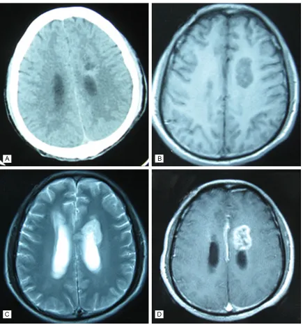
[image:2.612.92.522.370.703.2]T2 fluid-attenuated inversion recovery. Enha- nced MRI scanning showed a heterogeneous and annular enhancement (Figure 3). The lesion was not treated. He also didn’t receive any adjuvant therapy. At 3 months of follow-up, the muscle strength and sensation of both legs did not rehabilitate.
Discussion
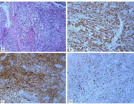
PXA is a rare glial neoplasm, originating from subpial astrocytes, which has relative favorable outcomes due to WHO grade II designation. The overall survival rate at 5 years was 74% [5]. PXA often occurs in the first three decades of life. There is no sex predilection. PXA commonly (hematoxylin and eosin; original magnification, ×200); B: Immunohistochemical examination shows that GFAP was positive (original magnification, ×200); C: Immunohistochemical examination shows that S-100 was positive (origi
[image:3.612.90.525.118.585.2]-nal magnification, ×200); D: Approximately 2% of tumor cells are positive for Ki-67 (origi-nal magnification, ×200).
occurs in supratentorial location, especially the temporal lobe [5]. Seizure is the most common symptom, and headache may also present. Histologically, PXA is characterized by spindle cells, multinucleated giant cells and foamy lip-id-laden xanthomatous cells. PXA is positive for GFAP by immunohistochemistry. Mitosis and necrosis is commonly absent. Typical radiologic findings of supratentorial lesions are cystic neoplasm companied by mural nodule. On CT scanning, PXA shows iso-dense to hypodense. Calcification is uncommon. The cystic mass shows hypointense on T1 weighted imaging, and hyperintense on T2 weighted imaging. Mural nodule is strongly enhanced after the administration of gadolinium. Peritumor edema was minimal.
Supratentorial PXAs are the most common. Lesions of other locations were also reported, such as cerebellum, spine and pineal body. Spinal PXAs are extremely rare. Herpers firstly reported a 66-year-old patient with spinal PXA in 1994 [6].Only six patients were presented in the previous literatures until now [2, 5-9]. The age distribution was 12.9-66 years old with an average age of 42.0 years old. Most of the lesions were located in cervical or thoracic cord.
Single tumor is the most common. Most of the reports thought that surgery should be the first choice. The patients could have a favorable outcome after gross total resection [6]. However, some patients suffered recurrence and anaplastic transformation. And some patients suffered dissemination by meninges or cerebrospinal fluid.
Multicentric PXAs were reported rarely at diag-nosis. In 2005, Saikali firstly reported multicen-tric PXA in a 30-year-old patient with NF1 [10]. At the same year, McNatt reported a 13-year-old synchronous multicentric PXA [11]. In 2012 Gardiman firstly reported a multicentric PXA with cerebrospinal dissemination to cauda equine [3]. It is impossible to confirm that the two lesions represent the same histopathologi-cal entity in our patient. But the intracranial lesion is similar to PXA by radiology, and the intracranial lesion was periventricular. We con-clude that this case can be considered as mul-ticentric tumor. To our knowledge, this is the first case with dissemination to cauda equine at first diagnosis in an aged patient.
Conclusion
Though poor growth in biological behavior and relative favorable outcome, physicians should be realized multicentric dissemination by meninges or cerebrospinal fluid in PXA patients. It is important to describe the particular case in order to better understanding of clinical and radiological features.
Disclosure of conflict of interest
None.
Address correspondence to: Dr, Xintong Zhao, Department of Neurosurgery, Yijishan Hospital, Wannan Medical College, Wuhu, Anhui Province, P.R. China. Tel: +86 5535739594; E-mail: zhaoxin- tong30@163.com
References
[1] Kepes JJ, Rubinstein LJ, Eng LF. Pleomorphic
xanthoastrocytoma: a distinctive meningocer-ebral glioma of young subjects with relatively favorable prognosis. A study of 12 cases. Cancer 1979; 44: 1839-1852.
[2] Das S, Yip S, Hukin J, Cochrane D, Dunham C. Pleomorphic xanthoastrocytoma of the spinal cord: case report and literature review. Clin Neuropathol 2014; 33: 190-196.
[3] Gardiman MP, Fassan M, Orvieto E, Iaria L, Calderone M, Mardari R, DAavella D, Perilongo G. A 14-year-old girl with multiple tumors. Brain Pathol 2012; 22: 865-868.
[4] Katayama K, Asano K, Shimamura N, Ogasawara Y, Naraoka M, Ohkuma H, Kurose
A. Case of pleomorphic xanthoastrocytoma with anaplastic features in the pineal gland. Brain Tumor Pathol 2013; 30: 242-246. [5] Fouladi M, Jenkins J, Burger P, Langston J,
Merchant T, Heideman R, Thompson S, Sanford
A, Kun L, Gajjar A. Pleomorphic xanthoastrocy-toma: favorable outcome after complete surgi-cal resection. Neuro Oncol 2001; 3: 184-192. [6] Herpers MJ, Freling G, Beuls EA. Pleomorphic
xanthoastrocytoma in the spinal cord. Case re-port. J Neurosurg 1994; 80: 564-569. [7] Gill M, Pathak HC, Madan R, Bhattacharya S,
Choudhary GS. Primary spinal pleomorphic xanthoastrocytoma. Neurol India 2010; 5: 771-773.
[8] Nakamura M, Chiba K, Matsumoto M, Ikeda E, Toyama Y. Pleomorphic xanthoastrocytoma of the spinal cord. Case report. J Neurosurg Spine 2006; 5: 72-75.
pleomorphic xantoastrocytoma. Case report. Neurocirugia (Astur) 2010; 21: 390-395. [10] Saikali S, Le Strat A, Heckly A, Stock N,
Scarabin JM, Hamlat A. Multicentric pleomor-phic xanthoastrocytoma in a patient with
neu-rofibromatosis type 1. Case report and review
of the literature. J Neurosurg 2005; 102: 376-81.