Starch Nanoparticles – Two Ways of their Preparation
David CHENA ALDAO 1, Evžen ŠÁRKA1*, Pavel ULBRICH 2 and Eva MENŠIKOVÁ1
1Department of Carbohydrates and Cereals and 2Department of Biochemistry and Microbiology, Faculty of Food and Biochemical Technology, University of Chemistry
and Technology Prague, Prague, Czech Republic *Corresponding author: sarkae@vscht.cz
Abstract
Chena Aldao D., Šárka E., Ulbrich P., Menšiková E. (2018): Starch nanoparticles – two ways of their preparation. Czech J. Food Sci., 36: 133–138.
Starch nanoparticles (SNP) originate from the disruption of the semi-crystalline structure of starch granules. They are very useful in food packaging technology because they increase the mechanical and water vapour resistance of the matrix as well as hinder its recrystallisation during storage in high humidity atmospheres. In medicine, SNP are suitable as carriers in modulated drug delivery for immobilized bioactive or therapeutic agents. Depending on the method of preparation, nanoparticles with different physicochemical, technical or mechanical properties can be obtained. Two different methods of preparation were characterized and compared in this work: the first involving acid hydrolysis of the amorphous part of a starch molecule and the second focusing on the debranching of starch by enzymatic treatment with pullulanase.
Keywords: acid hydrolysis; crystallisation; enzymolysis; nanobiotechnology; short-chain amylose
Supported by the Ministry of Agriculture of the Czech Republic, Project No. QJ1310219, and from specific uni-versity research, Project No. MSMT 20-SVV/2017.
Starch is a polysaccharide that serves as a supply of energy in plants in the form of semicrystalline starch granules which are insoluble in water at normal temperatures. Starch is composed of two polymers of d-glucose, normally 20–30% of amylose with linear structure and 70–80% of branched amylopectin.
The properties and synthesis of starch-based na-noparticles have been extensively studied in recent years due to great interest in biomedical applications, e.g., their use as drug delivery carriers. Nanocarrier drug delivery systems can increase drug solubility and lead to controlled release and/or drug targeting.
Another industrial application is in their employment as filler in food packaging, as they are environmen-tally friendly because the amount of required plastics would then be reduced. One of the advantages of food packaging with bioactive materials is the possibility to use them for nanoencapsulation, which permits the controllable release of active compounds. Furthermore,
they can help control oxidation of foodstuffs and prevent the formation of off-flavours and undesirable food textures (Sozer & Kokini 2009). According to Li et al. (2014) starch nanocrystals prepared from waxy maize starch could adsorb at the oil-water interface to stabilize oil-in-water emulsions containing liquid paraffin with high coalescence stability.
Under specific conditions, amylopectin can be debranched in the presence of debranching enzymes e.g. pullulanase. The chain length distribution of the debranched amylopectin is strongly related to its crystalline structures and influences tempera-ture sensitivity, which differs from species to spe-cies. Angellier et al. (2004) used acid hydrolysis of waxy maize starch granules in order to optimize the preparation of aqueous suspensions of starch nanocrystals.
B- type starches with relatively short and long chains are less sensitive to temperature than the C- type starches, and they maintain their own crystalline structures even when subjected to a relatively large temperature change (Hizukuri 1985). Native starch granules may give A-, B-, C-, and V-type X-ray dif-fraction patterns. The A-type crystalline structure is characteristic of cereal starches, where hydrogen bonds between hydroxyl groups of the amylopectin chains lead to the formation of an outer double helix, whereas the B-type structure is found in potato starch where the amylopectin chains form a closed circle, with water molecules inside the cavity (Hizukuri 2006). The C-type polymorph is a mixture of the A- and B- type polymorphs and occurs in some tuber and legume starches (Bogracheva et al. 1998). The V-type diffraction pattern occurs in fully ungelatinised starch granules; for example, it is characteristic of amylose bound in different complexes, or it can be induced by ther-mal and wet processing (18–45% humidity, 90–130°C) like in the extrusion process (Biliaderis 2009).
MATERIAL AND METHODS
Material used for the preparation of nanoparticles consisted of native wheat starch supplied by the Amylon Havlíčkův Brod starch company (Czech Republic) and maize waxy starch supplied by Ro-quette Italia S.P.A. Pullulanase Optimax L-1000 (E.C.3.2.1.41, 1000–1260 ASPU/g) was provided by Genencor (Belgium).
Preparation of starch nanoparticles by the acid hydrolysis method. A mixture of 117.5 g of wheat or waxy maize starch and 800 ml of sulfuric acid 3.16 mol/l was placed in a reactor submerged in an oil bath and heated to a temperature of 40°C with a stirring speed of 100 rpm. After 7 days the reac-tion was stopped, and the suspension was washed with distilled water by successive centrifugation at 5000 rpm (2236 g) for 10 min until neutralization was achieved. The final suspension was refrigerated at 6°C for one week (Angellier et al. 2004).
Preparation of starch nanoparticles by enzymatic treatment with pullulanase. A suspension of 5% w/v of starch in a sodium acetate buffer (pH 4.5) was pre-pared and heated at a temperature of approximately 80–90°C with constant stirring within 15 minutes. After the heating process, the suspension was transferred to an autoclave and incubated at 121°C for 60 min, where the process of starch gelatinization took place.
After gelatinization, the suspension was cooled and 1 ml of pullulanase (1000 ASPU/g of dry starch) was added at 55°C. After 24 hours the reaction was stopped and the suspension was put into a boiling water bath for 30 min to deactivate the enzyme, followed by 2 h spontaneous cooling to laboratory temperature. Subsequently, the samples EM1, EM2 and EM3 were stored in the refrigerator at 4°C. The samples were repeatedly treated as follows:
EM1: refrigeration (4°C, 24 h), autoclaving (121°C, 60 min), cooling to laboratory temperature (2 h), refrigeration (4°C, 24 h); EM2: refrigeration (4°C, 48 h), autoclaving (121°C, 60 min), cooling to labo-ratory temperature (2 h), refrigeration (4°C, 48 h), autoclaving, cooling (2 h), refrigeration (4°C, 48 h), autoclaving, cooling (2 h), refrigeration (4°C, 24 h); EM3: refrigeration (4°C, 72 h), autoclaving, cooling to laboratory temperature (2 h), refrigeration (4°C, 72 h), autoclaving, cooling (2 h), refrigeration (4°C, 72 h), autoclaving, cooling (2 h), refrigeration (4°C, 24 h).
Morphology and average particle size. The starch nanoparticle suspension, observed using a trans-mission electron microscope (TEM) operating at 100 kV, was further diluted to a 0.2% concentration, and then ultrasonic treatment was applied for 5 min prior to brief sonication by needle, which consider-ably reduced the aggregation of the nanoparticles. Subsequently, the suspension of starch nanoparticles was deposited onto a glow discharged carbon-coated microscopy grid and allowed to partially dry. The excess liquid was immediately blotted with filter paper and the remaining film was negatively stained for 2–3 min with a drop of 2% (w/v) aqueous solution of uranyl acetate for observation and photographing.
X-ray diffraction. The crystalline structure of the starch nanoparticles was analysed using an X’Pert Pro θ-θ powder diffractometer (PANalytical, The Netherlands) with parafocusing Bragg-Brentano geometry using Cu radiation (U = 40 kV, I = 30 mA). Data evaluation was performed by the HighScore Plus software package.
RESULTS AND DISCUSSION
After the acid-treatment process, the starch granules were destroyed and degraded to nanoparticles with the size range of 30–80 nm (Figure 1), which coincides with the results published by Chen et al. (2008).
Starch nanoparticles obtained after enzymatic treatment with pullulanase. Waxy maize starch nanoparticles had globular shapes with diameters of approximately 5–25 nm (Figure 2). The results were similar to those of Qiu et al. (2016).
As can be seen in the TEM images of starch nano-particles (Figure 3), most of the SNP showed irregular shaped particles as platelets of 15–45 nm that were well-dispersed without aggregation.
The formation of nanoparticles was mainly due to the recrystallisation of linear glucans after the enzymolysis of native starch. The linear glucans were easily susceptible to retrogradation. Therefore, the formation process of the SNP through
crystalliza-tion consisted of two steps: firstly, associating linear glucans into double helices and forming clusters with hydrogen bonds; and secondly, the rearrangement of clustering into the SNP (Sun et al. 2014).
X-ray diffraction pattern. The X-ray diffraction patterns characteristic of native waxy starch and EM samples are presented in Table 1. Native waxy starch showed a typical A-type crystalline structure, with peaks at 2θ of about 15, 17, and 23°. This observa-tion is in agreement with the data of Cai and Shi (2010). All EM samples displayed a typical V-type crystalline structure with main diffraction peaks at 2θ of 19 and 22°.
[image:3.595.67.533.97.280.2]Similar findings were also previously reported by Kim and Lim (2009), who found that the SNP prepared by complex formation with n-butanol and enzymatic hydrolysis exhibited a V-type XRD pattern with four sharp reflections at 2θ = 7.4, 13.1, 19.8, and 22.5°. Figure 1. Transmission electron microscopic images of starch nanoparticles prepared by acid hydrolysis of (A) waxy maize starch (WK1) and (B) wheat starch
Figure 2. Transmission electron microscopic images and the particle size of short-chain amylose (A) EM1 and (B) EM2
(A) (B)
[image:3.595.67.534.574.746.2]As shown in Figure 4, the crystallinity of the EMx samples obviously decreased compared to their na-tive counterparts, indicating that a more amorphous crystalline structure was formed after the pullulanase hydrolysis (Liu et al. 2015).
Pullulanase can selectively cleave 1,6-α-d-glycosidic bonds, leading to the formation of short linear glucan chains. After the pullulanase hydrolysis, the contents of short linear glucan chains visibly increased in all of the starch samples.
XRD patterns of the native starch and the acid hy-drolysed starch nanoparticles are shown in Figure 5. Native starches showed the typical A- and V-patterns of crystalline structure as reported. Waxy and nor-mal maize starches exhibited diffraction peaks at Bragg angles (2θ) of 15, 17 and 23° representing the A-type crystalline structure, whereas the peak at 19.9° represents the V-type crystalline structure.
For waxy maize starch, a weak peak appeared at 19° (2θ), which was characterised as a V-type crystal. The nanoparticles of the WK1 maize starch showed a slight
increase in peak intensity, mainly in characteristic peaks of A-type crystalline structure, including the mean peak that characterises a V-type crystal. The main diffraction peaks at 17° and 23° (2θ) of the waxy maize starch still remained after acid hydrolysis (Kim et al. 2012).
Acid hydrolysis may increase the degree of crystal-linity because it is assumed to preferentially cleave the starch chains situated in the amorphous regions (Kim
et al. 2013).
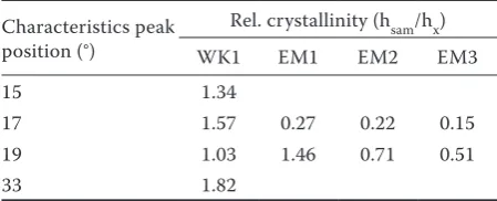
The relative crystallinity and the type of diffraction pattern of native waxy maize starch and the SNP are summarized in Table 2.
Compared with native starch, the relative crystal-linity of starch after the acid hydrolysis increased, but the crystallinity of the waxy starch after the enzymatic hydrolysis decreased. One way to increase the crystal-linity of starch after debranching the amylopectin bonds by enzyme hydrolysis is through its recrystallisation (Sun et al. 2014).
[image:4.595.64.532.137.268.2]The crystallinity was evaluated using a method based on the intensity of the heights of the relevant peaks by searching for their local minima. The latter were inter-connected by a straight line on which the intensity of the peak height was measured. The sample values were compared with the main responses of the RTG wheat starch and the RTG waxy starch using the following Equation (1):
[image:4.595.65.291.566.737.2]Figure 3. Transmission electron microscopic images and the particle size of platelets
Table 1. Characteristics of crystallinity of native waxy maize starch after acid hydrolysis (WK1) and short-chain amylose (EMx)
Crystallinity Characteristics peak position (°) Sample (intensity)
waxy maize starch WK1 EM1 EM2 EM3
A-type 15 2572 3449
A-type 17 5569 8775 1524 1260 850
V-type 19 490 508 716 351 250
V-type 22 478 501 442
A-type 23 6243 9266
A-type 30 805 1155 358
B-type 33 964 1756
Table 2. Degree of crystallinity of native waxy maize starch after acid hydrolysis (WK1) and short-chain am-ylose (EMx)
Characteristics peak position (°)
Rel. crystallinity (hsam/hx)
WK1 EM1 EM2 EM3
15 1.34
17 1.57 0.27 0.22 0.15
19 1.03 1.46 0.71 0.51
[image:4.595.304.529.673.764.2]Dc = hsam/hx (1)
where: Dc – degree of crystallinity; hsam/hx – ratio of the sample response height to the response of native wheat starch or waxy starch (Šárkaet al. 2011)
CONCLUSIONS
This study indicated that the short-chain amylose obtained by hydrolysis with pullulanase presented mainly the V-type crystallinity, and its crystallinity was less resistant than that of the starch nanoparti-cles obtained by acid hydrolysis. The recrystallisa-tion increased the crystallinity of the nanoparticles. However, the longer the recrystallisation period (48 and 72 h), the lower the number of crystals obtained. Thanks to the relatively high crystal-linity of the starch nanoparticles obtained by acid
hydrolysis, they can withstand high temperatures encountered in food processing because they are highly stable.
References
Angellier H., Choisnard L., Molina-Boisseau S., Ozil P., Dufresne A. (2004): Optimization of the preparation of aqueous suspensions of waxy maize starch nanocrystals using a response surface methodology. Biomacromol-ecules, 5: 1545–1551.
Biliaderis C. (2009): Structural transitions and related physical properties of starch. In: BeMiller J.N., Whistler R.L. (eds): Starch Chemistry and Technology. London, Academic Press: 293–372.
[image:5.595.80.503.93.289.2]Bogracheva T.Y., Morris V.J., Ring S.G., Hedley C.L. (1998): The granular structure of C-type pea starch and its role in gelatinization. Biopolymers, 45: 323–332.
[image:5.595.81.496.316.502.2]Figure 5. X-ray diffraction of native waxy maize starch and WK1 after acid hydrolysis
Cai L., Shi Y.C. (2010): Structure and digestibility of crystal-line short-chain amylose from debranched waxy wheat, waxy maize, and waxy potato starches. Carbohydrate Polymers, 79: 1117–1123.
Chen Y., Cao X., Chang P., Huneault M.A. (2008): Compara-tive study on the films of poly(vinyl alcohol)/pea starch nanocrystals and poly(vinyl alcohol)/native pea starch. Carbohydrate Polymers, 73: 8–17.
Hizukuri S. (1985): Relationship between the distribution of the chain length of amylopectin and the crystalline structure of starch granules. Carbohydrate Research, 141: 295–306.
Hizukuri S., Abe J., Hanashiro I. (2006): Starch: analytical aspects. In: Eliasson A. (ed.): Carbohydrates in Food. London, New York, CRC Press: 366–367.
Kim J.Y., Lim S.T. (2009): Preparation of nano-sized starch particles by complex formation with n-butanol. Carbo-hydrate Polymers, 76: 110–116.
Kim H.Y., Lee J.H., Kim J.Y., Lim W.J., Lim S.T. (2012): Char-acterization of nanoparticles prepared by acid hydrolysis of various starches. Starch/Starke, 64: 367–373.
Kim H.Y., Han J.A., Kweon D.K., Park J.D., Lim S.T. (2013): Effect of ultrasonic treatments on nanoparticle prepara-tion of acid-hydrolyzed waxy maize starch. Carbohydrate Polymers, 93: 582–588.
Li C., Li Y., Sun P., Yang C. (2014): Starch nanocrystals as particle stabilisers of oil-in-water emulsions Journal of the Science of Food and Agriculture, 94: 1802–1807. Liu G., Hong Y., Gu Z., Li Z., Cheng L. (2015):
Pullula-nase hydrolysis behaviors and hydrogel properties of debranched starches from different sources. Food Hy-drocolloids, 45: 351–360.
Qiu C., Qin Y., Zhang S., Xiong L., Sun Q. (2016): A com-parative study of size-controlled worm-like amylopectin nanoparticles and spherical amylose nanoparticles: Their characteristics and the adsorption properties of polyphe-nols. Food Chemistry, 213: 579–587.
Sozer N., Kokini J.L. (2009): Nanotechnology and its applica-tions in the food sector. Trends in Biotechnology, 27: 82–89. Sun Q., Li G., Dai L., Ji N., Xiong L. (2014): Green prepara-tion and characterisaprepara-tion of waxy maize starch nanopar-ticles through enzymolysis and recrystallisation. Food Chemistry, 162: 223–228.
Šárka E., Maixner J., Weider M., Smrčková P., Bubník Z. (2011): Shape and crystallinity of potato starch granules after wet ultrafine grinding. In: Proceedings 7th Interna-tional Conference on Polysaccharides-Glycoscience, Nov 2–4, 2011, Prague, Czech Republic: 177–179.