Original Article
Exposure to cigarette smoke alters AgNOR number
and HIF-1alpha expression in colorectal
tubular adenocarcinoma in rats
Leonardo Oliveira Trivilin1, Diego Camuzi Cassiano2, Suzanny Oliveira Mendes3, Aline Ribeiro Borçoi3,
Anderson Barros Archanjo3, Ester Ribeiro Cunha4, José Zago Pulido5, Jankerle Neves Boeloni1, Adriana
Madeira Alvares da Silva Conforti4
Departments of 1Veterinary, 4Biology, Universidade Federal do Espírito Santo (UFES), Alegre, Espírito Santo, Brazil; 2Instituto Federal do Espírito Santo (IFES), Alegre, Espírito Santo, Brazil; 3RENORBIO Graduate Program, Vitória, Espírito Santo, Brazil; 5Evangelic Hospital of Cachoeiro de Itapemirim, Cachoeiro de Itapemirim, Espírito Santo, Brazil
Received October 29, 2016; Accepted January 6, 2017; Epub March 1, 2017; Published March 15, 2017
Abstract: The relationship between exposure to tobacco and colorectal cancer development emerges in the long-term, leading to hypoxia and changes in the number of nucleolar organizer regions. There is evidence that this
exposure influences on tumor characteristics, culminating in malignancy and poorer prognosis. This article aims at evaluating the influence of cigarette smoke exposure in HIF-1alpha hypoxia marker expression and AgNOR count, as
well as the relationship between these two markers and tubular adenocarcinoma differentiation level in an experi-mental colorectal cancer model. Rats were induced colorectal cancer through 1, 2-dimethylhydrazine and randomly allocated into two groups: exposed and control. Exposed group was then, directly exposed to burning cigarette
smoke. Tubular adenocarcinoma obtained was subjected to AgNOR counting technique and immunoblotted for HIF-1alpha protein. Smoke exposed groups had lower AgNOR numbers (P = 0.00017) and HIF-HIF-1alpha highest score (P = 0.00017). The average relationship between AgNOR and HIF-1alpha is weak in both groups and the differentiation level is not influenced by the AgNOR count in both groups. Notwithstanding this weak relationship, well-differentiat
-ed tumors in the control group have shown HIF-1alpha higher scores. In the smoke expos-ed group, as HIF-1alpha score increases, the tumor differentiation grade decreases. Thus, HIF-1alpha and AgNOR have shown themselves
as biomarker study targets for diagnosis, prognosis and treatment response in colorectal tubular adenocarcinomas, since they tended to be related to the degree of tumor malignancy.
Keywords: NOR, carcinogenesis, neoplasia, hypoxia, rat, smoking
Introduction
According to World Health Organization [1], smoking is the leading cause of preventable death in the world and it is estimated that one third of the adult population, more than one bil-lion people, are smokers. Tobacco is responsi-ble for one death every six seconds and one in every ten deaths in adults, totaling 5.4 million deaths each year worldwide [1].
Furthermore, smoking is a risk factor for de- veloping gastrointestinal cancer, including the oral cavity, esophagus, stomach, ileum and colon [2-6]. Specifically, colorectal cancer is the fourth most common cause of death worldwide with 694,000 incidents a year [1].
The influence of tobacco in colorectal cancer is clearer in the long term, since smoking for at least 20 years is significantly related to the emergence of small polyps. When that expo-sure exceeds 20 years, it is associated with large polyp’s appearance and over 35 years exposure contributes to colorectal carcinoma onset [6].
Scientific Use of Animals (#11794/2008). The experimental procedures were reviewed and approved by the internal Ethics Committee under protocol #003/2014.
Experimental design
The experiment used 24 male Wistar adult rats, kept in the Centre for Development of Experimental Models of Universidade Federal do Espírito Santo. Animals were housed in the number of 6 per cage under controlled temper-ature (21-24°C), humidity (45-55%) and lighting (12 h light, 12 h dark; lights on at 6:45 AM). Food and water were available ad libitum throughout the experiments.
All animals were induced colorectal cancer through 1, 2-dimethylhydrazine (DMH). DMH was dissolved in 0.9% NaCl containing 1.5% EDTA as a vehicle, adjusted to a final pH of 6.5 with 1 N NaOH solution and administered sub-cutaneously once a week for five weeks at a dose of 65 mg/kg body weight. This protocol was chosen based on previous data [23].
The 24 animals were then randomly divided into 2 groups, one directly exposed to smoke from burning cigarettes (exposed group) and another unexposed (control group). Exposure to cigarette smoke was started simultaneously with the induction of carcinogenesis and was carried out for 20 weeks in inhalation cham-bers equipped with trademark smoke puff. After exposure phase, the animals were eutha-nized by anesthetic induction with a ketamine and xylazine association followed by the injec-tion of supersaturated potassium chloride solu-tion. They were then subjected to necropsy and their intestines were removed from the ceccum to the anus and opened with scissors at the mesenteric insertion for the removal of tumors. The collected tissue was fixed in 10% buffered formalin solution and processed according to paraffin embedding routine. Following this stage, the blocks were cut in 3 μm thick histo-logical sections, stained with hematoxylin and eosin and diagnosed according to Perše and Cerar [24].
For assessing the differentiation degree, AgNOR count and HIF1-alpha expression, the slides were analyzed in an optical microscope In mice with colorectal cancer induced by 1,
2-dimethylhydrazine (DMH), a greater number of AgNOR (Argyrophilic Nucleolar Organizer Region) was found in tumors when compared to normal mucosa [9]. In this regard, DMH experi-mental models are widely used and share many similarities with colorectal cancer in humans, including response to inductive and preventive agents [10].
AgNOR count can be related to cell proliferation and differentiation. Specifically, a larger num-ber of AgNOR in cell nucleus is linked to a lower cell differentiation degree and poorer tumor prognosis [11, 12].
Smoking for 10 minutes reduces the oxygen tension in tissues for approximately one hour [13]. In addition, a significant increase in hypox-ia-inducing factors in cells exposed to cigarette smoke components were pointed out [14]. Cells undergoing hypoxia tend to decrease their divi-sion rate [15-17] and even shutdown the cell cycle in some tumors [18]. Low oxygen tension also alters cellular homeostasis, leading to HIF-1alpha protein activation, which expression is highly regulated by oxygen concentration and, for this reason, has been widely used as a main hypoxia marker [19-21]. HIF-1alpha expression in solid tumors may contribute to malignancy and aggressive behavior [22].
Given that exposure to cigarette smoke may induce changes in AgNOR and HIF-1alpha markers and these affect tumor characteristics such as malignancy and prognosis, this study aims to evaluating the influence of exposure to smoke on HIF-1alpha expression and the AgNOR count. It also intends to verify the rela-tionship between these two markers, as well as the degree of colorectal tubular adenocarci-noma differentiation in an experimental model for colorectal cancer with DMH in order to assist other studies that use biomarkers as a diagnostic and prognostic tool.
Materials and methods
Ethical aspects
cells, in at least three fields of the neoplastic region, ignoring the edge and its adjacent zones.
Immunohistochemistry
The same tumors used for the AgNOR stain- ing technique were used for immunohistochem-istry. Paraffin-embedded blocks were sec-tioned and mounted on silanized slides. The 3-µm sections were deparaffinized in xylene and rehydrated through three baths in absolute alcohol. Slides were rinsed with deionized wa- ter and subjected to antigen retrieval with sodi-um citrate at high temperature for 15 minutes. Next, the slides were washed in 1× TRIS and endogenous peroxidases were blocked with 30% hydrogen peroxide in 1× TRIS for 20 min-utes at 25°C. After three 5-minute washes in 1× TRIS, slides were incubated in nonspecific protein blocking solution (3% milk powder dilut-ed in 1× TRIS) for 60 minutes at 25°C and sub-jected to more three 5-minute washes in 1× TRIS.
Control (no primary antibody) and experimental slides were incubated at room temperature, respectively, in 1× TRIS or antibody diluting solution with Anti-HIF-1alpha antibody (1:2000, [EP1215Y], AB51608, Abcam, Cambridge MA) for 60 minutes. After three 5-minute washes in 1× TRIS, slides were incubated with detection system for rat tissue (N-Histofine-Simple Stain Max PO Rat, 414191F, NICHIREI®) at 25°C for
30 minutes. After another three washes with onex TRIS, staining was visualized with peroxi-dase-sensitive Sigma fast 3, 3’-Diaminoben- zidine (DAB, Sigma, St. Louis, MO). All slides were counterstained with Harris Hematoxylin for 1 minute, dehydrated in ethanol, cleared in xylene and mounted with Permount (Fisher Scientific, Pittsburgh, PA).
The slides were analyzed in an optical micro-scope and sample scoring was performed by semi quantitative analysis, considering the number of stained cells and signal intensity. Considering the percentage of HIF-1alpha immune-positive cells, a score of 0 was given when all cells were negative; 1. when 1-25% of cells were positive, 2. when 25-50% of cells were positive and 3. when > 50% of cells were positive. Signal intensity was scored as nega-tive (0), weak (1), moderate (2) and strong (3). Both scores were multiplied according to Soini by two pathologists, who received the same
slides for analysis independently, without previ-ous discussion of the findings. Their index of agreement was 99.1%, calculated according to the following formula: [AGREEMENT/(AGREED + DISAGREED)] × 100.
The degree of differentiation of each sample was determined by subjective analysis, consid-ering the number of glands according to Fleming and cols. [25] to human colorectal tumors. That scale considers tumors formed by over 95% of glands as well differentiated; moderately differ-entiated tumors display between 50-95% of glands; and poorly differentiated tumors are composed mostly of solid parts with less than 50% of glandular formation. The lesions were then classified as benign or malignant neo-plasm depending on their differentiation grade (well differentiated - 1, moderately differentiat-ed - 2 and poorly differentiatdifferentiat-ed - 3).
Staining and AgNOR count
From the selected sample, 3 μm thick histologi-cal sections were cut and then subjected to AgNOR staining technique as described by Ploton and cols. [26] with modifications.
[image:3.612.91.299.134.290.2]The NOR count with silver staining (AgNOR) was performed with an optical microscope at 100× magnification under immersion. On each slide, AgNOR number was counted in 100 mucosa
Table 1. Number and percentage of intestinal mucosal lesions in an experimental model of colorectal cancer between exposed and not exposed to tobacco smoke groups according to their diagnoses
Lesion diagnosis
Control
Group Exposed Group
n % n %
Mild dysplasia - - 1 3.03
Moderate dysplasia 2 8.0 3 9.09
High grade dysplasia - - 1 3.03
Carcinoma in situ 1 4.0 5 15.15
Tubular adenoma 2 8.0 -
-Signett-ring cell adenocarcinoma 2 8.0 3 9.09
Tubular adenocarcinoma 17 68.0 20 60.61
Mucinous adenocarcinoma 1 4.0 - -Total 25 100 33 100
compared to the control group (P < 0.0001), with an average score of 2.94 ± 1.57 on control group and 5.57 ± 1.72 on exposed groups. All differences are illustrated in Figure 1.
Relationship between HIF-1alpha and AgNOR count and between both and the differentia-tion grade in colorectal tubular adenocarci-noma
In the control group, relationship between AgNOR average and HIF-1alpha score is weak negative (r = -0.2211; P = 0.3937), as well as in the exposed group (r = -0.1680; P = 0.4667), but not significant (Figure 2A and 2D), as only 4.89% (control group) and 2.82% (exposed and cols. [27] and Campos and cols. [28] and
the resulting score was used in our analyses. Statistical analysis
The data were submitted to analysis of vari-ance, and the means of the treated samples and controls were compared using two-tailed Student’s t-test (Program R©, 2015). Differences
[image:4.612.90.376.71.415.2]were considered significant for P < 0.05. This test was used for AgNOR and HIF-1alpha score. The Pearson Correlation test was used to eval-uate the relationship between AgNOR and HIF-1alpha score, and then, its relationship with differentiation grade. Software GraphPad Prism 6 Demo was used for the drawing of graphics.
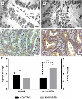
Figure 1. AgNOR average and HIF-1alpha score in colorectal adenocarcino -ma tubular from experimental model for colorectal cancer exposed and no exposed to smoke cigarette. (A) Sample derived from control group and (B) sample belonging to exposed group showing the difference in the AgNOR number on cells (arrow). AgNOR staining. (C) Sample from control group
clas-sified with HIF-1alpha score 3. (D) Sample from exposed group clasclas-sified with HIF-1alpha score 6. IHQ, DAB. (E) Significative difference between con -trol and exposed groups by Student t test (**AgNOR average, P = 0.00017;
***HIF-1alpha score, P < 0.0001), α = 0.05.
Results
All animals used in this exper-iment showed lesions in colorectal mucosa. In the exposed group, the minimum number of lesions was one and the maximum number of lesions found was nine, with an average of 2.75 injuries per animal. In the control group, 25 lesions were found with a mean of 2.08 per ani-mal injuries. In this group, the minimum and maximum num-ber of lesions was one and eight, respectively. Table 1 shows the number of lesions and their diagnoses from which only Tubular Adenocar- cinomas were selected, given their homogeneous charac-teristics, and due to the fact of being the most frequent injuries.
AgNOR number and HIF-1al-pha expression in colorectal tubular adenocarcinoma
predicting the prognosis of smokers who have this type of tumor. Thus, our study evaluated HIF-1alpha expression and AgNOR count in an experimental model of colorectal cancer ex- posed to smoke from direct burning cigarette. A previous study showed increased average number of AgNOR in colorectal cancer experi-mental model induced by DMH compared to the uninduced control group [9]. Our results showed that the average AgNOR count in colorectal adenocarcinoma induced by DMH group followed this increasing trend. However, when exposed to cigarette smoke, colorectal adenocarcinomas had lower average AgNOR count. We believe that the reduction in the mean number of AgNOR found in colorectal adenocarcinoma experimental model exposed to cigarette smoke in our study is related to the tolerogenic influence that exposure to cigarette smoke exerts on colorectal mucosa. That influ-ence was mentioned in a review published by Cabral and Barbosa [30]. Thus, different car-cinogens and co-carcar-cinogens, time and type of exposure, like cigarette smoke, can affect the AgNOR analysis.
Cytological studies with AgNOR count in smok-ers and nonsmoksmok-ers’ oral mucosa showed an increase count in tobacco-exposed group [7, group) of change in AgNOR number can be
explained by HIF-1alpha score.
The colorectal tubular adenocarcinoma differ-entiation grade in control group has shown little influence from AgNOR number (r = 0.1530, P = 0.5576), that is, only 2.34% of AgNOR is relat- ed to the worst degree of tumor differentiation. In the exposed group, relationship between AgNOR and tumor differentiation degree is much lower (r = 0.0861, P = 0.7106) and only 0.74% of AgNOR count can be related to the worst tumor differentiation degree in this group (Figure 2B and 2E).
In the control group, tumors seem well differen-tiated according to HIF-1alpha score increases, but this ratio was only 1.27% (r = -0.1126, P = 0.6670). However, in the exposed group, 4.07% of HIF-1alpha score is related to differentiation grade of analyzed tumors, and, the higher the HIF-1alpha score, the lower the tumor differen-tiation grade (r = 0.2017, P = 0.3806) (Figure 2C and 2F).
Discussion
[image:5.612.89.522.71.311.2]The correlation between smoking and colorec-tal cancer has been recently described by Gross and Baranauskas [29] as being impor-tant for selecting diagnostic biomarkers and
Figure 2. AgNOR and HIF-1alpha score relationship and its influence on colorectal tubular adenocarcinoma degree
differentiation in smoke cigarette exposed group and no exposed group in colorectal cancer experimental model.
rats exposed to cigarette smoke underwent hypoxia and that this information should be considered in diagnostic and prognostic issues of colorectal cancer.
Therefore, HIF-1alpha score expression rela-tionship with colorectal tubular adenocarcino-ma differentiation grade was evaluated. It was noticed that in the control group, whose HIF-1alpha expression was lower, there was a bet-ter tumor differentiation grade. However, poorly differentiated tumors were associated with higher scores of HIF-1alpha expression, as observed in the exposed group. Several patho-logical aspects of colorectal cancer as depth of the intestinal wall affected by the tumor, num-ber of lymph nodes containing metastasis and metastasis to distant organs, may interfere with the survival of patients, and are usually related to a poorer prognosis [39, 40]. It has also been reported that high expression of HIF-1alpha in human colorectal cancer is associat-ed with a poor prognosis [41] and as well asso-ciated with increased mortality, regardless of patient clinical characteristics and molecular variables [42]. In this sense, the expression of HIF-1alpha could provide important answers regarding the prognosis of smokers who devel-op colorectal cancer.
In conclusion, both AgNOR and HIF-1alpha have presented themselves as biomarkers, targets for study of diagnosis, prognosis and treatment response in colorectal tubular adenocarcino-mas since they tended to be related to tumor malignancy grade.
Acknowledgements
This work was not supported by operating research grants.
Disclosure of conflict of interest
None.
Address correspondence to: Leonardo Oliveira Tri-
vilin, Department of Veterinary, Universidade Fed-eral do Espírito Santo (UFES), s/n, Alto Universitário,
Guararema, 29500-000, Alegre, Espírito Santo,
Brazil. Tel: +552835528649; Fax: +552835528649;
E-mail: leotrivilin@gmail.com
References
[1] WHO (2016). World Health Organization. [On
-line] Available at: http://www.who.int [Ac
-cessed 21 Feb. 2016].
31]. These results differ from those found in our study in terms of AgNOR count by cigarette smoke exposure, because our assessment was carried out in colorectal mucosa. However, increased AgNOR count in tobacco exposed group oral mucosa is probably related to type of cigarette smoke exposure, which is different from oral mucosa (direct exposure) to colorec-tal mucosa (indirect exposure).
It is known that hazard ratio between smoking and colon cancer is conditioned to the expo-sure period so that as higher the expoexpo-sure, higher the malignancy degree [6]. The differ-ence between the AgNOR averages in groups may be related to tumor malignancy. Yang and cols [32] showed differences in AgNOR average in benign and malignant human colon tumors, as well as Joyce and cols. [33] who reported dif-ference between malignancy degrees of human’s colorectal cancer according to its AgNOR count. In these works, the worst diagno-ses are accompanied by increased AgNOR average. On the other hand, Rüschoff and cols. [34], Rayter and cols. [35] and Yamaguchi and cols [36] have found no difference between AgNOR average and the degree of malignancy in human colorectal tumors. Our results showed that there is a weak relationship between degree of colorectal tubular adenocarcinoma differentiation and AgNOR count in both control group and exposed group.
There is then a similarity between studies us- ing AgNOR quantification as a characterization tool for malignancy and tumor prognosis of colorectal carcinomas. Therefore, a low AgNOR average in tubular adenocarcinomas in the exposed group may not be linked to a low malig-nancy compared to control group, but to other factors such as hypoxia.
[15] Schmaltz C, Hardenbergh PH, Wells A and Fisher DE. Regulation of proliferation-survival
decisions during tumor cell hypoxia. Mol Cell Biol 1998; 18: 2845-2854.
[16] Rohwer N and Cramer T. Hypoxia-mediated
drug resistance: novel insights on the
function-al interaction of HIFs and cell death pathways. Drug Resist Updat 2011; 14: 191-201. [17] Carmeliet P, Dor Y, Herbert JM, Fukumura D,
Brusselmans K, Dewerchin M, Neeman M,
Bono F, Abramovitch R, Maxwell P, Koch CJ,
Ratcliffe P, Moons L, Jain RK, Collen D and
Keshert E. Role of HIF-1alpha in hypoxia-medi -ated apoptosis, cell proliferation and tumour angiogenesis. Nature 1998; 394: 485-490.
[18] Lal A, Peters H, St Croix B, Haroon ZA, Dewhirst MW, Strausberg RL, Kaanders JH, van der Ko -gel AJ and Riggins GJ. Transcriptional response to hypoxia in human tumors. J Natl Cancer Inst 2001; 93: 1337-1343.
[19] Huang LE and Bunn HF. Hypoxia-inducible fac -tor and its biomedical relevance. J Biol Chem 2003; 278: 19575-19578.
[20] Chowdhury R, Hardy A and Schofield CJ. The
human oxygen sensing machinery and its ma-nipulation. Chem Soc Rev 2008; 37: 1308-1319.
[21] Brahimi-Horn C, Mazure N and Pouyssegur J.
Signalling via the hypoxia-inducible
factor-1al-pha requires multiple posttranslational modifi -cations. Cell Signal 2005; 17: 1-9.
[22] Semenza GL. Targeting HIF-1 for cancer thera -py. Nat Rev Cancer 2003; 3: 721-732.
[23] Laranjeira LLS, Taha MO, Ferme A, Lemos R, Plapler H. Localização de lesões tumorais in -duzidas pela 1,2-dimetilhidrazina e seu grau de atipia no cólon de ratos. Acta Cir Bras 1998; 13.
[24] Perse M and Cerar A. Morphological and mo-lecular alterations in 1, 2-dimethylhydrazine and azoxymethane induced colon carcinogen-esis in rats. J Biomed Biotechnol 2011; 2011: 473964.
[25] Fleming M, Ravula S, Tatishchev SF and Wang HL. Colorectal carcinoma: Pathologic aspects.
J Gastrointest Oncol 2012; 3: 153-173.
[26] Ploton D, Menager M, Jeannesson P, Himber G, Pigeon F and Adnet JJ. Improvement in the
staining and in the visualization of the argyro-philic proteins of the nucleolar organizer
re-gion at the optical level. Histochem J 1986; 18:
5-14.
[27] Soini Y, Kahlos K, Puhakka A, Lakari E, Saily M,
Paakko P and Kinnula V. Expression of induc-ible nitric oxide synthase in healthy pleura and in malignant mesothelioma. Br J Cancer 2000; 83: 880-886.
[28] Campos AH, Aldred VL, Ribeiro KC, Vassallo J and Soares FA. Role of immunoexpression of [2] Chen C, Ricks S, Doody DR, Fitzgibbons ED,
Porter PL and Schwartz SM. N-Acetyltransfer-ase 2 polymorphisms, cigarette smoking and alcohol consumption, and oral squamous cell cancer risk. Carcinogenesis 2001; 22: 1993-1999.
[3] Gallus S, Bosetti C, Franceschi S, Levi F, Si -monato L, Negri E and La Vecchia C. Oesopha-geal cancer in women: tobacco, alcohol, nutri-tional and hormonal factors. Br J Cancer 2001; 85: 341-345.
[4] Giovannucci E. An updated review of the epi- demiological evidence that cigarette smoking increases risk of colorectal cancer. Cancer Epi-demiol Biomarkers Prev 2001; 10: 725-731.
[5] Neugut AI, Jacobson JS, Suh S, Mukherjee R and Arber N. The epidemiology of cancer of the small bowel. Cancer Epidemiol Biomarkers Prev 1998; 7: 243-251.
[6] You WC, Zhang L, Gail MH, Chang YS, Liu WD, Ma JL, Li JY, Jin ML, Hu YR, Yang CS, Blaser MJ, Correa P, Blot WJ, Fraumeni JF Jr and Xu GW.
Gastric dysplasia and gastric cancer: helico-bacter pylori, serum vitamin C, and other risk factors. J Natl Cancer Inst 2000; 92: 1607-1612.
[7] Omar GAS, Shamssain M and McGarry K. Mon-itoring histological changes in oral mucosa us-ing AgNORs as biomarkers for oxygenic stress in smokers and COPD patients. IOSR J Pharm 2015; 5: 24-29.
[8] Goodpasture C and Bloom SE. Visualization of nucleolar organizer regions im mammalian chromosomes using silver staining. Chromo-soma 1975; 53: 37-50.
[9] Zhang ZG, Wu JY, Fu XD, Gu DK and Fang F.
P21 and CEA expression and AgNOR counts in dimethylhydrazine-induced colon carcinoma in rats. World J Gastroenterol 1997; 3: 163-165.
[10] Corpet DE and Pierre F. How good are rodent models of carcinogenesis in predicting
effi-cacy in humans? A systematic review and me-ta-analysis of colon chemoprevention in rats, mice and men. Eur J Cancer 2005; 41: 1911-1922.
[11] Pich A, Chiusa L and Margaria E. Prognostic relevance of AgNORs in tumor pathology. Mi-cron 2000; 31: 133-141.
[12] Derenzini M. The AgNORs. Micron 2000; 31: 117-120.
[13] Jensen JA, Goodson WH, Hopf HW and Hunt
TK. Cigarette smoking decreases tissue oxy-gen. Arch Surg 1991; 126: 1131-1134.
[14] Yu H, Li Q, Kolosov VP, Perelman JM and Zhou X. Regulation of cigarette smoke-mediated mu
-cin expression by hypoxia-inducible factor-1α
[36] Yamaguchi A, Tsukioka Y, Kurosaka Y, Nishimu
-ra G, Kanno M, Yonemu-ra Y and Miyazaki I.
Prognostic value of nucleolar organizer regions in endoscopically biopsied tissues of colorec-tal cancers. Oncology 1993; 50: 121-126.
[37] Fahling M. Surviving hypoxia by modulation of
mRNA translation rate. J Cell Mol Med 2009; 13: 2770-2779.
[38] Mekhail K, Rivero-Lopez L, Khacho M and Lee
S. Restriction of rRNA synthesis by VHL main -tains energy equilibrium under hypoxia. Cell Cycle 2006; 5: 2401-2413.
[39] Pereira T Jr, Torres RA, Nogueira AM.
Acometi-mento metastático linfonodal no câncer color -retal. Arq Gastroenterol 2006; 43: 89-93.
[40] Campos FG, Calijuri-Hamra MC, Imperiale AR,
Kiss DR, Nahas SC and Cecconello I. Locally advanced colorectal cancer: results of surgical treatment and prognostic factors. Arq Gastro-enterol 2011; 48: 270-275.
[41] Rasheed S, Harris AL, Tekkis PP, Turley H, Sil -ver A, McDonald PJ, Talbot IC, Glynne-Jones R,
Northover JM and Guenther T. Hypoxia-induc -ible factor-1alpha and -2alpha are expressed in most rectal cancers but only hypoxia-induc-ible factor-1alpha is associated with prognosis. Br J Cancer 2009; 100: 1666-1673.
[42] Baba Y, Nosho K, Shima K, Irahara N, Chan AT,
Meyerhardt JA, Chung DC, Giovannucci EL,
Fuchs CS and Ogino S. HIF1A overexpression
is associated with poor prognosis in a cohort of 731 colorectal cancers. Am J Pathol 2010; 176: 2292-2301.
nitric oxide synthases by Hodgkin and
Reed-Sternberg cells on apoptosis deregulation and
on clinical outcome of classical Hodgkin lym -phoma. Mol Cell Biochem 2009; 321: 95-102.
[29] Gross JL and Baranauskas MVB. Tabaco e
câncer. In: Lopes A, Chammas R, Iyeyasu H, editors. Oncologia para a Graduação. 3. São
Paulo: Lemar; 2013. pp. 196-201.
[30] Cabral P and Barbosa E. Tabacco and inflam -matory bowel disease. Rev Port Coloproctol 2014; 14-22.
[31] Kadivar M and Attar M. Argyrophilic nucleolar organizer region counts in exfoliative cytology of buccal mucosa from opium addicts,
smok-ers and nonsmoksmok-ers. Anal Quant Cytol Histol
2008; 30: 274-278.
[32] Yang P, Huang GS and Zhu XS. Role of nucleo -lar organiser regions in differentiating malig-nant from benign tumours of the colon. J Clin Pathol 1990; 43: 235-238.
[33] Joyce WP, Fynes M, Moran KT, Gough DB, Dervan P, Gorey TF and Fitzpatrick JM. The
prognostic value of nucleolar organiser regions in colorectal cancer: a 5-year follow-up study. Ann R Coll Surg Engl 1992; 74: 172-176; dis-cussion 176-177.
[34] Ruschoff J, Bittinger A, Neumann K and
Schmitz-Moormann P. Prognostic significance
of nucleolar organizing regions (NORs) in carci-nomas of the sigmoid colon and rectum. Pathol Res Pract 1990; 186: 85-91.