Original Article
MiR-194 suppresses human gastric cancer cell
proliferation and induces apoptosis by targeting GGCT
Yi Zhang1, Cong Chen2, Ke Hu3
Departments of 1Colorectal Surgery, 2Neurology, The First Affiliated Hospital of Nanjing Medical University, Nan -jing, Jiangsu, China; 3School of Stomatology, Nanjing Medical University, Nanjing, Jiangsu, China
Received December 19, 2016; Accepted December 27, 2016; Epub March 1, 2017; Published March 15, 2017
Abstract: Background: Gamma-glutamylcyclotransferase (GGCT) has been confirmed to be involved in many kinds
of cancers, while the biological function of GGCT in gastric cancer is still largely unknown. Methods: The expression level of miR-194 was detected in gastric cancer tissues and the adjacent non-tumor tissues as well as the gastric cancer cell lines. The proliferation and apoptosis of MGC-803 cells were detected after transfected with miR-194 mimics or miR-194 mimic control. qRT-PCR and Western blot were performed to detect the expression of related proteins in MGC-803 cells. The target gene of miR-194 was investigated by luciferase assay and Western blot.
Results: The expression level of miR-194 was markedly decreased in gastric cancer patients compared to the
non-tumor people. Up regulation of miR-194 significantly inhibited the proliferation and induced apoptosis of MGC-803
cells. What’s more, the luciferase assay demonstrated that GGCT was a target gene of miR-194, restoration of GGCT could reverse the inhibiting effect of miR-194 on tumor cells proliferation. Conclusions: MiR-194 suppresses tumor development by regulating GGCT expression in gastric cancer and may thus be a potential prognostic marker and a therapeutic target in gastric cancer.
Keywords: Gastric cancer, miR-194, proliferation, apoptosis, GGCT
Introduction
Gastric cancer, as a common cancer, is the fourth common malignant tumor and the sec-ond leading cause of cancer-related death worldwide, especially in East Asia [1-3]. About 951,600 new gastric cancer cases and 723,100 deaths came about in 2012 [4]. The prevalence of gastric cancer is generally about twice as high in men than those in women and varies widely all over the countries. Despite great efforts have been made to improve early diagnosis rate and afford synthesized and advanced treatment methods for patients with gastric cancer, the prognosis of patients with gastric cancer is still very poor as they often experience post-treatment cancer relapses and metastasis [5, 6]. Therefore, a better understanding of the molecular mechanisms underlying gastric cancer development and
metastasis is essential for finding novel thera -peutic strategies.
MicroRNAs, a kind of small endogenous
non-that negatively regulate the post-transcriptio- nal regulation of target genes through their binding to 3’-untranslated regions (UTRs) [7-9].
Currently, many miRNAs has been verified to
play critical roles in regulating the development of tumor processes and tumor metastasis [10-12]. Previous studies reported that miR-194 was dramatically decreased in gastric cancer tissues, and up-regulation of miR-194 could
significantly inhibit migration, invasion and pro -liferation of gastric cancer cells by targeting RBX1 [13]. While, a single miR may have many targets, whether there have other targets need-ed further study.
GGCT, as an important enzyme in the regulation
of a γ-glutamyl cycle by regulating glutathione degradation, can specifically change γ-Glu-AA
absorbance at 450 nm was measured using an electroluminescence immunosorbent assay
reader (Thermo Fisher Scientific, Waltham,
MA).
Apoptosis of gastric cancer cells wasdetected by flow cytometry
Cells were collected and washed twice with cold phosphate-buffered saline solution (PBS)
to remove floating cells then labeled with
Annexin V-FITC (BD Biosciences, San Jose, CA).
Apoptosis were evaluated with a flow cytometry
analyzer. Data were analyzed by Flowjo 7.6 software.
Terminal deoxynucleotidyl transferase (TdT) dUTP nick-end labeling assay
Terminal deoxynucleotidyl transferase (TdT) dUTP Nick-End Labeling (TUNEL) (Roche, Shanghai, China) assay has been used to detect the late stages of apoptosis. Cells were
fixed with 4% paraformaldehyde, permeabilized in 0.2% Triton X-100, and incubated with the
TUNEL detection kit from Roche at 37°C for 1 hour. After that, the samples were mounted in mounting media containing DAPI. Fluorescent
images were captured using fluorescence
microscope at 20× magnification. The total
number of DAPI positive cells and total number of TUNEL positive cells were counted.
RNA extraction and real-time quantitative PCR (qRT-PCR)
Total RNA was extracted from cell lines and clinical samples by using TRIzol Reagent (Invitrogen, Carlsbad, CA, USA) according to
the operating instructions. RNA was quantified
by using UV absorbancies at 260 and 280 nm (A260/280). Subsequently the RNA was reverse-transcribed into cDNA using reverse
transcription system (Thermo Scientific, CA,
USA). The expression level of miR-194 was detected by the ABI PRISM 7500 Sequence Detection System (ABI) using the TaqMan MicroRNA assay kits (Applied Biosystems, California, USA). U6 small nuclear RNA (snRNA) was used as the control normalize. The ex- pression level of GGCT also analyzed by SYBR Green and normalized to GAPDH. The judgment
of primer sequences’ specificity was based on
dissociation curve, 2-ΔΔCt (cycle threshold) was used to calculate the relative gene expression levels.
In this study, we investigated the effect of miR-194 on gastric cancer cell proliferation and apoptosis. In addition, we also found that GGCT is a target gene of miR-194.
Materials and methods
Clinical tissues and cell culture
We recruited 52 gastric cancer patients (47±
12 years, 46% males) who underwent D2 radi -cal resection surgery at our hospital from 2014 to 2015. None of them had received radiother-apy or chemotherradiother-apy prior to surgery. The use of tissues for this study has been approved by the ethics committee of our hospital and all of the patients agreed to participate in this study and gave written informed consent. Once the clinical samples surgically collected from patients, they were immediately frozen in liquid nitrogen and stored at -80°C for RNA extrac-tion. The human gastric cancer cell lines, NCI-N87, SGC-7901, MGC-803 were purchased from American Type Culture Collection (ATCC) and human gastric epithelial GES-1 cell line were purchased from the Institute of Bio- chemistry and Cell Biology of the Chinese Academy of Sciences (Shanghai, China). All
cells were incubated at 37°C in 5% CO2 and at saturation humidity in RPMI-1640 medium
con-taining 10% fetal bovine serum and penicillin/
streptomycin (100 U/ml and 100 mg/ml, respectively).
Plasmid transfection
The miR-194 mimics and miR-194 mimics con-trol were synthesized from Gene-Chem (Gene-Chem, Shanghai, China). Human GGCT gene was constructed into pcDNA3.1+HA empty vec-tor by Life Technologies (Invitrogen, CA) and the empty vector were acted as the negative con-trol. Cells were transfected by using Lipo- fectamine 2000 reagent (Invitrogen, Carlsbad, California, USA) according to the manufactur-er’s protocols.
Cell proliferation assay
The proliferation of cells was detected by Cell Counting Kit-8 (CCK-8, Dojindo, Japan) follow-ing the manufacturer’s instruction. MGC-803 cells were seeded at a density of 5×10³ cells
Lentivirus transfection and luciferase assays The miR-194 mimics and miR-194 mimics con-trol were purchased from Gene-Chem (Gene-Chem, Shanghai,China). Cells were transfect- ed by using Lipofectamine 2000 reagent (Invitrogen, Carlsbad, California, USA) accord-ing to the manufacturer’s protocols. Cells were incubated in the 24-well plate for 24 h before transfection. The GGCT 3’UTR of GGCT cDNA including putative site for the miR-194 was syn-thesized and inserted into the Renilla lucifearse plasmid (Promega, Madison, WI, USA). Cells
Western blot
Protein was collected by using RIPA buffer which contain a protease inhibitor cocktail and phosphatase inhibitors (Sigma, St. Louis, MO, USA) according to the operating instructions.
30 μg of protein samples were separated by
SDS-PAGE and then transferred to
polyvinyli-dene difluoride (PVDF, Millipore, Bedford, MA,
USA) membranes using the Bio-Rad transfer system. The protein levels were detected with
[image:3.612.96.520.73.245.2]an ECL kit (Thermo Scientific, CA, USA) follow -ing the manufacturer’s instructions.
Figure 1. MiR-194 is down-regulated in gastric cancer tissues and cell lines. A. The expression level of miR-194 in gastric cancer tissues are decreased when compared with the adjacent non-cancer tissues. B. The expression of
miR-194 was significantly decreased in gastric cancer cell lines (*P<0.05 when compared with the GES-1, **P<0.01
when compared with the GES-1).
[image:3.612.93.521.321.519.2]increased when the expression of miR-194 was up-regulated (Figure 3A and 3B). The apoptosis of MGC-803 cells were also detected by using TUNEL assay, over-expression the level of miR-194 could dramatically increase percentage of TUNEL positive cells when compared with the control group (Figure 3C and 3D). At the same time, the protein expression levels of Bcl-2 decreased and Bax markedly increased in MGC-803 cells after transfected with miR-194 mimics (Figure 3E). All these results illustrated that over-expression of miR-194 facilitate gas-tric cancer cells apoptosis.
GGCT is a direct target gene of miR-194
Targetscan was used to search for target genes of miR-194 in human cells. Among mRNAs involving miR-194 recognition sites in their 3’-UTRs, we focused on GGCT (Figure 4A). Then GGCT wild-type (WT) or mutant 3’-UTR was sub-cloned into a luciferase reporter vector and co-transfected with miR-194 mimics or mimics control into MGC-803 cells. Results demon-strated that miR-194 dramatically inhibited the luciferase activity of the wild type (WT) but not the mutant 3’-UTR of GGCT (Figure 4B). Furthermore, both the qPCR and Western blot analyses discovered that over-expression of miR-194 markedly decreased the expression level of GGCT in MGC-803 cells (Figure 4C-E). These results revealed that GGCT is a direct target gene of miR-194 in gastric cancer cells. GGCT contributes to miR-194 increased prolif
-eration of MGC-803 cells
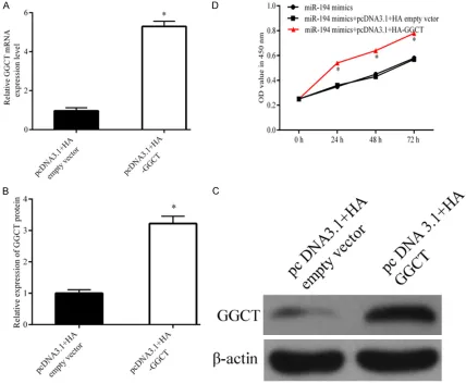
In order to increase the expression of GGCT in MGC-803 cells pcDNA3.1+HA-GGCT was trans-fected intothe cells which stable over-expressed miR-194. Western blot and qPCR revealed that
the expression level of GGCT was significantly
gained in the cells transfected with pcDNA3.1+ HA-GGCT when compared with the empty vec-tor (Figure 5A-C). The Figure 5D showed that
over-expression of miR-194 significantly
inhi-bit MGC-803 cells proliferation, while GGCT restoration reversed the anti-proliferation of miR-194.
were co-transfected with miR-194 mimics or mimic control and WT 3’UTR or MUT 3’UTR for 48 h and detected by the Dual-Luciferase Reporter Assay System (Promega, Madison, USA) according to the manufacturer’s ins- truction.
Statistical analysis
All the results analyses were performed by SPSS 19.0. Dates were presented as the means ± SD (standard deviation). One-way ANOVA test and Student’s t-test were used to measure the differences between the groups. P<0.05 was considered to be statistically
sig-nificant differences.
Results
Expression level of miR-194 was down-regulat
-ed in gastric cancer tissues and cells
In order to investigate the role of miR-194 in tumor metastasis and growth, we subsequen- tly researched the level of miR-194 in gastric cancer tissues. As shown in Figure 1A, miR-194 expression level was down-regulated in gastric cancer tissues when compared with the adjacent normal tissues. In addition, the expression level of miR-194 in gastric cancer cell lines was lower than in human gastric epi-thelial GES-1 cell line (Figure 1B).
Upregulation of miR-194 suppresses the prolif
-eration of MGC-803 cells
As shown in the Figure 2A, the expression level of miR-194 was obviously increased in MGC-803 cells after treatment with miR-194 mimi- cs (P<0.05). CCK8 results demonstrated that
upregulation of miR-194 could significantly sup -press the proliferation ability of MGC-803 cells at 48 h and 72 h (P<0.05), while when
com-pared with the control there was no significant
difference at 24 h (P>0.05) (Figure 2B). Upregulation of miR-194 promotes MGC-803
cells apoptosis
Flow cytometry results showed that the
apopto-sis rates of MGC-803 cells were significantly
targeting RAP2B in human bladder cancer [20]. Zhao et al found that miR-194 functioned as a prognostic marker and regulated the develop-ment of HCC through directly suppressing the expression level of MAP4K [21]. Even more important, Li et al revealed that Exogenous expression of miR-194 inhibited cell migration, invasion, and the epithelial-mesenchymal tran-sition phenotype in gastric cancer cells and that miR-194 acted as a tumor inhibitor through targeting FoxM1 [22].
In our present study, we illustrated that miR-194 was dramatically downregulated in gastric cancer tissues when compared with the adja-cent normal tissues in vivo. Moreover, we also discovered that miR-194 markedly decreased
in gastric cancer cell lines in vitro. These finding
is consistent with the previous research show-ing that miR-194 is downregulated in many kinds of cancers [23, 24]. In addition, we stud-ied the role of miR-194 in gastric cancer cells. First, we demonstrated that up-regulation the GGCT contributes to miR-194 suppressed
apoptosis of MGC-803 cells
The pro-apoptosis role of miR-194 also revers- ed by GGCT restoration. As shown in Figure 6A-D. Both the Flow cytometry and TUNEL an- alysis revealed that the apoptosis of MGC-803 cells were markedly decreased when transfect-ed with miR-194 mimics+pcDNA3.1+HA-GGCT. What’s more, the levels of Bcl-2 increased and Bax level declined in MGC-803 cells after trans-fected with miR-194 mimics+pcDNA3.1+HA-GGCT (Figure 6E).
Discussion
Previous studies have demonstrated that miR-NAs can be act as a tumor regulator, either as a cancer suppressor or an oncogene [18, 19].
The functions of miR-194 have been confirmed
[image:6.612.364.521.70.409.2]in a variety of kinds of human cancer. For instance, Zhang et al illustrated that miR-194 inhibits migration, proliferation and invasion by
knockdown of GGCT inhibits cell proliferation and induces late apoptosis in human gastric
cancer [25]. These findings are consistent with
our results. Bcl-2 and Bax are apoptosis-relat-ed genes. In this study, we found that when recovery of GGCT, it regulates Bcl-2 family members, promoting the BCL-2 resistance to apoptosis and inhibition Bax promoting apopto-sis, which is consistent with the previous research.
According to our results, we showed that the expression level of miR-194 was down-regulat-ed in gastric cancer patients and in the cell
lines. Over-expression of miR-194 significantly
inhibited proliferation and induced apoptosis of gastric cancer cells by targeting GGCT. Our research provided a better understanding of miR-194 in gastric cancer development, which
[image:7.612.91.519.74.427.2]level of miR-194 significantly suppressed gas -tric cancer cell proliferation and induced apop-tosis than the cells transfected with miR-194 mimic control. Moreover, we used luciferase activity assay and western blot showed that GGCT is a potential target gene of miR-194. Then both the western blot and qPCR showed that the expression level of GGCT can be nega-tively regulated by miR-194, which was play the role by binding with a site in the GGCT 3’-UTR. Additionally, recovery of GGCT partly changed the function of miR-194 in MGC-803 cells. So we concluded that GGCT may be involved in miR-194 regulated gastric cells proliferation and apoptosis. In previously, KS et al found that knockdown of GGCT dramatically suppressed the growth of many kinds of cancer cells includ-ing lung, prostate, bladder, and breast cancer cells [16]. Zhang et al also demonstrated that
FN, Herrmanns K, Bosco D, Kerr-Conte J, Pat-tou F, Rulicke T and Stoffel M. The microR-NA-200 family regulates pancreatic beta cell survival in type 2 diabetes. Nat Med 2015; 21: 619-627.
[9] Liu X, Zheng Q, Vrettos N, Maragkakis M, Alex-iou P, Gregory BD and Mourelatos Z. A microR-NA precursor surveillance system in quality control of MicroRNA synthesis. Mol Cell 2014; 55: 868-879.
[10] Li X, Wang F and Qi Y. MiR-126 inhibits the in-vasion of gastric cancer cell in part by targeting Crk. Eur Rev Med Pharmacol Sci 2014; 18: 2031-2037.
[11] Wang P, Li Z, Liu H, Zhou D, Fu A and Zhang E. MicroRNA-126 increases chemosensitivity in drug-resistant gastric cancer cells by targeting EZH2. Biochem Biophys Res Commun 2016; 479: 91-96.
[12] Yoo JO, Kwak SY, An HJ, Bae IH, Park MJ and Han YH. miR-181b-3p promotes epithelial-mesenchymal transition in breast cancer cells through snail stabilization by directly targeting YWHAG. Biochim Biophys Acta 2016; 1863: 1601-1611.
[13] Chen X, Wang Y, Zang W, Du Y, Li M and Zhao G. miR-194 targets RBX1 gene to modulate proliferation and migration of gastric cancer cells. Tumour Biol 2015; 36: 2393-2401. [14] Oakley AJ, Yamada T, Liu D, Coggan M, Clark
AG and Board PG. The identification and struc -tural characterization of C7orf24 as gamma-glutamyl cyclotransferase. An essential en-zyme in the gamma-glutamyl cycle. J Biol Chem 2008; 283: 22031-22042.
[15] Gromov P, Gromova I, Friis E, Timmermans-Wielenga V, Rank F, Simon R, Sauter G and
Moreira JM. Proteomic profiling of mammary carcinomas identifies C7orf24, a gamma-glu -tamyl cyclotransferase, as a potential cancer biomarker. J Proteome Res 2010; 9: 3941-3953.
[16] Kageyama S, Iwaki H, Inoue H, Isono T, Yuasa T, Nogawa M, Maekawa T, Ueda M, Kajita Y, Ogawa O, Toguchida J and Yoshiki T. A novel
tumor-related protein, C7orf24, identified by
proteome differential display of bladder uro-thelial carcinoma. Proteomics Clin Appl 2007; 1: 192-199.
[17] Takemura K, Kawachi H, Eishi Y, Kitagaki K, Negi M, Kobayashi M, Uchida K, Inoue J, Inaza-wa J, KaInaza-wano T and Board PG. gamma-Glutam-may also be benefit for the process of miRNA
controlled diagnostic and therapeutic against gastric cancer.
Acknowledgements
This study was founded by the Science and Technology Agency project of Jiangsu province. (No. 2015-JS-41).
Disclosure of conflict of interest
None.
Address correspondence to: Yi Zhang, Department
of Colorectal Surgery, The First Affiliated Hospital of
Nanjing Medical University, 300 Guangzhou Road, Gulou District, Nanjing 211166, Jiangsu, China. Tel: +86-25-83718836; Fax: +86-25-83718836; E-mail: njzhangyi55@126.com; Ke Hu, School of Stoma- tology, Nanjing Medical University, 101 Longmian Road, Nanjing 211166, Jiangsu, China. Tel: +86- 25-86862654; Fax: +86-25-86862654; E-mail: 1402469333@qq.com
References
[1] McLean MH and El-Omar EM. Genetics of gas-tric cancer. Nat Rev Gastroenterol Hepatol 2014; 11: 664-674.
[2] Van Cutsem E, Sagaert X, Topal B, Hauster-mans K and Prenen H. Gastric cancer. Lancet 2016; 388: 2654-2664.
[3] Chen W, Zheng R, Baade PD, Zhang S, Zeng H, Bray F, Jemal A, Yu XQ and He J. Cancer statis-tics in China, 2015. CA Cancer J Clin 2016; 66: 115-132.
[4] Torre LA, Bray F, Siegel RL, Ferlay J, Lortet-Tieu-lent J and Jemal A. Global cancer statistics, 2012. CA Cancer J Clin 2015; 65: 87-108. [5] Kanda M and Kodera Y. Recent advances in
the molecular diagnostics of gastric cancer. World J Gastroenterol 2015; 21: 9838-9852. [6] Li T, Chen J, Liu QL, Huo ZH and Wang ZW.
Me-ta-analysis: E-cadherin immunoexpression as a potential prognosis biomarker related to gas-tric cancer metastasis in Asian patients. Eur Rev Med Pharmacol Sci 2014; 18: 2693-2703. [7] Ambros V. The functions of animal microRNAs.
Nature 2004; 431: 350-355.
[8] Belgardt BF, Ahmed K, Spranger M, Latreille M, Denzler R, Kondratiuk N, von Meyenn F, Villena
Figure 6. GGCT contributes to miR-194 inhibited apoptosis of MGC-803 cells. A and B. Flow cytometry showed that the apoptosis of MGC-803 cells decreased after treated with miR-194 mimics+pcDNA3.1+HA-GGCT. C and D. TUNEL assay demonstrated that the apoptosis of MGC-803 cells decreased after treated with miR-194 mimics+pcDNA3.1+HA-GGCT. E. The protein expression levels of Bcl-2 and Bax. (**P<0.01 when compared with the
[23] Zhu X, Li D, Yu F, Jia C, Xie J, Ma Y, Fan S, Cai H, Luo Q, Lv Z and Fan L. miR-194 inhibits the proliferation, invasion, migration, and enhanc-es the chemosensitivity of non-small cell lung cancer cells by targeting forkhead box A1 pro-tein. Oncotarget 2016; 7: 13139-13152. [24] Zhao HJ, Ren LL, Wang ZH, Sun TT, Yu YN,
Wang YC, Yan TT, Zou W, He J, Zhang Y, Hong J and Fang JY. MiR-194 deregulation contrib-utes to colorectal carcinogenesis via targeting AKT2 pathway. Theranostics 2014; 4: 1193-1208.
[25] Zhang W, Chen L, Xiang H, Hu C, Shi W, Dong P and Lv W. Knockdown of GGCT inhibits cell pro-liferation and induces late apoptosis in human gastric cancer. BMC Biochem 2016; 17: 19. ylcyclotransferase as a novel
immunohisto-chemical biomarker for the malignancy of esophageal squamous tumors. Hum Pathol 2014; 45: 331-341.
[18] Barger JF and Nana-Sinkam SP. MicroRNA as tools and therapeutics in lung cancer. Respir Med 2015; 109: 803-812.
[19] Kang SM and Lee HJ. MicroRNAs in human lung cancer. Exp Biol Med (Maywood) 2014; 239: 1505-1513.
[20] Zhang M, Zhuang Q and Cui L. MiR-194 inhib-its cell proliferation and invasion via repres-sion of RAP2B in bladder cancer. Biomed Phar-macother 2016; 80: 268-275.
[21] Zhao Y, Li F, Zhang X, Liu A, Qi J, Cui H and Zhao P. MicroRNA-194 acts as a prognostic marker and inhibits proliferation in hepatocellular car-cinoma by targeting MAP4K4. Int J Clin Exp Pathol 2015; 8: 12446-12454.