Original Article
MiR-506 suppresses proliferation and invasion of
bladder cancer by targeting FOXQ1
Xiang Sun, Longhua Luo, Liang Feng, Kezhao Zhong
Department of Urology, The First Affiliated Hospital of Nanchang University, Nanchang, Jiangxi, China Received December 5, 2015; Accepted February 15, 2016; Epub April 1, 2017; Published April 15, 2017
Abstract: FOXQ1 (Forkhead box protein Q1), a member of the forkhead family of transcription factors, is implicated in progression of various cancers. However, the modulation of FOXQ1 expression in human bladder cancer at post-transcriptional levels remains unclear. In this study, luciferase assay revealed that miR-506 was able to target 3’-untranslated region (3’-UTR) of FOXQ1. Furthermore, quantitative PCR and western blot assay demonstrated that miR-506 was able to suppress the expression of FOXQ1 at the levels of mRNA and protein. miR-506 expression was downregulated in bladder cancer tissue and cell lines, and overexpressed miR-506 significantly attenuated cellular proliferation and invasion. The rescue experiment confirmed that miR-506 exerted its biological functions by target-ing FOXQ1. Collectively, our study demonstrates that miR-506 can impair the proliferation or metastasis of human bladder cancer cells by downregulation of FOXQ1.
Keywords: miR-506, FOXQ1, bladder cancer, proliferation, invasion
Introduction
Bladder cancer is a urological malignancy with particularly high morbidity worldwide [1]. Many molecules involved in these alterations and serve as diagnostic markers of tumor growth and disease progression. Despite improve-ments of diagnosis and treatment strategies, bladder cancer remains a highly prevalent and lethal malignancy [2]. Therefore, it is urgent to elucidate the underlying molecular mecha-nisms of osteosarcoma and find novel diagnos-tic and prognosdiagnos-tic biomarkers for improving the clinical outcome of bladder cancer patients. MicroRNA (miRNA), an abundant group of small (22-nucleotide) noncoding RNA molecules that regulate gene expression by binding to the 3’ untranslated region (UTR) of mRNA by partial sequence homology and play an important role in variety of biological processes including cell proliferation, apoptosis, invasion, migration, differentiation and so on [3-6]. Accumulated evidences prove that miRNAs function as either oncogenes or tumor suppressors and are aber-rantly expressed in many human cancers [7]. miRNAs have been recognized as critical regu-lators in development and progression of
can-cer including bladder cancan-cer [8-10]. miR-506 functions as an tumor suppressor and was downregulated in various cancers including esophageal cancer [11], colon cancer [12, 13], cervical cancer [14], liver cancer [15], gastric Cancer [16], ovarian cancer [17] and so on. However, the expression levels and mechanism of miR-506 in bladder cancer tumorigenesis is still unclear.
Human tissue specimens
Twenty paired tissue specimens of bladder can-cer and matched normal tissues were obtained from the First Affiliated Hospital of Nanchang University. The matched normal tissues were further confirmed by pathologist for their nor-mal origin that they do not have tumor cells. All the tissues were obtained at the time of surgery and immediately stored in liquid nitrogen until use. The Institute Research Medical Ethics Committee of Nanchang University granted approval for this study.
Cell culture and transfection
Human bladder cancer cell lines (BIU-87, EJ and MGH-U1) and non-malignant bladder cell line (SV-HUC1) were maintained in Hyclon 1640 medium supplemented with 10% fetal bovine serum (HyClone, Logan, Utah, USA) and 1% penicillin/streptomycin and incubated in a humidified (37°C, 5% CO2) incubator.
Human bladder cancer cell lines BIU-87 cells were seeded in 12-well plates and incubated overnight, then transiently transfected with miR-506 mimic, miR-506 inhibitor, negative control (miR-con), and inhibitor negative control (anti-miR-con) sequences using Lipofectamine 2000 (Invitrogen) according to the manufactur-er’s instructions. The full-length FOXQ1 cDNA (which included the ORF and 3’ UTR) was PCR amplified and cloned into the pcDNA3.1 vector to generate the pcDNA-FOXQ1 constructs that were used in the rescue assays. BIU-87 cells in 12-well plates were con-transfected with miR-506 mimic and the pcDNA-FOXQ1 plasmid DNA.
Luciferase assay
Cells at 70% confluence in a 96-well plate were
TaqMan miRNA Reverse Transcription Kit (Applied Biosystems, Foster City, CA, USA) and TaqMan Human MiRNA Assay Kit (Applied Biosystems, Foster City, CA, USA). The relative expression of miR-506 was shown as fold dif-ference relative to U6. The PCR amplification for the quantification of the FOXQ1 and GAPDH mRNAs was performed using an ABI PRISM 7300 Sequence Detection System (Applied Biosystems, Foster City, CA, USA) and a SYBR®Premix Ex Taq™ ii (Perfect Real Time) Kit (Takara Bio, Shiga, Japan).
Western blot
Whole cell extracts were prepared with a cell lysis reagent (Sigma-Aldrich, St. Louis, MO, USA) according to the manual, and then, the protein was quantified by a BCA assay (Pierce, Rockford, IL, USA). Then, the protein samples were separated by SDS-PAGE (10%) and detect-ed by Western blot using polyclonal (rabbit) anti-FOXQ1 antibody (Santa Cruz Bio-tech- nology, Santa Cruz, CA, USA). Goat anti-rabbit IgG (Pierce, Rockford, IL, USA) secondary anti-body conjugated to horseradish peroxidase and ECL detection systems (SuperSignal West Femto, Pierce) were used for detection.
Cell proliferation
Cell invasion assay
At 48 hours after transfection, cells were seed-ed onto the basement Matrigel-coatseed-ed mem-brane matrix (BD Biosciences) present in the insert of a 24-well culture plate. Fetal bovine serum was added to the lower chamber as a chemoattractant. After a further 48 hours, the
noninvading cells were gently removed with a cotton swab. Invasive cells located on the lower side of the chamber were stained with Crystal Violet and counted.
Statistical analysis
Each experiment was repeated at least three times. Data were shown as mean ± S.D and analyzed using SPSS 18.0. Statistical compari-sons between groups were analyzed using Student’s t-test and a two-tailed P < 0.05 was considered to indicate statistical significance.
Results
MiR-506 expressed was downregulated in bladder cancer specimens and cell lines
[image:3.612.98.519.76.226.2]To evaluate the expression of miR-506 in human bladder carcinoma, we first quantita-tively analyzed the levels of reverse transcrip-tion-PCR in 20 cases of clinical bladder carci-noma tissues and matched adjacent non- neoplastic tissues. The result showed that miR-506 expression levels were generally reduced in bladder cancerous specimens, compared with corresponding adjacent non-neoplastic tissues (Figure 1A). We also determined the expression level of miR-506 in different blad-der cancer cell lines by quantitative RT-PCR. Comparing with the non-malignant bladder cell line (SV-HUC1), the expression level of miR-506 was consistently down-regulated in three blad-Figure 1. The expressions of miR-506 in bladder cancer tissue and cell lines. A. miR-506 expression levels were examined by qRT-PCR in bladder cancer tissue (Cancer) and adjacent non-neoplastic tissues (Normal). Each bar represents the mean of three independent experiments. *P < 0.01 versus adjacent non-neoplastic tissues (Nor-mal). B. miR-506 expression levels were examined by qRT-PCR in non-malignant bladder cell line (SV-HUC1) and three bladder cell lines (BIU-87, EJ and MGH-U1). Each bar represents the mean of three independent experiments. *P < 0.01 versus SV-HUC1 cell line.
[image:3.612.93.286.318.491.2]der cell lines (BIU-87, EJ and MGH-U1) (Figure 1B).
FOXQ1 is a direct target gene of miR-506 in bladder cancer cells
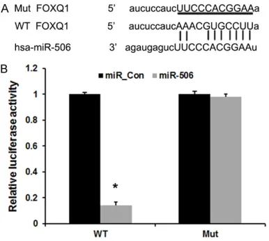
In this study, the miRNA target prediction web-sites www.microRNA.org and TargetScan were used and identified a conserved miR-506-bind-ing site in the 3’-UTR of FOXQ1 mRNA. We then cloned WT or Mut target region sequence of the FOXQ1 3’-UTR, which was inserted into a lucif-erase reporter vector (Figure 2A). Subsequently, these reporter vectors were cotransfected with miR-506 mimics and miR-506mimics control
(miR_con) into the bladder cancer cell line. As shown in Figure 2B, co-transfection of miR-506 mimics suppressed the luciferase activity of the reporter containing wild-type FOXQ1 3’ UTR sequence, but failed to inhibit that of mutated FOXQ1by dual-luciferase reporter assay. These data indicate that FOXQ1 is one of the direct targets of miR-506 in bladder cancer.
miR-506 represses FOXQ1 expression in blad-der cancer cells
[image:4.612.93.524.76.447.2]miR-506 andpcDNA-FOXQ1 in bladder cancer cells. qRT-PCR and Western blot analysis revealed FOXQ1 expression were significantly increased by transfecting with pcDNA-FOXQ1 (Figure 3A). miR-506 expression was signifi-cantly increased by transfecting with miR-506 mimics and decreased by transfecting with miR-506 inhibitors, compared with negative control group (Figure 3B). In addition, miR-506 mimics markedly increased the expression of FOXQ1 (Figure 3B), which was rescued by ecto-pic FOXQ1 (Figure 3C). These results demon-strated that FOXQ1 expression is regulated by miR-506 in bladder cancer cells.
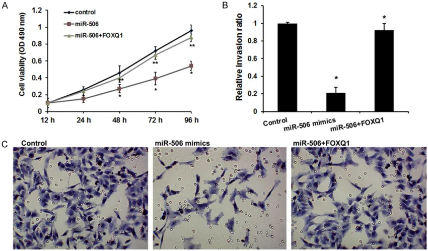
MiR-506 suppresses proliferation and invasion of bladder cancer cells via FOXQ1
It has been reported that FOXQ1 is implicated in bladder cancer progression [24]. Therefore, we are interested in whether miR-506 inhibits proliferation and migration of bladder cancer cells through FOXQ1 (Figure 4). The MTT assay revealed that miR-506 could remarkably reduce migration ability of bladder cancer cells, which was reversed by ectopic FOXQ1 (Figure
4A). The transwell invasion assay manifested that cell invasion was decreased when the cells were treated with miR-506, but FOXQ1 overex-pression could rescue inhibition of cell invasion mediated by miR-506 in the cells treated with both miR-506 and FOXQ1 (Figure 4B and 4C). Collectively, we conclude that miR-506 is able to suppress cell proliferation and invasion through targeting FOXQ1 in bladder cancer.
Discussion
[image:5.612.93.518.75.324.2]sion of miR-506 was commonly down-regulated in breast cancer cells and breast cancer speci-mens, and up-regulation of miR-506 inhibited cellular proliferation, migration and invasion as well as disrupt the cell cycle of breast cancer cells [28]. miR-506 is also downregulated in hepatocellular carcinoma, and enforced expres-sion of miR-506 inhibits proliferation, migration and invasion in vitro, and suppresses tumor growth in vivo [29]. In our study, we identified that the expression of miR-506 was significant-ly downregulated in bladder cancer tissues and cell lines. Overexpressed miR-506 significantly repressed cell proliferation and invasion in bladder cancer cell lines. These data suggest miR-506 as a tumor suppressor in bladder cancer.
Foxq1, a member of the FOX gene family, has been reported to be involved in various biologi-cal processes including embryonic develop-ment, cell cycle regulation, tissue-specific gene expression, cell signaling, and tumorigenesis [30]. Recent studies have clearly showed that Foxq1 was implicated in tumor proliferation and metastasis in various cancers including breast cancer, gastric cancer, colorectal cancer, hepa-tocellular carcinoma, gastric cancer and glio-blastoma [19-21, 23, 31-33]. Recently, high expression levels of FOXQ1 were observed in bladder cancer tissues [24]. However, the potential mechanism remains unclear. Here, we found that FOXQ1 was a direct target of miR-506, and miR-506 could regulate the expres-sion of FOXQ1 in bladder cancer. Moreover, FOXQ1 overexpression reversed miR-506-in-duced repression of the proliferation and inva-sion of bladder cancer.
In conclusion, our results suggest that the expression of miR-506 is down-regulated in bladder cancer tissues and cell lines, and
func-References
[1] Torre LA, Bray F, Siegel RL, Ferlay J, Lortet-Tieu-lent J and Jemal A. Global cancer statistics, 2012. CA Cancer J Clin 2015; 65: 87-108. [2] Shirodkar SP and Lokeshwar VB. Potential new
urinary markers in the early detection of blad-der cancer. Curr Opin Urol 2009; 19: 488-493. [3] Li Z, Lei H, Luo M, Wang Y, Dong L, Ma Y, Liu C,
Song W, Wang F, Zhang J, Shen J and Yu J. DNA methylation downregulated mir-10b acts as a tumor suppressor in gastric cancer. Gastric Cancer 2015; 18: 43-54.
[4] Xiao X, Tang C, Xiao S, Fu C and Yu P. Enhance-ment of proliferation and invasion by MicroR-NA-590-5p via targeting PBRM1 in clear cell renal carcinoma cells. Oncol Res 2013; 20: 537-544.
[5] Yin WZ, Li F, Zhang L, Ren XP, Zhang N and Wen JF. Down-regulation of microRNA-205 pro-motes gastric cancer cell proliferation. Eur Rev Med Pharmacol Sci 2014; 18: 1027-1032. [6] Yang X, Ni W and Lei K. miR-200b suppresses
cell growth, migration and invasion by target-ing Notch1 in nasopharyngeal carcinoma. Cell Physiol Biochem 2013; 32: 1288-1298. [7] Garzon R, Calin GA and Croce CM. MicroRNAs
in cancer. Annu Rev Med 2009; 60: 167-179. [8] Zhao X, Li J, Huang S, Wan X, Luo H and Wu D.
MiRNA-29c regulates cell growth and invasion by targeting CDK6 in bladder cancer. Am J Transl Res 2015; 7: 1382-1389.
[9] Zhang X, Zhang Y, Liu X, Fang A, Li P, Li Z, Liu T, Yang Y, Du L and Wang C. MicroRNA-203 is a prognostic indicator in bladder cancer and en-hances chemosensitivity to cisplatin via apop-tosis by targeting bcl-w and survivin. PLoS One 2015; 10: e0143441.
[10] Yao K, He L, Gan Y, Zeng Q, Dai Y and Tan J. MiR-186 suppresses the growth and metasta-sis of bladder cancer by targeting NSBP1. Di-agn Pathol 2015; 10: 146.
[12] Zhang Y, Lin C, Liao G, Liu S, Ding J, Tang F, Wang Z, Liang X, Li B, Wei Y, Huang Q, Li X and Tang B. MicroRNA-506 suppresses tumor pro-liferation and metastasis in colon cancer by directly targeting the oncogene EZH2. Oncotar-get 2015; 6: 32586-32601.
[13] Tong JL, Zhang CP, Nie F, Xu XT, Zhu MM, Xiao SD and Ran ZH. MicroRNA 506 regulates ex-pression of PPAR alpha in hydroxycamptothe-cin-resistant human colon cancer cells. FEBS Lett 2011; 585: 3560-3568.
[14] Wen SY, Lin Y, Yu YQ, Cao SJ, Zhang R, Yang XM, Li J, Zhang YL, Wang YH, Ma MZ, Sun WW, Lou XL, Wang JH, Teng YC and Zhang ZG. miR-506 acts as a tumor suppressor by directly tar-geting the hedgehog pathway transcription factor Gli3 in human cervical cancer. Onco-gene 2015; 34: 717-725.
[15] Lu Z, Zhang W, Gao S, Jiang Q, Xiao Z, Ye L and Zhang X. MiR-506 suppresses liver cancer an-giogenesis through targeting sphingosine ki-nase 1 (SPHK1) mRNA. Biochem Biophys Res Commun 2015; 468: 8-13.
[16] Li Z, Liu Z, Dong S, Zhang J, Tan J, Wang Y, Ge C, Li R, Xue Y, Li M, Wang W, Xiang X, Yang J, Ding H, Geng T, Yao K and Song X. miR-506 Inhibits Epithelial-to-Mesenchymal Transition and Angiogenesis in Gastric Cancer. Am J Pathol 2015; 185: 2412-2420.
[17] Liu G, Sun Y, Ji P, Li X, Cogdell D, Yang D, Parker Kerrigan BC, Shmulevich I, Chen K, Sood AK, Xue F and Zhang W. MiR-506 suppresses pro-liferation and induces senescence by directly targeting the CDK4/6-FOXM1 axis in ovarian cancer. J Pathol 2014; 233: 308-318.
[18] Christensen J, Bentz S, Sengstag T, Shastri VP and Anderle P. FOXQ1, a novel target of the Wnt pathway and a new marker for activation of Wnt signaling in solid tumors. PLoS One 2013; 8: e60051.
[19] Sun HT, Cheng SX, Tu Y, Li XH and Zhang S. FoxQ1 promotes glioma cells proliferation and migration by regulating NRXN3 expression. PLoS One 2013; 8: e55693.
[20] Liang SH, Yan XZ, Wang BL, Jin HF, Yao LP, Li YN, Chen M, Nie YZ, Wang X, Guo XG, Wu KC, Ding J and Fan DM. Increased expression of FOXQ1 is a prognostic marker for patients with gastric cancer. Tumour Biol 2013; 34: 2605-2609.
[21] Wang W, He S, Ji J, Huang J, Zhang S and Zhang Y. The prognostic significance of FOXQ1 oncogene overexpression in human hepatocel-lular carcinoma. Pathol Res Pract 2013; 209: 353-358.
[22] Feng J, Zhang X, Zhu H, Wang X, Ni S and Huang J. FoxQ1 overexpression influences poor prognosis in non-small cell lung cancer, associates with the phenomenon of EMT. PLoS One 2012; 7: e39937.
[23] Kaneda H, Arao T, Tanaka K, Tamura D, Aomat-su K, Kudo K, Sakai K, De Velasco MA, MatAomat-su- Matsu-moto K, Fujita Y, Yamada Y, Tsurutani J, Oka-moto I, Nakagawa K and Nishio K. FOXQ1 is overexpressed in colorectal cancer and en-hances tumorigenicity and tumor growth. Can-cer Res 2010; 70: 2053-2063.
[24] Zhu Z, Zhu Z, Pang Z, Xing Y, Wan F, Lan D and Wang H. Short hairpin RNA targeting FOXQ1 inhibits invasion and metastasis via the rever-sal of epithelial-mesenchymal transition in bladder cancer. Int J Oncol 2013; 42: 1271-1278.
[25] Liu B, Che W, Xue J, Zheng C, Tang K, Zhang J, Wen J and Xu Y. SIRT4 prevents hypoxia-in-duced apoptosis in H9c2 cardiomyoblast cells. Cell Physiol Biochem 2013; 32: 655-662. [26] Iorio MV and Croce CM. MicroRNA
dysregula-tion in cancer: diagnostics, monitoring and therapeutics. A comprehensive review. EMBO Mol Med 2012; 4: 143-159.
[27] Koutsaki M, Spandidos DA and Zaravinos A. Epithelial-mesenchymal transition-associated miRNAs in ovarian carcinoma, with highlight on the miR-200 family: prognostic value and prospective role in ovarian cancer therapeu-tics. Cancer Lett 2014; 351: 173-181. [28] Hua K, Yang W, Song H, Song J, Wei C, Li D and
Fang L. Up-regulation of miR-506 inhibits cell growth and disrupt the cell cycle by targeting YAP in breast cancer cells. Int J Clin Exp Med 2015; 8: 12018-12027.
[29] Dai W, Huang HL, Hu M, Wang SJ, He HJ, Chen NP and Li MY. microRNA-506 regulates prolif-eration, migration and invasion in hepatocel-lular carcinoma by targeting F-spondin 1 (SPON1). Am J Cancer Res 2015; 5: 2697-2707.
[30] Zhan HX, Xu JW, Wang L, Wu D, Zhang GY and Hu SY. FoxQ1 is a Novel Molecular Target for Pancreatic Cancer and is Associated with Poor Prognosis. Curr Mol Med 2015; 15: 469-477. [31] Abba M, Patil N, Rasheed K, Nelson LD,
Mud-duluru G, Leupold JH and Allgayer H. Unravel-ing the role of FOXQ1 in colorectal cancer me-tastasis. Mol Cancer Res 2013; 11: 1017-1028. [32] Zhang H, Meng F, Liu G, Zhang B, Zhu J, Wu F,
Ethier SP, Miller F and Wu G. Forkhead tran-scription factor foxq1 promotes epithelial-mes-enchymal transition and breast cancer metas-tasis. Cancer Res 2011; 71: 1292-1301. [33] Peng XH, Huang HR, Lu J, Liu X, Zhao FP, Zhang