Case Report
Large polypoid angiomyofibroblastoma
of the vulva: report of a case
Masayuki Shintaku1, Hirotaka Noda2, Hiromi Hashimoto3
1Department of Pathology, Shiga General Hospital, Moriyama, Shiga, Japan; 2Department of Pathology,
Amagasaki Central Hospital, Amagasaki, Hyogo, Japan; 3Uehonmachi Hiromi Women’s Clinic, Osaka, Japan
Received August 27, 2018; Accepted September 16, 2018; Epub November 1, 2018; Published November 15, 2018
Abstract: We report a case of angiomyofibroblastoma which arose in the vulva of a 46-year-old woman. The tumor
formed a large pedunculated polypoid mass, measuring 14 cm in maximal dimension, which hung down from the
right labium majus. It consisted of a dense or loose proliferation of fibroblastic and myofibroblastic cells on an
edematous background, and the tumor cells occasionally exhibited an increased cellularity around well-developed, medium-sized or small blood vessels. In small areas, conglomerates of capillaries exhibited an appearance
resem-bling that of capillary hemangioma. Tumor cells were immunoreactive for vimentin, desmin, estrogen receptor, and progesterone receptor, but not for α-smooth muscle actin, CD34, CD10, S-100 protein, calretinin, podoplanin, or cytokeratin. Angiomyofibroblastoma usually appears as a small subcutaneous nodule, and the formation of a large pedunculated polypoid mass is rare. The differential diagnosis from aggressive angiomyxoma and other mesenchy
-mal tumors which preferentially involve the vulvo-vaginal region was briefly discussed.
Keywords: Angiomyofibroblastoma, vulva, polypoid lesion
Introduction
Angiomyofibroblastoma is a relatively rare, be-nign soft tissue tumor that was first described
by Fletcher et al. [1], preferentially arising in the subcutaneous tissue of the vulvo-vaginal region of young to middle-aged women [1-6]. It usually appears as a small nodular lesion mea-suring less than 5 cm in diameter and is often diagnosed clinically as Bartholin’s gland cyst, but rare examples in which the tumor formed a large polypoid mass have also been report- ed [7-10]. In this article, we report an example
of angiomyofibroblastoma of the vulva which
formed a large pedunculated polypoid mass,
and briefly discuss some pathological diagnos -tic problems.
Clinical history
The patient was a 46-year-old, nulligravid
wo-man, who had noticed a small subcutaneous nodule measuring about 2 cm in diameter in
the vulvar region about five years ago.
Alth-ough no subjective symptoms were present, the nodule gradually enlarged from about one
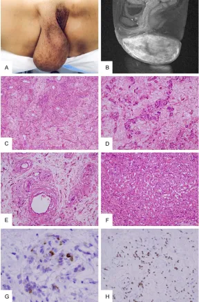
year before. On gynecological examination, a large pedunculated polypoid mass was found hanging down from the right labium majus (Figure 1A). The mass was soft in consistency
and covered by skin with a normal color. Nei-
ther pain nor tenderness was noted. The uter -ine cervix was unremarkable, but the uter-ine corpus was slightly enlarged. Bilateral adnexa showed no remarkable change.
Magnetic resonance imaging (MRI) revealed a well-circumscribed, large polypoid mass with a relatively wide pedicle and measuring about 14 by 8 by 6 cm, in the subcutaneous tissue of the vulva. It showed a homogeneously low intensity
on a T1-weighted image and heterogeneously high intensity on a T2-weighted image, suggest
-ing remarkable intralesional edema. The lesion
was irregularly enhanced by the administration of a contrast medium (Figure 1B). A few small nodules suggestive of leiomyoma were present within the myometrium.
Based on these clinical and MRI findings, a diagnosis of angiomyofibroblastoma of the
was performed. No signs suggestive of recur-rence have been noted as of eight years after the operation.
foot” appearance, and some vessels exhibited
[image:2.612.89.377.72.509.2]mural hyalinization or chronic inflammatory cell infiltration within the walls (Figure 1E). Organiz- Figure 1. A: A large polypoid tumor hung down from the right labium
ma-jus. B: T1-weighted MRI with contrast enhancement demonstrated a large,
well-circumscribed subcutaneous tumor showing an intense and irregular
enhancement. C: The tumor was composed of alternating hypercellular and
hypocellular areas and consisted of a proliferation of spindle cells on the edematous and highly vascularized stroma. (Hematoxylin-eosin stain, × 10).
D: Plump spindle cells surrounded small vessels and occasionally formed
small cellular trabeculae or cords. (Hematoxylin-eosin stain, × 25). E: Some blood vessels showed hyalinized thickening of the walls and intramural
chronic inflammatory cell infiltration. (Hematoxylin-eosin stain, × 25). F: In
small areas, capillary-sized anastomosing blood vessels formed dense ag-gregates resembling capillary hemangioma. (Hematoxylin-eosin stain, × 25).
G: The cytoplasm of some tumor cells was immunoreactive for desmin (× 100). H: The nuclei of most tumor cells were immunoreactive for estrogen
receptor (× 25).
Pathological findings
The extirpated tumor
weigh-ed 495 grams and, although
no fibrous capsule was
form-ed, it was well-circumscribed from the surrounding tissue.
The tumor was soft, and the
cut surface was homogene- ous and yellowish-white. No foci of hemorrhage or necro-sis were found.
Histopathologically, the tumor was composed of alternating hypercellular and hypocellular areas and consisted of a den- se or loose proliferation of plump spindle cells with an edematous background, be- ing accompanied by an accu-mulation of delicate, curling
collagen fibers and a prolifer -ation of abundant medium-sized or small blood vessels (Figure 1C). The deposition of
acid mucopolysaccharide was not demonstrated in the ma- trix with colloidal iron or alcian-blue (at pH 2.5) stain.
The characteristic perivascu -lar arrangement of plump spindle cells was evident, and some tumor cells also formed small cellular trabeculae or cords reminiscent of epitheli-al tumors (Figure 1D). There
was little tendency for tumor cells to form long, sweeping cellular fascicles. No lipoma-tous component was found within the tumor. Individual tumor cells had elliptical, ch- romatin-rich nuclei and am-
phophilic, fibrillary or plump
cytoplasm. Nuclear atypism and pleomorphism were ab-
“chicken-ed thrombi were infrequently found within the vascular lumina. In small areas, capillary-sized, anastomosing blood vessels formed dense ag- gregates and exhibited an appearance resem-bling capillary hemangioma (Figure 1F). Many mast cells were scattered throughout the lesion.
An immunohistochemical study was performed
on paraffin sections using the Envision Plus detection system (Dako, Glostrup, Denmark)
and employing monoclonal primary antibodies against the following substances: vimentin
(clone vim384, Dako, 1:100), α-smooth muscle actin (α-SMA) (clone 1A4, Dako, 1:100), desmin (clone D33, Dako, prediluted), CD34 (clone QBEnd, Dako, 1:100), CD10 (clone 56C6, Novo-castra, Newcastle-upon-Tyne, UK, 1:80), S-100 protein (clone ER-PR8, Dako, 1:500), calretinin (clone DC8, Zymed Labs, San Francisco, CA, USA, prediluted), podoplanin (clone D2-40, Ni-chirei, Tokyo, Japan, prediluted), cytokeratin (clone AE1/AE3, Dako, 1:500), estrogen recep
-tor (ER) (clone 1D5, Dako, 1:75), progesterone receptor (PgR) (clone PgR636, Dako, 1:500), and Ki67 (clone MIB-1, Dako, 1:100).
Heat-in-duced epitope retrieval using a hot bath was
performed prior to the immunostaining. The
cytoplasm of tumor cells was uniformly immu-noreactive for vimentin, and some of the tu- mor cells were also immunoreactive for des- min (Figure 1G). Nuclei of the vast majority of tumor cells were immunoreactive for both ER (Figure 1H) and PgR. Tumor cells were not im-munoreactive for α-SMA, CD34, CD10, S-100
protein, calretinin, podoplanin, or cytokeratin.
The Ki67 labeling index of tumor cell nuclei
was less than 1%. Discussion
Angiomyofibroblastoma was first described by
Fletcher et al. as a benign soft tissue tumor of the vulvar region which should be distin-guished from aggressive angiomyxoma [1]. It
consists of a diffuse proliferation of myofibro -blasts in association with a proliferation of ab- undant medium-sized or small blood vessels [1-6]. Mesenchymal tumors which preferential- ly arise in the vulvar region of young to middle-aged women include, in addition to the above two tumors, various pathologic entities, such
as cellular angiofibroma [11], superficial myofi
-broblastoma [12], and superficial angiomyxo
-ma [13]. In most of these tumors, the nuclei of tumor cells express ER and PgR [3, 5, 6, 11, 12, 14], and they are presumed to be derived from
site-specific and hormonally active mesenchy -mal cells (or perivascular stem cells) which are distributed in the female lower genital tract [1,
5, 6, 12, 14]. Tumors closely resembling angio
-myofibroblastoma also arise in the inguinal
region or scrotum of male patients [3, 15, 16],
and some examples of angiomyofibroblastoma
contain mature adipocytes as an integral com-ponent of the tumor (lipomatous variant) [4, 5, 7, 15, 16]. Nielsen et al. reported an extremely rare example of the sarcomatous
transforma-tion of angiomyofibroblastoma [17]. Although angiomyofibroblastoma shares some morpho
-logical characteristics with cellular angiofibro
-ma, mammary type myofibroblasto-ma, and sp-indle cell lipoma, a fluorescence in situ hybrid -ization study demonstrated absence of the ch- romosomal aberration which is commonly seen in these neoplasms, namely, monoallelic loss of 13q14 [6].
Among mesenchymal tumors involving the vul-var region, aggressive angiomyxoma forms a
large mass lesion which infiltrates the pelvic cavity, and, because of the difficulty in total
removal of the lesion, it is prone to recurrence [14, 18, 19]. It is therefore important, especial-ly when the lesion is large, to differentiate
angi-omyofibroblastoma from aggressive angiomyx -oma. However, because both tumors
predomi-nantly consist of a proliferation of fibroblasts and myofibroblasts and accompany a marked
proliferation of medium-sized or small blood vessels, their distinction is not always straight-forward [3]. Some tumors have been known to show a composite morphology with pathologi-cal features of these two tumors [19]. How- ever, a recent study showed that aggressive
angiomyxoma exhibits specific rearrangements
of the HMGA2 gene located on chromosome 12q15, demonstrating that the tumor occupies a biologically distinct position among many kinds of mesenchymal tumors of the female lower genital tract [20]. Aberrant immunohisto-chemical expression of HMGA2 protein is ob- served in the nuclei of tumor cells of most aggressive angiomyxomas [21]. In the present
case, the MRI findings, that is, a well-circum -scribed subcutaneous tumor showing a high
intensity on a T2-weighted image and irregular
for clinical differential diagnosis. The useful -ness of MRI for the correct preoperative
diag-nosis of angiomyofibroblastoma has been
po-inted out by several investigators [8, 22]. In the present case, aggressive angiomyxoma was also histopathologically excluded by the rela-tively high cellularity, absence of the deposi- tion of acid mucopolysaccharide in the stroma, lack of arterial vessels with abnormal morphol-ogies, and the presence of the characteristic perivascular arrangement of tumor cells.
Angiomyofibroblastoma typically represents as
a well-circumscribed, relatively small tumor no- dule in the subcutaneous tissue of the vulva, and only a few examples which formed a large polypoid mass have been previously reported.
These examples, which were reported by Omori
et al. [7] (48-year-old, tumor of the labium ma- jus weighing 360 grams), Nagai et al. [8] (48- year-old, tumor of the labium majus weighing 4,534 grams), Ito-Miyazaki et al. [9] (45-year-old, tumor of the labium majus weighing 714 grams), and by Wang et al. [10] (20-year-old, tumor of the labium majus measuring 18 cm in maximal dimension), very closely resembled the tumor in our case on the gross appearan- ce. In these cases including our own, marked
stromal edema seems to have significantly con -tributed to an increase of the tumor volume.
The stromal edema and degenerative changes
of the vascular wall with thrombus formation in
our case probably reflect the long preoperative
clinical course.
In small areas of the present tumor, capillary-sized blood vessels formed dense aggregates and exhibited an appearance resembling
capil-lary hemangioma. The proliferation of capillar -ies reminiscent of capillary hemangioma within the tumor has rarely been described in
angio-myofibroblastoma [4, 7]. Whether this finding
represents the endothelial differentiation of tumor cells or merely an exaggerated vascular reaction is unknown. Whereas Fukunaga et al. interpreted it as an expression of the diverse differentiation potential of primitive mesenchy-mal cells [4] and some of the tumor cells in the case of Omori et al. were immunoreactive for
CD34 [7], in our case the tumor cells were neg
-ative for CD34 and no evidence of the endothe -lial differentiation of tumor cells was obtained. In previous reports, the immunohistochemical
profiles of tumor cells have been repeatedly
emphasized as useful for the differential
diag-nosis of angiomyofibroblastoma from aggres
-sive angiomyxoma. Tumor cells in the former
are characteristically immunoreactive for
des-min and negative for α-SMA [1, 2], whereas
tumor cells in the latter are usually
immunore-active for α-SMA [14, 19]. The immunopheno -type of tumor cells in the present case well
coincided with these previous findings.
How-ever, because the immunohistochemical
phe-notypes of myofibroblasts are versatile among
various pathologic conditions [23, 24] and the immunophenotypes of tumor cells in angiomy-
ofibroblastoma and aggressive angiomyxoma
are also varying among cases [14, 15, 19], it is not prudent to depend solely upon the immu-
nohistochemical findings of tumor cells in the pathological differential diagnosis of fibroblas
-tic or myofibroblas-tic tumors of the lower geni -tal tract [3, 19].
In conclusion, we report a rare case of
angio-myofibroblastoma which formed a large pedun -culated polypoid mass in the vulva. In the case
of a large angiomyofibroblastoma, distinction
from aggressive angiomyxoma is important,
and MRI is a useful clinical tool. Various fibro
-blastic or myofibro-blastic tumors of the lower
genital tract share clinico-pathological and im- munohistochemical characteristics, and their histogenesis is probably similar as well.
Disclosure of conflict of interest
None.
Address correspondence to: Dr. Masayuki Shintaku, Department of Pathology, Shiga General Hospital, Moriyama 524-8524, Shiga, Japan. Tel:
077-582-5031; Fax: 077-582-5426; E-mail: [email protected]
References
[1] Fletcher CD, Tsang WY, Fisher C, Lee KC, Chan JK. Angiomyofibroblastoma of the vulva. A be -nign neoplasm distinct from aggressive angio-myxoma. Am J Surg Pathol 1992; 16: 373-382. [2] Hisaoka M, Kouho H, Aoki T, Daimaru Y, Hashi
-moto H. Angiomyofibroblastoma of the vulva. A
clinicopathologic study of seven cases. Pathol Int 1995; 45: 487-492.
[3] Ockner DM, Sayadi H, Swanson PE, Ritter JH, Wick MR. Genital angiomyofibroblastoma.
[4] Fukunaga M, Nomura K, Matsumoto K, Doi K, Endo Y, Ushigome S. Vulvar angiomyofibroblas -toma. Clinicopathologic analysis of six cases. Am J Clin Pathol 1997; 107: 45-51.
[5] Laskin WB, Fetsch JF, Tavassoli FA. Angiomyofi -broblastoma of the female genital tract. Analy-sis of 17 cases including a lipomatous variant. Hum Pathol 1997; 28: 1046-1055.
[6] Magro G, Righi A, Caltabiano R, Casorzo L, Mi
-chal M. Vulvovaginal angiomyofibroblastomas. Morphologic, immunohistochemical, and fluo -rescence in situ hybridization analysis for de- letion of 13q14 region. Hum Pathol 2014; 45: 1647-1655.
[7] Omori M, Toyoda H, Hirai T, Ogino T, Okada S. Angiomyofibroblastoma of the vulva. A
lar-ge pedunculated mass formation. Acta Med Okayama 2006; 60: 237-242.
[8] Nagai K, Aadachi K, Saito H. Huge peduncu
-lated angiomyofibroblastoma of the vulva. Int
J Clin Oncol 2010; 15: 201-205.
[9] Ito-Miyazaki R, Uchi H, Chiba T, Moroi Y, Kiryu H, Furue M. Large pedunculated angiomyofi
-broblastoma of the vulva. Eur J Dermatol
2013; 23: 520-522.
[10] Wang Y, Zhang Y, Lv B, Feng Y. Large-sized pe
-dunculated and polypoidal angiomyofibroblas -toma of the vulva. A case report and literature review. J Obstet Gynecol Res 2018; 44: 1492-1497.
[11] Iwasa Y, Fletcher CD. Cellular angiofibroma.
Clinicopathologic and immunohistochemical analysis of 51 cases. Am J Surg Pathol 2004; 28: 1426-1435.
[12] Laskin WB, Fetsch JF, Tavassoli FA. Superficial cervicovaginal myofibroblastoma. Fourteen
ca-ses of a distinctive mesenchymal tumor arising from the specialized subepithelial stroma of the lower female genital tract. Hum Pathol 2001; 32: 715-725.
[13] Fetsch JF, Laskin WB, Tavassoli FA. Superficial
angiomyxoma (cutaneous myxoma). A clinico-pathologic study of 17 cases arising in the genital region. Int J Gynecol Pathol 1997; 16: 325-334.
[14] Fetsch JF, Laskin WB, Lefkowitz M, Kindblom LG, Meis-Kindblom JM. Aggressive angiomyxo -ma. A clinicopathologic study of 29 female patients. Cancer 1996; 78: 79-90.
[15] Laskin WB, Fetsch JF, Mostofi FK. Angiomyofi -broblastomalike tumor of the male genital tract. Analysis of 11 cases with comparison
to female angiomyofibroblastoma and spindle
cell lipoma. Am J Surg Pathol 1998; 22: 6-16. [16] Shintaku M, Naitou M, Nakashima Y. Angio
-myofibroblastoma-like tumor (lipomatous vari -ant) of the inguinal region of a male patient. Pathol Int 2002; 52: 619-622.
[17] Nielsen GP, Young RH, Dickersin GR, Rosen
-berg AE. Angiomyofibroblastoma of the vulva
with sarcomatous transformation
(“angiomyo-fibrosarcoma”). Am J Surg Pathol 1997; 21:
1104-1108.
[18] Steeper TA, Rosai J. Aggressive angiomyxoma
of the female pelvis and perineum. Report of nine cases of a distinctive type of gynecologic soft-tissue neoplasm. Am J Surg Pathol 1983; 7: 463-475.
[19] Granter SR, Nucci MR, Fletcher CDM. Aggres -sive angiomyxoma. Reappraisal of its
relation-ship to angiomyofibroblastoma in a series of
16 cases. Histopathology 1997; 30: 3-10. [20] Medeiros F, Erickson-Johnson MR, Keeney GL,
Clayton AC, Nascimento AG, Wang X, Oliveira AM. Frequency and characterization of HMGA2 and HMGA1 rearrangements in mesenchymal tumors of the lower genital tract. Genes Chro-mosomes Cancer 2007; 46: 981-990. [21] McCluggage WG, Connolly L, McBride HA.
HM-GA2 is a sensitive but not specific
immuno-histochemical marker of vulvovaginal aggres-sive angiomyxoma. Am J Surg Pathol 2010; 34: 1037-1042.
[22] Geng J, Hu S, Wang F. Large paravaginal angio
-myofibroblastoma. Magnetic resonance imag
-ing find-ings. Jpn J Radiol 2011; 29: 152-155.
[23] Skalli O, Schürch W, Seemayer T, Lagacé R, Montandon D, Pittet B, Gabbiani G. Myofibro -blasts from diverse pathologic settings are het-erogeneous in their content of actin isoforms
and intermediate filament proteins. Lab Invest
1989; 60: 275-285.
[24] Schmitt-Gräff A, Desmoulière A, Gabbiani G. Heterogeneity of myofibroblast phenotypic fea
-tures. An example of fibroblastic cell plasticity.