Original Article
MiR-122-3p sensitizes breast cancer
cells to ionizing radiation via controlling
of cell apoptosis, migration and invasion
Junhua Zhang, Yong Cui, Xiaomeng Lin, Gang Zhang, Zhong Li
Department of Breast Surgery, Affiliated Hospital of Hebei University, Baoding 071000, Hebei, China
Received September 5, 2016; Accepted October 24, 2016; Epub January 1, 2017; Published January 15, 2017
Abstract: Nowadays, radio-resistance remains a grim problem occurs in patients with breast cancer. This study was aimed to explore the role of microRNA-122-3p (miR-122-3p) on breast cancer cells radio-sensitivity. Human breast cancer cells line MDA-MB-231 was used and transfected with miR-122-3p mimic, inhibitor or control. Transfected cells were exposed to various doses of irradiations, and then the surviving rate, apoptosis, cells migration and in-vasion were detected by clonogenic survival assay, flow cytometry, and transwell assay respectively. Furthermore, Western blot analysis was used to detect the expression changes of phosphatase and tensin homolog/phospha-tidylinositol-3-kinase/serine/threonine kinas (PTEN/PI3K/AKT) pathway and epithelial-mesenchymal transition (EMT) related proteins in transfected cells after irradiation. The surviving rate of MDA-MB-231 cells was significantly decreased by miR-122-3p overexpression (P < 0.05 or P < 0.01). Apoptosis was induced by miR-122-3p overex-pression (P < 0.001) while cells migration and invasion were inhibited by miR-122-3p overexpression (P < 0.001). Besides, miR-122-3p overexpression significantly down-regulated the expressions of p-AKT, AKT, Vimentin and Snail 1 (P < 0.01 or P < 0.001), whereas significantly up-regulated the levels of PTEN and E-Cadherin (P < 0.01). MiR-122-3p suppression showed the inversed impacts on the surviving rate, apoptosis, cells migration and invasion, as well as these proteins expressions. MiR-122-3p enhanced the radio-sensitivity of breast cancer cells via controlling of cell apoptosis, migration and invasion. PTEN/PI3K/AKT pathway and ETM related proteins were implicated in this enhancement impacts.
Keywords: MicroRNA-122-3p, breast cancer, radio-sensitivity, apoptosis, migration, invasion
Introduction
Breast cancer is the most common cancer and it is the leading cause of cancer death in fe- males worldwide [1]. To date, the exact cause of breast cancer is unclear, while it is believed genetics and environmental factors contribute to the carcinogenesis and progression of breast cancer [2, 3]. For the treatment, multimodal therapy often includes surgery, radiotherapy, chemotherapy, hormone therapy and biologic therapy [4]. Radiotherapy is always given after surgery to destroy microscopic tumor cells that may have escaped surgery, and this therapeu-tic strategy can greatly reduce the risk of recur-rence [5, 6]. However, radio-resistance remains a serious obstacle to kill tumor cells success-fully [7]. Thus, finding effective radio-sensitiz-ers will be helpful for us to improve the treat-ment, as well as improve the outcomes of pa- tients with breast cancer.
The role of miR-122-3p in breast cancer
diseases, including hepatocellular carcinoma, idiopathic asthenospermia, rheumatoid arthri-tis and hepatiarthri-tis C [15-18]. However, to our knowledge, none literature has reported the role of miR-122-3p in the radio-resistance of breast cancer cells. In the present study, we aimed to examine the role of miR-122-3p in breast cancer. Human breast cancer cells line MDA-MB-231 was used and transfected with miR-122-3p mimic, inhibitor or control. Trans- fected cells were exposed to various doses of ionizing radiations, and then the surviving rate, apoptosis, migration and invasion were deter-mined respectively, aiming to explore the func-tional impacts of miR-122-3p on the radio- sensitivity of breast cancer cells. In addition, the expressions of phosphatase and tensin homolog/phosphatidylinositol-3-kinase/ser-ine/threonine kinas (PTEN/PI3K/AKT) pathway and EMT related proteins in cells were detect- ed to reveal the possible underlying mecha-nism of miR-122-3p in breast cancer cells. Materials and methods
Cell culture and transfection
Human breast cancer cells line MDA-MB-231 was purchased from the American Type Culture Collection (ATCC; Manassas, VA, USA). Cells were cultured in Dulbecco’s Modified Eagle’s Medium (DMEM; Sigma-Aldrich, St Louis, MO, USA) supplemented with 10% fetal bovine se- rum (FBS; Hyclone, Logan, UT, USA), 100 U/mL penicillin, and 100 U/mL streptomycin (Invitro-
gen, Carlsbad, CA, USA) [19]. Cells were incu-bated in a humidified 5% CO2 incubator at 37°C. Cells were seeded into 6-well culture dishes at a density of 2 × 10⁵ cells/well and cultured for 24 h at 37°C. The cells were then transfect-ed with miR-122-3p mimic, inhibitor or control (RiboBio, Guangzhou, China) by using Lipofe- ctamine 2000 (Invitrogen Life Technologies, Carlsbad, CA, USA), according to the manufa- cturer’s instructions [20]. After 48 h of trans-fection, cells were collected for the forthcom- ing analyses.
Clonogenic survival assay
After transfection, cells were seeded into 6-well plants at a density of 1 × 10⁵ cells/well and fol-lowed by exposure to the indicated doses of radiations (2, 4, 6, 8 or 10 Gy), by using 6 MV X-rays generated by linear accelerators (Varian 2300EX; Varian, Palo Alto, CA) at a dose rate of 3 Gy/min [21]. After 14 days of incubation at 37°C, colonies were fixed with ice-cold etha-nol/acetone (1:2) for 10 min, and then stained with 1% crystal violet (Sigma-Aldrich, St. Louis, MO, USA) for 5 min. Microscopic inspection (Olympus IX71; Olympus, Tokyo, Japan) was used for the colonies observation. Colonies containing ≥ 50 normal-looking cells were counted and clonogenic survival was calculat-ed as previous describcalculat-ed [22].
Apoptosis assay
Apoptotic cells were quantified by using an Annexin V-FITC/PI Kit (4 A Biotech Co. Ltd., Beijing, China) according to the manufacturer’s protocol. Transfected cells were exposed to 10 Gy X-rays for 12 h at 37°C, and then cells were collected and re-suspended in 200 μL binding buffer containing 10 μL Annexin V-FITC and 5 μL PI. After incubate in the dark at room temperature for 20 min, the samples were analyzed on the flow cytometry (FACS Calibur, Bec-ton Dickson, San Jose, CA, USA) [23]. The number of intact cells, apoptotic cells and necrotic cells were discriminated by counting the cells directly.
Migration and invasion assays
[image:2.612.92.280.73.230.2]Cell migration and invasion were measured by transwell assay. Briefly, transfected cells were harvested and seeded into the upper cham- ber of transwell inserts (8-μm pore size and 24-well insert; BD Biosciences). DMEM media containing 10% FBS was added to the lower
chamber as the attractant. For cell invasion assay, each upper chamber was coated with Matrigel (Becton-Dickinson Labware, Bedford, MA, USA) before cells were seeded. Cells were treated with continuous 10 Gy X-rays for 12 h at 37°C. Afterward, cells on the upper surface of the filter were removed by cotton swabs. Then the migrated and invaded cells in the lower surface were permeabilized with 100% methanol for 15 min and stained with Giem- sa (Leagene, Beijing, China) for 30 min [24]. Mi- croscopic inspection (Olympus IX71; Olympus, Tokyo, Japan) was used for viewing and pho- tographing stained cells. Cell numbers were counted form ten different fields.
Western blot
[image:3.612.94.500.75.512.2]Transfected cells were exposed to 10 Gy dose of radiation for 12 h at 37°C. Cellular protein was extracted by lysis buffer (Beyo- time, Shanghai, China) and proteins in each sample were quantified using BCA Protein As- say Kit (Beyotime), according to the manufac-turer’s instructions. Equal amounts of protein were loaded in sodium dodecyl sulfate-poly-acrylamide gel electrophoresis (SDS-PAGE) and transferred into polyvinylidene fluoride (PV- DF) membranes. The membranes were block- ed within 5% nonfat dry milk for 1 h at room temperature. Then membranes were incuba-
The role of miR-122-3p in breast cancer
ted overnight in primary antibodies for E-cad- herin (sc-8426), Vimentin (sc-373717), Snail 1 393172), p-AKT 514032), AKT (sc-5298), PTEN (sc-7974) or Actin (sc-8432) (Santa Cruz Biotechnology, Santa Cruz, CA) at 4°C. The secondary antibody horseradish peroxidase (HRP) conjugate (sc-516087; San- ta Cruz Biotechnology) was used to incubate membranes for 1 h at room temperature. Blots were visualized by ECL Plus Western Blotting Substrate (Thermo Scientific).
Statistical analysis
All data were expressed as mean ± standard deviations (SD) from three independent experi-ments. Statistical analysis was performed us- ing Student t tests analysis of variance in GraphPad Prism 5 software (GraphPad Soft- ware Inc., San Diego, CA, USA). A value of P < 0.05 was considered as statistical difference. Results
MiR-122-3p sensitized MDA-MB-231 cells to ionizing radiation
To evaluate the role of miR-122-3p in the radio-sensitivity of breast cancer cells, MDA-MB-231 cells were transfected with miR-122-3p mimic, inhibitor or control, and the transfected cells were exposed to various doses of ionizing radi-ations, and then surviving rate was calculated. As results showed in Figure 1, overexpression
of miR-122-3p significantly decreased the surviving rate when cells were exposed to 4- 10 Gy doses of radiations (P < 0.05 or P < 0.01). However, miR-122-3p suppression dis-played the inverse results, that it significantly increased the surviving rate at the same ra- diation doses (P < 0.05). These data eviden- ced that miR-122-3p could sensitize breast cancer cells to ionizing radiation.
MiR-122-3p induced apoptosis while sup
-pressed migration and invasion in MDA-MB-231 cells after irradiation
In order to investigate the detailed function of miR-122-3p on breast cancer cells, MDA-MB-231 cells were first transfected with miR-122-3p mimic, inhibitor or control and then exposed to 10 Gy dose of irradiation. Cells apoptosis, migration and invasion were de- termined respectively. As results showed in Figure 2A and 2B, miR-122-3p overexpression significantly increased the apoptotic cells rate after irradiation (P < 0.001), while miR-122-3p suppression displayed the inverse effect (P < 0.05). Besides, transwell assay showed that (Figure 3A and 3B), miR-122-3p overexpres-sion significantly inhibited cells migration and invasion after irradiation (P < 0.001). Unsur- prisingly, miR-122-3p suppression significantly enhanced cells migration and invasion (P < 0.05 or P < 0.001). Thus, we inferred that miR-122-3p sensitized breast cancer cells to
[image:4.612.101.519.76.259.2]ing radiation might be via controlling of cell apoptosis, migration and invasion.
MiR-122-3p sensitized MDA-MB-231 cells to ionizing radiation via regulating PTEN/PI3K/ AKT pathway and EMT related proteins
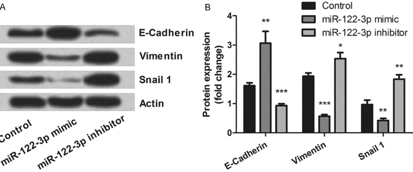
To explore the possible molecular mechanisms of miR-122-3p on breast cancer cells, miR-tr- ansfected MDA-MB-231 cells were exposed to 10 Gy dose of irradiation, and then the
[image:5.612.93.518.74.254.2]expres-sion levels of PTEN/PI3K/AKT pathway and EMT related proteins in cells were detected. As results showed in Figure 4A and 4B, miR-122-3p overexpression significantly down-regulated the levels of p-AKT (P < 0.001) and AKT (P < 0.01), whereas up-regulated the level of PTEN (P < 0.01). However, miR-122-3p suppression showed the contrary regulatory impacts (P < 0.01). Figure 5A and 5B showed that, up-regu-lation of E-Cadherin (P < 0.01) and down-regu-lation of Vimentin (P < 0.001) and Snail 1 (P <
Figure 4. PTEN/PI3K/AKT pathway was involved in the effects of miR-122-3p on MDA-MB-231 cells apoptosis. After MDA-MB-231 cells were transfected with miR-122-3p mimic, inhibitor or control, and exposed to 10 Gy of irradia-tion, the proteins expression changes of p-AKT, AKT and PTEN in cells were determined by Western blot analysis. PTEN/PI3K/AKT, phosphatase and tensin homolog/phosphatidylinositol-3-kinase/serine/threonine kinase; miR-122-3p, microRNA-122-3p; **, P < 0.01; ***, P < 0.001.
[image:5.612.97.517.340.515.2]The role of miR-122-3p in breast cancer
0.01) were found in miR-122-3p overexpress- ed cells. As expected, miR-122-3p suppression regulated these proteins expression in the re- versed direction (P < 0.05, P < 0.01 or P < 0.001). These data evidenced that miR-122- 3p sensitized breast cancer cells to ionizing ra- diation might be via regulating these protein expressions.
Discussion
Radiation is a part of therapy for breast cancer and is often provided following surgery. Never- theless, radio-resistance remains a grim pro- blem occurs in patients with breast cancer, and the underlying mechanism of radio-resis-tant remains unclear. In this study, we found that after ionizing radiation, miR-122-3p over-expression significantly decreased the surviv-ing rate of MDA-MB-231 cells. In addition, miR-122-3p overexpression induced apoptosis, and suppressed migration and invasion after cells were exposed to irradiation. Further, down-reg-ulation of p-AKT, AKT, Vimentin and Snail 1, as well as up-regulation of PTEN and E-Cadherin was found in miR-122-3p overexpressed cells. The role of several miRNAs in the radio-resis-tance of breast cancer has been elucidated. Anastasov et al. reported that miR-21 expres-sion in breast cancer cells contributed to ra- diation resistance by compromising cell cycle progression [25]. Sun et al. demonstrated that miR-200c enhanced radio-sensitivity in breast cancer cells by targeting Ubiquilin 1 (UBQLN1) [26]. Here, our results showed that overexpre- ssion of miR-122-3p significantly suppressed the surviving rate, after breast cancer cells were exposed to irradiation. These data provid-ed the first evidence that miR-122-3p func-tioned as a radio-sensitizer in breast cancer.
Cancer cells tend to be resistant to radiothe- rapy partly due to defects in apoptosis [27]. Moreover, studies have showed that altered miRNA expression may play an important role in the radiotherapy of cancer cells by impair- ing cellular responses that affect apoptosis [28]. Gwak et al. found that silencing of miR- 21 conferred radio-sensitivity in malignant glio-ma cells through the regulation of apoptosis [29]. Studies in vivo and in vitro have reveal- ed that overexpression of miR-34a could sig- nificantly enhance the radio-sensitivity by in- ducing cell apoptosis [30]. Similar with these previous studies, our study displayed that
miR-122-3p overexpression increased the apopto- tic cells rate after irradiation, suggesting that miR-122-3p enhanced radio-sensitivity of bre- ast cancer cells via controlling of apoptosis. PTEN is widely known as both tumor suppres-sor gene and apoptosis regulator that is impli-cated in breast cancer [31]. Besides, negative regulation of PI3K/AKT has been identified as a crucial downstream signaling pathway of PTEN [32]. Previous studies haverevealed that deregulation of PTEN/PI3K/AKT pathway was associated with the trastuzumab resistance of breast cancer [33] via blocking apoptosis [34]. However, in this study, miR-122-3p over- expression notably up-regulated PTEN while down-regulated p-AKT and AKT after irradia-tion. Thus, we hypothesized that PTEN/PI3K/ AKT pathway might be involved in the effects of miR-122-3p on irradiation induced apopto- sis in breast cancer cells.
In conclusion, we demonstrated that miR-122-3p sensitized breast cancer cells to ionizing radiation via controlling of cell apoptosis, migra-tion and invasion. PTEN/PI3K/AKT pathway and ETM-related proteins were implicated in the effects of miR-122-3p on breast cancer cells radio-sensitivity. However, further studies still needed to investigate whether the impacts of miR-122-3p on MDA-MB-231 cells can be reproduced in other types of breast cancer cells.
Disclosure of conflict of interest
None.
Address correspondence to: Dr. Zhong Li, Depart- ment of Breast Surgery, Affiliated Hospital of He-bei University, No. 212, East Yuhua Road, Baoding 071000, Hebei, China. E-mail: lizhong9637@126. com
References
[1] Hui Z, Yiling C, Wenting Y, Xuqun H, Chuanyi Z and Hui L. miR-491-5p functions as a tumor suppressor by targeting JMJD2B in ERalpha-positive breast cancer. FEBS Lett 2015; 589: 812-821.
[2] Schoemaker MJ, Jones ME, Wright LB, Griffin J, McFadden E, Ashworth A and Swerdlow AJ. Psychological stress, adverse life events and breast cancer incidence: a cohort investigation in 106,000 women in the United Kingdom. Breast Cancer Res 2016; 18: 72.
[3] Xu M, Ren Z, Wang X, Comer A, Frank JA, Ke ZJ, Huang Y, Zhang Z, Shi X, Wang S and Luo J. ErbB2 and p38gamma MAPK mediate alcohol-induced increase in breast cancer stem cells and metastasis. Mol Cancer 2016; 15: 52. [4] Courneya KS, McNeely ML, Culos-Reed SN,
Vallance JK, Bell GJ, Mackey JR, Matthews CE, Morielli AR, Cook D, MacLaughlin S, Farris MS, Voaklander S, O’Reilly R and Friedenreich CM. The alberta moving beyond breast cancer (AMBER) cohort study: recruitment, baseline assessment, and description of the first 500 participants. BMC Cancer 2016; 16: 481. [5] Belletti B, Vaidya JS, D’Andrea S, Entschladen
F, Roncadin M, Lovat F, Berton S, Perin T, Candiani E, Reccanello S, Veronesi A, Can-zonieri V, Trovo MG, Zaenker KS, Colombatti A, Baldassarre G and Massarut S. Targeted intraoperative radiotherapy impairs the stimu-lation of breast cancer cell proliferation and invasion caused by surgical wounding. Clin Cancer Res 2008; 14: 1325-1332.
[6] Vaidya JS, Joseph DJ, Tobias JS, Bulsara M, Wenz F, Saunders C, Alvarado M, Flyger HL, Massarut S, Eiermann W, Keshtgar M, Dewar J, Kraus-Tiefenbacher U, Sutterlin M, Esserman L, Holtveg HM, Roncadin M, Pigorsch S, Metaxas M, Falzon M, Matthews A, Corica T, Williams NR and Baum M. Targeted intraopera-tive radiotherapy versus whole breast radio-therapy for breast cancer (TARGIT-A trial): an international, prospective, randomised, non-inferiority phase 3 trial. Lancet 2010; 376: 91-102.
[7] Liang Z, Ahn J, Guo D, Votaw JR and Shim H. MicroRNA-302 replacement therapy sensitizes breast cancer cells to ionizing radiation. Pharm Res 2013; 30: 1008-1016.
[8] Li Y, Deng X, Zeng X and Peng X. The Role of Mir-148a in Cancer. J Cancer 2016; 7: 1233-1241.
[9] He L and Hannon GJ. MicroRNAs: small RNAs with a big role in gene regulation. Nat Rev Genet 2004; 5: 522-531.
[10] Bartel DP. MicroRNAs: target recognition and regulatory functions. Cell 2009; 136: 215-233.
[11] Gomes BC, Martins M, Lopes P, Morujao I, Oliveira M, Araujo A, Rueff J and Rodrigues AS. Prognostic value of microRNA-203a expres-sion in breast cancer. Oncol Rep 2016; 36: 1748-56.
[12] Damavandi Z, Torkashvand S, Vasei M, Soltani BM, Tavallaei M and Mowla SJ. Aberrant ex-pression of breast development-related mi-croRNAs, miR-22, miR-132, and miR-212, in breast tumor tissues. J Breast Cancer 2016; 19: 148-155.
[13] Samaeekia R, Adorno-Cruz V, Bockhorn J, Chang YF, Huang S, Prat A, Ha N, Kibria G, Huo D, Zheng H, Dalton R, Wang Y, Moskalenko G and Liu H. MicroRNA-206 inhibits stemness and metastasis of breast cancer by targeting MKL1/IL11 pathway. Clin Cancer Res 2016; [Epub ahead of print].
[14] Raza U, Saatci O, Uhlmann S, Ansari SA, Eyüpoğlu E, Yurdusev E, Mutlu M, Ersan PG, Altunda AM, Zhang JD, Doğan HT, Guler G and Şahin Ö. The miR-644a/CTBP1/p53 axis sup -presses drug resistance by simultaneous inhi-bition of cell survival and epithelial-mesenchy-mal transition in breast cancer. Oncotarget 2016; [Epub ahead of print].
The role of miR-122-3p in breast cancer
[16] Zhang X, Wei R, Lou J and Zhou J. [Seminal plasma miR-122-3p and miR-141-5p stability and its diagnosis value for idiopathic astheno-spermia]. Zhonghua Yi Xue Yi Chuan Xue Za Zhi 2016; 33: 320-323.
[17] Wang W, Zhang Y, Zhu B, Duan T, Xu Q, Wang R, Lu L and Jiao Z. Plasma microRNA expression profiles in Chinese patients with rheumatoid arthritis. Oncotarget 2015; 6: 42557-42568. [18] Van Keuren-Jensen KR, Malenica I, Courtright
AL, Ghaffari LT, Starr AP, Metpally RP, Beecroft TA, Carlson EW, Kiefer JA, Pockros PJ and Rakela J. microRNA changes in liver tissue as-sociated with fibrosis progression in patients with hepatitis C. Liver Int 2016; 36: 334-343. [19] Oner C, Turgut Cosan D and Colak E. Estrogen
and androgen hormone levels modulate the expression of PIWI interacting RNA in prostate and breast cancer. PLoS One 2016; 11: e0159044.
[20] Potenza N, Castiello F, Panella M, Colonna G, Ciliberto G, Russo A and Costantini S. Human miR-544a modulates SELK expression in he-patocarcinoma cell lines. PLoS One 2016; 11: e0156908.
[21] Kang M, Xiao J, Wang J, Zhou P, Wei T, Zhao T and Wang R. MiR-24 enhances radiosensitiv-ity in nasopharyngeal carcinoma by targeting SP1. Cancer Med 2016; 5: 1163-73.
[22] Patties I, Kortmann RD, Menzel F and Glasow A. Enhanced inhibition of clonogenic survi- val of human medulloblastoma cells by multi-modal treatment with ionizing irradiation, epi -genetic modifiers, and differentiation-inducing drugs. J Exp Clin Cancer Res 2016; 35: 94. [23] Pan XW and Zhao XH. In vitro proliferation
and anti-apoptosis of the papain-generated casein and soy protein hydrolysates towards osteoblastic cells (hFOB1.19). Int J Mol Sci 2015; 16: 13908-13920.
[24] Alaee M, Danesh G and Pasdar M. Plakoglobin reduces the in vitro growth, migration and in- vasion of ovarian cancer cells expressing N- cadherin and mutant p53. PLoS One 2016; 11: e0154323.
[25] Anastasov N, Hofig I, Vasconcellos IG, Rappl K, Braselmann H, Ludyga N, Auer G, Aubele M and Atkinson MJ. Radiation resistance due to high expression of miR-21 and G2/M check-point arrest in breast cancer cells. Radiat Oncol 2012; 7: 206.
[26] Sun Q, Liu T, Yuan Y, Guo Z, Xie G, Du S, Lin X, Xu Z, Liu M, Wang W, Yuan Q and Chen L. MiR-200c inhibits autophagy and enhances radio-sensitivity in breast cancer cells by targeting UBQLN1. Int J Cancer 2015; 136: 1003-1012. [27] Chen J, Shen Z, Zheng Y, Wang S and Mao W.
Radiotherapy induced Lewis lung cancer cell apoptosis via inactivating beta-catenin
medi-ated by upregulmedi-ated HOTAIR. Int J Clin Exp Pathol 2015; 8: 7878-7886.
[28] Ma H, Rao L, Wang HL, Mao ZW, Lei RH, Yang ZY, Qing H and Deng YL. Transcriptome ana-lysis of glioma cells for the dynamic response to gamma-irradiation and dual regulation of apoptosis genes: a new insight into radiothera-py for glioblastomas. Cell Death Dis 2013; 4: e895.
[29] Gwak HS, Kim TH, Jo GH, Kim YJ, Kwak HJ, Kim JH, Yin J, Yoo H, Lee SH and Park JB. Silencing of microRNA-21 confers radio-sensi-tivity through inhibition of the PI3K/AKT path-way and enhancing autophagy in malignant glioma cell lines. PLoS One 2012; 7: e47449. [30] Liu C, Zhou C, Gao F, Cai S, Zhang C, Zhao L,
Zhao F, Cao F, Lin J, Yang Y, Ni J, Jia J, Wu W, Zhou L, Cui J, Zhang W, Li B and Cai J. MiR-34a in age and tissue related radio-sensitivity and serum miR-34a as a novel indicator of radia-tion injury. Int J Biol Sci 2011; 7: 221-233. [31] Li J, Yen C, Liaw D, Podsypanina K, Bose S,
Wang SI, Puc J, Miliaresis C, Rodgers L, McCombie R, Bigner SH, Giovanella BC, Ittmann M, Tycko B, Hibshoosh H, Wigler MH and Parsons R. PTEN, a putative protein tyro-sine phosphatase gene mutated in human brain, breast, and prostate cancer. Science 1997; 275: 1943-1947.
[32] Chalhoub N and Baker SJ. PTEN and the PI3-kinase pathway in cancer. Annu Rev Pathol 2009; 4: 127-150.
[33] Gallardo A, Lerma E, Escuin D, Tibau A, Munoz J, Ojeda B, Barnadas A, Adrover E, Sanchez-Tejada L, Giner D, Ortiz-Martinez F and Peiro G. Increased signalling of EGFR and IGF1R, and deregulation of PTEN/PI3K/Akt pathway are related with trastuzumab resistance in HER2 breast carcinomas. Br J Cancer 2012; 106: 1367-1373.
[34] Stemke-Hale K, Gonzalez-Angulo AM, Lluch A, Neve RM, Kuo WL, Davies M, Carey M, Hu Z, Guan Y, Sahin A, Symmans WF, Pusztai L, Nolden LK, Horlings H, Berns K, Hung MC, van de Vijver MJ, Valero V, Gray JW, Bernards R, Mills GB and Hennessy BT. An integrative ge-nomic and proteomic analysis of PIK3CA, PTEN, and AKT mutations in breast cancer. Cancer Res 2008; 68: 6084-6091.
[35] Flores-Perez A, Marchat LA, Rodriguez-Cuevas S, Bautista VP, Fuentes-Mera L, Romero-Za-mora D, Maciel-Dominguez A, de la Cruz OH, Fonseca-Sanchez M, Ruiz-Garcia E, la Vega HA and Lopez-Camarillo C. Suppression of cell migration is promoted by miR-944 through targeting of SIAH1 and PTP4A1 in breast can-cer cells. BMC Cancan-cer 2016; 16: 379.
-vasiveness of breast cancer cells by inducing epithelial-mesenchymal transition. Biochem Biophys Res Commun 2011; 412: 188-192. [37] Steinle M, Palme D, Misovic M, Rudner J,
Dittmann K, Lukowski R, Ruth P and Huber SM. Ionizing radiation induces migration of glioblastoma cells by activating BK K(+) chan-nels. Radiother Oncol 2011; 101: 122-126. [38] Pellegrino L, Stebbing J, Braga VM, Frampton
AE, Jacob J, Buluwela L, Jiao LR, Periyasamy M, Madsen CD, Caley MP, Ottaviani S, Roca-Alonso L, El-Bahrawy M, Coombes RC, Krell J and Castellano L. miR-23b regulates cyto-skeletal remodeling, motility and metastasis by directly targeting multiple transcripts. Nu- cleic Acids Res 2013; 41: 5400-5412.