Original Article
Ex-vivo expansion and function identification
of CD4
+CD25
+CD127
dimregulatory T
cells in severe aplastic anemia
Li Yan, Rong Fu, Huaquan Wang, Chunyan Liu, Hui Liu, Ting Wang, Weiwei Qi, Jing Guan, Zonghong Shao
Department of Hematology, General Hospital of Tianjin Medical University, Tianjin, China
Received February 19, 2016; Accepted July 12, 2016; Epub September 1, 2016; Published September 15, 2016
Abstract: Background: Regulatory T (Treg) cells play an important role in regulating immune responses. Successful ex-vivo expansion of Tregs has been achieved after T cell receptor (TCR) stimulation in the presence of T cell growth factors. In this study, we evaluated the proliferation ability and ex-vivo suppressive function of Tregs in severe
aplas-tic anemia (SAA). Methods: To define the optimum population for Treg cell therapy in SAA, CD4+ CD25+ CD127dim Treg subsets were sorted from the blood and bone marrow of SAA patients and normal controls, and expanded in
vitro in the presence of anti-CD3/CD28 beads with high concentration of recombinant human interleukin 2 (rhIL-2). Their phenotype was determined by flow cytometry. Mixed lymphocytes culture with Teff was performed to assess
the function of ex-vivo expanded Treg cells. Results: The Treg cells were expanded to approximately 10-fold in 1-2 weeks and retained the Treg cell phenotypic characteristics. Tregs from SAA patients even have more powerful proliferation ability than that from normal control. Moreover, the ex-vivo expanded Tregs have a high expression of CTLA-4. In ex-vivo immunosuppression experiment, the expansion rate of Teff from the four co-culture groups, including (control Teff: control Treg), (control Teff: SAA Treg), (SAA Teff: control Treg) and (SAA Teff: SAA Treg), did not
show any significant difference. The IFN-γ secretion by Teff was suppressible by expanded Tregs from SAA patients. Conclusion: Tregs from SAA patient can be sorted and expanded successfully in vitro, and used efficiently to sup -press effective T cells proliferation, paving the way for future clinical trials.
Keywords: Aplastic anemia, regulatory T cells, in vitro, expansion
Introduction
Acquired severe aplastic anemia (SAA) is a rare disease characterized by severe pancytopenia and bone marrow failure. Although the exact etiology is still unknown, previous studies have shown that it is an immune-mediated disease with destruction of hematopoietic cells by acti-vated T cells [1, 2]. In recent years, many stud-ies have demonstrated dysregulation of T cell function, especially that cytotoxic T lympho-cytes (CTLs) leads to bone marrow failure in SAA [3, 4]. However, it remains unclear that how T cell become activated. Regulatory T cells (Tregs) is a special subset of T cells reported first by Sakaguchi in 1995 [5]. One of the major characteristics of Tregs is immunosuppression, which performed as inhibiting the activation and proliferation of CD4+ and CD8+ T cells,
pre-venting the antigen presenting process of
anti-gen presenting cells (APC), and mediating tar-get cell death directly. Studies have shown that Tregs played a pivotal role in controlling adap-tive immune responses and maintenance of self-tolerance whether in mice or humans. Tregs express a variety of cell surface mole-cules, while some of them have a relatively close relationship with its function, such as CD25 (IL-2Rα chain), FoxP3 (forkhead box pro-tein 3), CTLA-4 (Cytotoxic T lymphocyte antigen, CD152), GITR (glucocorticoid-induced tumor ne- crosis factor receptor). Previous studies have shown that FoxP3 is required for Tregs to exert their suppressive ability and the ectopic expres-sion of this transcription factor by lymphoid and non-lymphoid cells is able to confer suppres-sive function, making FoxP3 a critical regulator of Tregs function [6-8]. These FoxP3+ cells
[9]. CD127 expression is associated with acqui-sition of Tregs function [10]. It inversely corre-lates with FoxP3 expression and Tregs suppres-sive activity [11]. With the gradual understand-ing of Tregs, now more and more researchers use CD4+ CD25+ CD127dim as the molecular
markers of Tregs.
There is accumulating evidence that impaired function of Tregs has been implicated in the development of several common autoimmune diseases [12, 13], including SAA [14, 15]. Until now, most reports of Treg abnormality in aplas-tic anemia have shown that the numbers of cir-culating Tregs decreased in most patients. Tregs can be expanded from patient blood and were safely used in recent phase 1 studies in graft versus host disease (GVHD) [16, 17] and type 1 diabetes [18]. Treg cell therapy is also conceptually attractive for SAA. However, barri-ers exist to this approach. The stability of Tregs expanded from SAA patient is unknown. In this study, we intended to investigate the in vitro proliferation ability and function of expanded CD4+ CD25+ CD127dim Tregs in SAA patients,
and then provide a theoretical basis for cell tar-geted therapy of SAA.
Materials and methods
Study subjects
A total of thirty (seventeen males, thirteen females) patients with a median age of 25 years (range 7-65 years) were included in the study. This cohort of patients included eleven (three males, eight females) SAA cases who were previously untreated and nineteen cases (fourteen males, five females) with recovering-SAA (R-recovering-SAA).
The diagnosis of SAA was established by bone marrow biopsy, bone marrow smear, and peripheral blood cell count according to In- ternational AA Study Group Criteria [2]. SAA was defined by following criteria: (1) bone mar-row cellularity <25% or 25-50% with <30% residual (hematopoietic) cells; (2) two of the following three: neutrophils <0.5 × 10⁹/L,
plate-lets <20 × 109/L, and reticulocytes <20 × 109/
L [19, 20]; (3) no evidence of malignant ease, no extensive iron deposition, storage dis-ease, myelofibrosis, and no other autoimmune diseases. Bone marrow cytogenetic studies
were performed in all patients, and the clinical characteristics of patients are listed in Table 1. Recovering SAA patients were defined as sub-stantial improvement after therapy with rabbit antithymocyte globulin (rATG) (Fresenius, Ger- many), decreasing dose of cyclosporine (CsA), glucocorticoid and hematopoietic stimulating factors (recombinant human erythropoietin, granulocyte colony- stimulating factor, recom- binant human thrombopoietin, and/or IL-11 in combination), and all the patients became completely transfusion independent. The dura-tion of erythrocytes/platelets transfusion in- dependency in 25%, 50% and 75% patients was 86/87, 140/126 and 196/183 days after ATG therapy, respectively. The median durat- ion that neutrophils, erythrocytes and plate- lets returned to normal level was 35 days, 11 months and 12.5 months after ATG therapy, respectively. Among all the R-SAA patients, eleven have achieved complete response (CR), while the other eight were partial response (PR) according to Camitta criteria (1976).
Normal values of regulatory T cells counts were determined by fifteen age-matched heal- thy volunteers aged 18-71 years old. The study was approved by the Ethics Committee of the Tianjin Medical University. Informed written consent was obtained from all patients or their parents in accordance with the Declaration of Helsinki.
Flow cytometry
PB and BM mononuclear cells were stain- ed with the following antibodies: CD4-PerCP, CD25-FITC, CD127-APC, CTLA-4-PE, and iso-typic control mouse IgG1-PE (all from BD Biosciences) according to the manufacturer’s instructions. Data acquisitions were perform- ed on a FACS-Calibur flow cytometer (BD Bio- sciences) and analyzed by FlowJo software (Version 7.6.1; TreeStar).
Purification of lymphocyte subpopulations
PB and BM mononuclear cells were separated by density-gradient centrifugation, respectively. Two T-cell subpopulations, including CD4+
CD25+ CD127dim Tregs and CD4+ CD25- effector
T cells (Teff) were purified using a commercial human CD4+ CD25+ CD127dim regulatory T cell
Table 1. Clinical and demographic parameters of patients participating in the study
Diagnosis Number (years)Age (× 10ANC 9/L) Hb (g/L) PLT (× 109/L) Ret % Bone marrow karyotype Therapy Duration (months) Untreated SAA 11 16 (7-65) 0.62±0.82 64.38±13.68 15.05±13.33 0.39±0.30 Male 46, XY [20]
Female 46, XX [20] except for transfusionsNot previously treated 2 (1-3) Recovering SAA 19 26 (8-59) 2.95±1.87 126.21±33.58 128.96±62.93 2.45±1.85 Male 46, XY [20]
Female 46, XX [20] combination indicatedTreated with 32 (6-116) Values are expressed as mean ± SD. Age and duration values are mid (min, max). ANC: Absolute neutrophil count; Hb: Hemoglobin; PLT: Platelet; Ret: Reticulocyte. Duration of “un
Treg proliferation
Once purified, Tregs were collected and cul-tured for 7-14 days at 37°C with 5% CO2 in TexMACS™ Medium (Miltenyi Biotec) (100 μl/105 cells) supplemented with 1% penicillin/
streptomycin, 1% 2-mercaptoethanol, 1% non-essential amino acids (NEAA), 1% HEPES, 1% sodium pyruvate, 10% fetal calf serum (FBS), and 1000 U/mL rIL-2. MACSiBead Particles pre-loaded with CD3 and CD28 antibodies (Miltenyi Biotec) was added at a bead-to-cell ratio of 4:1. Tregs were cultured in a 96-well round-bottom plate (1 × 10⁵ cells/well). Change
the culture medium, count the cells and save the supernatants one or two days. On day 7, the cells were re-stimulated with anti-CD3/ C28 beads.
Suppression assays
After cell proliferation, Tregs were mixed at a ratio of 1:4, and then added to autologous or allogeneic Teff cells seeded at 1 × 10⁵ cells/
well in a 96-well round-bottom plate. To ensure accurate results, the experiment was repeated three times. Cells were cocultured for 2-3 days at 37°C with 5% CO2 in TexMACS™ Medium (Miltenyi Biotec) (100 μl/105 cells)
supplement-ed with 1% penicillin/ streptomycin, 1% 2-mer-captoethanol, 1% NEAA, 1% HEPES, 1% sodium
pyruvate and 10% FBS. MACSiBead Particles pre-loaded with CD3 and CD28 antibodies (Miltenyi Biotec) was added at 5 × 10⁴/well.
CCK-8 reagent (Dojindo) was added at 10 μl/ well (final concentration) during the last 4 hours of culture. Evaluation of cell proliferation was assayed by microplate reader and absorbance (A450 nm) was taken as the ability of cell prolif-eration. The cell proliferation rate was calculat-ed using the formula: (absorbance of co-culture well/absorbance of blank well) × 100. For cross- culture experiments, Tregs from AA patients were mixed with autologous and healthy Teff at a ratio of 1:4, and vice versa.
Detection of levels of IL-10 and IFN-γ
Culture supernatants obtained from Tregs pro-liferation were analyzed for IL-10 by ELISA based on the manufacturer’s instructions (USCNK). Supernatants obtained from the sup-pression assays were analyzed for IFN-γ based on the manufacturer’s instructions (USCNK). Statistical analysis
[image:4.612.94.528.71.344.2]All analyses were performed using SPSS 21.0 software (SPSS Science). Data were presented as mean ± standard deviation (SD). The signifi-cance of the differences was assessed by One-way ANOVA. A value of P<0.05 was considered statistically significant.
Results
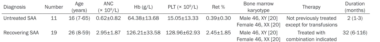
Purity of CD4+ CD25+ CD127dim Treg
CD4+ CD25+ CD127dim Treg cells comprised ap-
proximately 5-10% of all the CD4+ T cells in the peripheral blood of human, while SAA pa- tients had significantly decreased frequencies
of Tregs [21]. After sorting, the purity of CD4+
CD25+ CD127dim Treg and CD4+ CD25- Teff were
both more than 80% (Figure 1). Treg proliferation
Treg cells before and after culture is showed in Figure 2, and cell proliferation is obviously observed. Figure 3A shows the growth curves of viable cell counts at various time points, demonstrating approximately 10-fold expan-sion of the Treg cells. Tregs from SAA patients may have more powerful proliferation ability than that from normal control. The expansion rate of Treg cells from peripheral blood/bone marrow of SAA, R-SAA and normal control group were (17.54±10.34)/(9.21±8.54), (8.05±5.67)/ (11.29±18.28) and (6.14±8.80)/(3.71±2.84), respectively (Figure 3B).
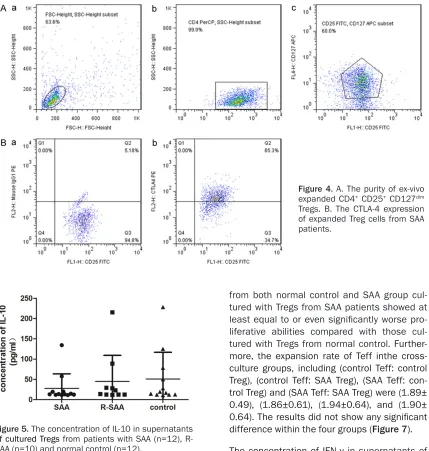
Cells were harvested according to their growth condition, and the purity was tested again by flow cytometry which still remain about 60% (Figure 4A). Based on the gate of CD4+ CD25+
CD127dim Tregs and isotype control used by
[image:5.612.90.524.73.233.2]mouse anti-human IgG antibody, we analyzed the expression of the functional molecular marker CTLA-4 (Figure 4B). The result showed that the CTLA-4 expression of expanded Treg cells from SAA patients was more than 60%, substantially and significantly increasing when compared with Tregs before expansion [21]. IL-10, one of the most important cytokine secreted by Tregs, is a key inhibitor of effec- tor T cell activation and a mediator of intes- tinal homeostasis [22]. The concentration of IL-10 in supernatants of cultured Tregs from
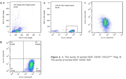
Figure 2. A. At low magnification, Treg cells in culture at day 1. B. At low magnification, Treg cells in culture at day 8.
Figure 3. A. The growth curves of viable cell counts. B. The expansion rate of Treg cells from PB/BM of SAA (n=3/8), R-SAA (n=6/13) and normal control
[image:5.612.93.286.282.555.2]SAA, R-SAA and normal control group were (27.82±35.92) pg/ml, (44.98±64.30) pg/ml and (50.94±65.91) pg/ml, without any signi- ficant difference (Figure 5).
Suppression assays
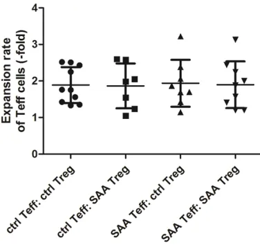
To address the efficiency of Treg-mediated im- munosuppression for effector T cells, we car-ried out co-cultures of Treg/Teff and analyzed the cell proliferation of Teff at different condi-tion, i.e. with or without autologous and alloge-neic Tregs. Figure 6 shows the growth curves of viable Teff cell counts at continuous time points of three days, demonstrating that Teff
from both normal control and SAA group cul-tured with Tregs from SAA patients showed at least equal to or even significantly worse pro- liferative abilities compared with those cul- tured with Tregs from normal control. Further- more, the expansion rate of Teff inthe cross-culture groups, including (control Teff: control Treg), (control Teff: SAA Treg), (SAA Teff: con- trol Treg) and (SAA Teff: SAA Treg) were (1.89± 0.49), (1.86±0.61), (1.94±0.64), and (1.90± 0.64). The results did not show any significant difference within the four groups (Figure 7).
The concentration of IFN-γ in supernatants of the four cross-culture groups were (25.87± 24.07) pg/ml, (21.89±18.65) pg/ml, (15.63± 9.35) pg/ml, and (10.16±5.52) pg/ml, respec-tively (Figure 8). The control Teff: control Treg group was significantly higher than SAA Teff: SAA Treg, demonstrating that expanded Tregs from SAA patients may successfully suppress the IFN-γ secretion by Teff.
Discussion
As one of the key lymphocyte subpopulation to induce immune tolerance, Tregs are belie- ved to control development and progression of autoimmunity by suppressing autoreactive T cells. Accumulating data provided evidence
Figure 4. A. The purity of ex-vivo
[image:6.612.95.524.73.524.2]expanded CD4+ CD25+ CD127dim Tregs. B. The CTLA-4 expression of expanded Treg cells from SAA patients.
Figure 5. The concentration of IL-10 in supernatants
of cultured Tregs from patients with SAA (n=12),
that impaired function of Tregs contributed to the development of autoimmunity diseases, such as autoimmune hepatitis [23, 24], type 1 diabetes [25], multiple sclerosis [26], rheuma-toid arthritis [27], and systemic lupus erythe-matosus. With the discovery that FoxP3 plays a central role in the differentiation and mainte-nance of Treg cells [6, 7], the use of flow
[image:7.612.326.518.73.245.2]cytom-etry-based analysis of FoxP3 expression in T cells became the gold standard for defining Treg cells. However, it then became evident that FoxP3 can also be expressed by effector T cells following activation [28], raising the possibility that any assessment of Treg cell number or function may include recently acti-vated effector T cells in the Treg cell popula- tion. Furthermore, as FoxP3 is a nuclear pro-tein, assessment of its expression in T cells requires fixation and permeabilization of the cells, resulting in an inability to obtain viable cells for further functional analysis. In the past few years, additional markers, such as CD127 (also known as IL-7Rα) [11], have been iden- tified that assist in the distinction of effector T cells from Treg cells and facilitate the ex- perimental purification of Treg cells [29]. An increasing number of studies have shown that SAA is an autoimmune disease caused by dysregulation of immune cell subsets, espe-cially T lymphocytes [1, 30, 31]. There are several immune abnormalities associated with pathogenesis of SAA, including imbalance of DC subsets (elevated DC1), enhancement of DC function, insufficiency of regulatory T cell, imbalance of Th1/Th2 subsets (enhanced Th1), increase of type I lymphoid factors (IL-2, IFN-γ), and decrease of NK cell proportion [32]. Previous data have shown inadequate num-bers of PB Tregs in patients with AA [14], and the quantities of Tregs in AA patients could be
Figure 6. The growth curves of viable Teff cell counts at different condition. A. Teff cells from normal con-trol. B. Teff cells from SAA patient.
Figure 7. The expansion rate of Teff in the four cross-culture groups, including (control Teff: control Treg)
[image:7.612.94.286.74.311.2](n=10), (control Teff: SAA Treg) (n=7), (SAA Teff: con-trol Treg) (n=87) and (SAA Teff: SAA Treg) (n=9).
Figure 8. The concentration of IFN-γ in supernatants
of the four cross-culture groups, including (control Teff: control Treg) (n=15), (control Teff: SAA Treg)
[image:7.612.93.281.377.554.2]improved substantially after IST. Nevertheless, little is known regarding the function of Tregs in AA because of the attack of autologous T cells on BM hematopoietic progenitors. Re- cent studies have revealed several impair-ments of Tregs in AA. Kordasti and his col-leagues have shown that sorted Tregs from AA patients were unable to suppress cytokine secretion (both IL-2 and IFN-γ) by autologous effector T cells [15]. Another study also re- ported the reversed ratio of Treg frequencies of BM versus PB, the abnormality of migra- tory potential, and defective immunosuppres-sion on Teff cells in vitro [33].
Physiologically, Tregs probably have multiple suppressive mechanisms, and the importance of each one may vary depending on the envi- ronment and the context of immunerespon- ses [34]. Tregs have powerful ability to sup-press proliferation of auto-reactive T cells th- rough contact-dependent mechanisms or in- hibitory cytokine production [13]. It is recog-nized that the fine immune homeostasis be- tween Tregs and effector T cells, regulating im- mune homeostasis, is often disrupted in auto-immune disorders [12, 35-37], including aplas-tic anemia [38].
As has been shown in animal models and human studies, such low-Treg numbers in rela-tion to effectors will not suppress immune re- sponses [39]. Therefore, it is of particular im- portance to expand these cells ex vivo prior to administration to the patient, ensuring their stability and function is maintained in vitro. Fortunately, a few efficient strategies for the large-scale expansion of Tregs have been de- veloped recently. Tregs are relatively refractory to T cell receptor (TCR) stimulation in vitro [40]; however, they can be driven to proliferate by combining TCR stimulation with high concen- tration of IL-2 [41] or IL-2 combined with IL-15 [42]. For example, large-scale ex-vivo expan-sion of human Tregs by stimulation with anti-CD3 and anti-CD28 monoclonal antibody-coat-ed beads and high-dose IL-2 has successful- ly been demonstrated [43]. Importantly, these expanded cells retain their suppressor funct- ion both in vitro [44] and in vivo in animal mod-els [45].
In this setting, we investigated the in vitro proliferation ability and function of expanded CD4+ CD25+ CD127dim Tregs in SAA patients.
The results suggest that Treg cells from SAA patient can be sorted and expanded success-fully with anti-CD3 and anti-CD28 monoclonal antibody-coated beads and high-dose IL-2 in vitro. Also it can be used efficiently to suppress effective T cells proliferation, paving the way for future clinical trials. Interestingly, the CTLA-4 expression of expanded Treg cells from SAA patients was substantially and significantly increasing when compared with Tregs before expansion. CTLA-4 is crucial for the suppres-sive function of Foxp3+ in vitro and in vivo [46].
It is expressed by Tregs which can modulate CD80 and CD86 expression by dendritic cells and there by inhibit the activation of effector T cells [47]. This provides us a hypothesis that there may be some kind of mechanism or abnormal microenvironment in SAA patient, which may somehow inhibit the function of Tregs in vivo. While once away from the morbid environment, Treg cells can be expanded and perform regular suppressive function in vitro. The further mechanism is under study and deserves to be investigated.
It has been hypothesized that an increased number of fully active Tregs could revert the imbalance of suppressor/effector cell ratio and may change the natural history of autoimmune diseases. Indeed, therapies that increase Treg numbers and activity have been shown to be effective at reversing autoimmune diseases in animal models such as experimental autoim-mune encephalitis [48] and diabetes [49]. Of note, ex vivo expanded regulatory T cells, adop-tively transferred in lupus-prone mice, reduced the rate of renal disease development, and a second transfer, in animals that already devel-oped proteinuria, further delayed the progres-sion of renal disease and significantly improved survival [50]. Moreover, a dose-escalation trial using autologous ex vivo expanded polyclonal Tregs (CD4+ CD25+ CD127dim/-) in diabetes pa-
tients is currently ongoing [51].
We believe that in vitro expanded autologous/ heterologous Tregs may help treating autoim-mune diseases, including SAA, and that CD4+
CD25+ CD127dim Treg cells are interesting from
ex-vivo-generated cells. There is at least a theo-retical risk that these Tregs have the potential to revert to T effector cells in vivo, especially if antigen-specific Tregs are infused. In support of this, it has been shown that a minority of adoptively transferred Tregs loses FoxP3 ex- pression and can differentiate into convention-al T cells [52]. Understanding why and how Tregs lose their suppressive phenotype, the relevance of this in the clinical setting and in particular how this can be prevented would improve both the safety and efficacy of Treg cell therapy strategies.
Acknowledgements
This work was supported by grants from the National Natural Science Foundation of China (81400085, 81400088, 81570106, 815701- 11), Tianjin Municipal Natural Science Found- ation (14JCYBJC25400, 15JCYBJC24300), He- alth Industry Research and Special Projects (201202017), Tianjin Cancer Research of Ma- jor Projects (12ZCDZSY17900, 12ZCDZSY180- 00), Tianjin Science and Technology support key project plan (20140109).
Disclosure of conflict of interest
None.
Address correspondence to: Dr. Zonghong Shao, Department of Hematology, General Hospital of
Tianjin Medical University, 154 Anshandao, Heping
District, Tianjin 300052, China. Tel: +86
226036-2086; Fax: +86 2260362085; E-mail: [email protected]
References
[1] Young NS, Scheinberg P and Calado RT.
Aplastic anemia. Curr Opin Hematol 2008; 15:
162-8.
[2] Marsh JC, Ball SE, Cavenagh J, Darbyshire P, Dokal I, Gordon-Smith EC, Keidan J, Laurie
A, Martin A, Mercieca J, Killick SB, Stewart R, Yin JA. British Committee for Standards in, Guidelines for the diagnosis and management
of aplastic anaemia. Br J Haematol 2009; 147: 43-70.
[3] Fogarty PF, Yamaguchi H, Wiestner A, Baer- locher GM, Sloand E, Zeng WS, Read EJ, Lansdorp PM and Young NS. Late presentation of dyskeratosis congenita as apparently ac-quired aplastic anaemia due to mutations in telomerase RNA. Lancet 2003; 362: 1628-30.
[4] Maciejewski JP and Risitano A. Hematopoietic stem cells in aplastic anemia. Arch Med Res
2003; 34: 520-7.
[5] Sakaguchi S, Sakaguchi N, Asano M, Itoh M and Toda M. Immunologic self-tolerance maintained by activated T cells expressing
IL-2 receptor alpha-chains (CD25). Breakdown
of a single mechanism of self-tolerance cau- ses various autoimmune diseases. J Immunol 1995; 155: 1151-64.
[6] Hori S, Nomura T and Sakaguchi S. Control of regulatory T cell development by the
transcrip-tion factor Foxp3. Science 2003; 299:
1057-61.
[7] Fontenot JD, Gavin MA and Rudensky AY.
Foxp3 programs the development and function
of CD4+CD25+ regulatory T cells. Nat Immunol
2003; 4: 330-6.
[8] Lipscomb MW, Taylor JL, Goldbach CJ, Watkins
SC, Wesa AK and Storkus WJ. DC expressing
transgene Foxp3 are regulatory APC. Eur J Immunol 2010; 40: 480-93.
[9] Hartigan-O’Connor DJ, Poon C, Sinclair E and McCune JM. Human CD4+ regulatory T cells express lower levels of the IL-7 receptor alpha chain (CD127), allowing consistent identifica -tion and sorting of live cells. J Immunol
Methods 2007; 319: 41-52.
[10] Seddiki N, Santner-Nanan B, Martinson J, Zaunders J, Sasson S, Landay A, Solomon M, Selby W, Alexander SI, Nanan R, Kelleher A, and Fazekas de St Groth B. Expression of
inter-leukin (IL)-2 and IL-7 receptors discriminates
between human regulatory and activated T
cells. J Exp Med 2006; 203: 1693-700.
[11] Liu W, Putnam AL, Xu-Yu Z, Szot GL, Lee MR, Zhu S, Gottlieb PA, Kapranov P, Gingeras TR, Fazekas de St Groth B, Clayberger C, Soper
DM, Ziegler SF and Bluestone JA. CD127 ex -pression inversely correlates with FoxP3 and
suppressive function of human CD4+ T reg cells. J Exp Med 2006; 203: 1701-11.
[12] Buckner JH. Mechanisms of impaired
regula-tion by CD4(+)CD25(+)FOXP3(+) regulatory T
cells in human autoimmune diseases. Nat Rev Immunol 2010; 10: 849-59.
[13] Campbell DJ and Koch MA. Phenotypical and functional specialization of FOXP3+ regulatory
T cells. Nat Rev Immunol 2011; 11: 119-30. [14] Solomou EE, Rezvani K, Mielke S, Malide D,
Keyvanfar K, Visconte V, Kajigaya S, Barrett AJ
and Young NS. Deficient CD4+ CD25+ FOXP3+
T regulatory cells in acquired aplastic anemia.
Blood 2007; 110: 1603-6.
Functional characterization of CD4+ T cells in
aplastic anemia. Blood 2012; 119: 2033-43. [16] Yang J, Fan H, Hao J, Ren Y, Chen L, Li G, Xie
R, Yang Y, Qian K and Liu M. Amelioration of acute graft-versus-host disease by adoptive transfer of ex vivo expanded human cord blood
CD4+CD25+ forkhead box protein 3+
regu-latory T cells is associated with the polariza-
tion of Treg/Th17 balance in a mouse model. Transfusion 2012; 52: 1333-47.
[17] Trzonkowski P, Bieniaszewska M, Juscinska J,
Dobyszuk A, Krzystyniak A, Marek N, Mysliwska
J and Hellmann A. First-in-man clinical results of the treatment of patients with graft versus host disease with human ex vivo expanded
CD4+CD25+CD127- T regulatory cells. Clin
Immunol 2009; 133: 22-6.
[18] Yi S, Ji M, Wu J, Ma X, Phillips P, Hawthorne WJ
and O’Connell PJ. Adoptive transfer with in
vitro expanded human regulatory T cells pro-tects against porcine islet xenograft rejection
via interleukin-10 in humanized mice. Diabetes
2012; 61: 1180-91.
[19] Camitta BM, Thomas ED, Nathan DG, Santos
G, Gordon-Smith EC, Gale RP, Rappeport JM and Storb R. Severe aplastic anemia: a pro-spective study of the effect of early ma- rrow transplantation on acute mortality. Blood
1976; 48: 63-70.
[20] Bacigalupo A, Hows J, Gluckman E, Nissen C,
Marsh J, Van Lint MT, Congiu M, De Planque
MM, Ernst P, McCann S, et al. Bone marrow transplantation (BMT) versus immunosuppres-sion for the treatment of severe aplastic anae-mia (SAA): a report of the EBMT SAA working party. Br J Haematol 1988; 70: 177-82.
[21] Yan L, Fu R, Liu H, Wang H, Liu C, Wang T, Qi W, Guan J, Li L and Shao Z. Abnormal quantity and function of regulatory T cells in peripheral blood of patients with severe aplastic anemia. Cell Immunol 2015; 296: 95-105.
[22] Seiffart V, Zoeller J, Klopfleisch R, Wadwa M,
Hansen W, Buer J, Riedel C and Westendorf
AM. IL10-Deficiency in CD4 T Cells Exacer-bates the IFNgamma and IL17 Response During Bacteria Induced Colitis. Cell Physiol Biochem 2015; 36: 1259-1273.
[23] Longhi MS, Hussain MJ, Mitry RR, Arora SK,
Mieli-Vergani G, Vergani D and Ma Y. Functional study of CD4+CD25+ regulatory T cells in
health and autoimmune hepatitis. J Immunol
2006; 176: 4484-91.
[24] Longhi MS, Ma Y, Bogdanos DP, Cheeseman P, Mieli-Vergani G and Vergani D. Impairment of CD4(+)CD25(+) regulatory T-cells in autoim
-mune liver disease. J Hepatol 2004; 41: 31-7.
[25] Long SA, Cerosaletti K, Bollyky PL, Tatum M, Shilling H, Zhang S, Zhang ZY, Pihoker C,
Sanda S, Greenbaum C and Buckner JH.
De-fects in IL-2R signaling contribute to
dimin-ished maintenance of FOXP3 expression in CD4(+)CD25(+) regulatory T-cells of type 1 diabetic subjects. Diabetes 2010; 59: 407-15.
[26] Viglietta V, Baecher-Allan C, Weiner HL and
Hafler DA. Loss of functional suppression by CD4+CD25+ regulatory T cells in patients with multiple sclerosis. J Exp Med 2004; 199:
971-9.
[27] Ehrenstein MR, Evans JG, Singh A, Moore S,
Warnes G, Isenberg DA and Mauri C.
Com-promised function of regulatory T cells in rheumatoid arthritis and reversal by
anti-TN-Falpha therapy. J Exp Med 2004; 200: 277-85.
[28] Walker MR, Kasprowicz DJ, Gersuk VH, Benard
A, Van Landeghen M, Buckner JH and Ziegler SF. Induction of FoxP3 and acquisition of T
regulatory activity by stimulated human CD4+ CD25- T cells. J Clin Invest 2003; 112:
1437-43.
[29] Sakaguchi S, Miyara M, Costantino CM and
Hafler DA. FOXP3+ regulatory T cells in the
human immune system. Nat Rev Immunol 2010; 10: 490-500.
[30] Young NS. Autoimmunity and its treatment in
aplastic anemia. Ann Intern Med 1997; 126:
166-8.
[31] Nakao S. Immune mechanism of aplastic
ane-mia. Int J Hematol 1997; 66: 127-34.
[32] Zonghong S, Meifeng T, Huaquan W, Limin X, Jun W, Rong F, Hong L and Yuhong W. Circu- lating myeloid dendritic cells are increased in individuals with severe aplastic anemia. Int J Hematol 2011; 93: 156-62.
[33] Shi J, Ge M, Lu S, Li X, Shao Y, Huang J, Huang Z, Zhang J, Nie N and Zheng Y. Intrinsic
impair-ment of CD4(+)CD25(+) regulatory T cells in
acquired aplastic anemia. Blood 2012; 120: 1624-32.
[34] Sakaguchi S, Yamaguchi T, Nomura T and Ono
M. Regulatory T cells and immune tolerance.
Cell 2008; 133: 775-87.
[35] Scheinecker C, Bonelli M and Smolen JS. Pa- thogenetic aspects of systemic lupus erythe-matosus with an emphasis on regulatory T
cells. J Autoimmun 2010; 35: 269-75.
[36] Alunno A, Bartoloni E, Bistoni O, Nocentini G,
Ronchetti S, Caterbi S, Valentini V, Riccardi C and Gerli R. Balance between regulatory T and
Th17 cells in systemic lupus erythematosus: the old and the new. Clin Dev Immunol 2012;
2012: 823085.
[37] Yang J, Chu Y, Yang X, Gao D, Zhu L, Yang X, Wan L and Li M. Th17 and natural Treg cell
population dynamics in systemic lupus
erythe-matosus. Arthritis Rheum 2009; 60: 1472-83.
[38] de Latour RP, Visconte V, Takaku T, Wu C, Erie
Th17 immune responses contribute to the
pathophysiology of aplastic anemia. Blood
2010; 116: 4175-84.
[39] Safinia N, Sagoo P, Lechler R and Lombardi G.
Adoptive regulatory T cell therapy: challenges
in clinical transplantation. Curr Opin Organ Transplant 2010; 15: 427-34.
[40] Sakaguchi S. Regulatory T cells: key controllers of immunologic self-tolerance. Cell 2000; 101: 455-8.
[41] Hoffmann P, Eder R, Kunz-Schughart LA, An- dreesen R and Edinger M. Large-scale in vitro
expansion of polyclonal human CD4(+)CD25
high regulatory T cells. Blood 2004; 104: 895-903.
[42] Karakhanova S, Munder M, Schneider M,
Bonyhadi M, Ho AD and Goerner M. Highly ef
-ficient expansion of human CD4+CD25+
re-gulatory T cells for cellular immunotherapy in patients with graft-versus-host disease. J Im- munother 2006; 29: 336-49.
[43] Godfrey WR, Ge YG, Spoden DJ, Levine BL,
June CH, Blazar BR and Porter SB. In
vitro-ex-panded human CD4(+)CD25(+) T-regulatory
cells can markedly inhibit allogeneic dendritic cell-stimulated MLR cultures. Blood 2004; 104: 453-61.
[44] Levings MK, Sangregorio R and Roncarolo MG. Human cd25(+)cd4(+) t regulatory cells sup-press naive and memory T cell proliferation and can be expanded in vitro without loss of function. J Exp Med 2001; 193: 1295-302. [45] Hoffmann P, Ermann J, Edinger M, Fathman
CG and Strober S. Donor-type CD4(+)CD25(+)
regulatory T cells suppress lethal acute graft-versus-host disease after allogeneic bone mar-row transplantation. J Exp Med 2002; 196: 389-99.
[46] Wing K, Onishi Y, Prieto-Martin P, Yamaguchi
T, Miyara M, Fehervari Z, Nomura T and Sa- kaguchi S. CTLA-4 control over Foxp3+
regula-tory T cell function. Science 2008; 322: 271-5. [47] Walker LS and Sansom DM. Confusing
sig-nals: Recent progress in CTLA-4 biology. Trends
Immunol 2015; 36: 63-70.
[48] Davidson TS and Shevach EM. Polyclonal Treg cells modulate T effector cell trafficking. Eur J Immunol 2011; 41: 2862-70.
[49] Tang Q, Henriksen KJ, Bi M, Finger EB, Szot G,
Ye J, Masteller EL, McDevitt H, Bonyhadi M,
and Bluestone JA. In vitro-expanded
antigen-specific regulatory T cells suppress autoim -mune diabetes. J Exp Med 2004; 199: 1455-65.
[50] Scalapino KJ, Tang Q, Bluestone JA, Bonyhadi
ML and Daikh DI. Suppression of disease in
New Zealand Black/New Zealand White lu- pus-prone mice by adoptive transfer of ex vivo expanded regulatory T cells. J Immunol 2006;
177: 1451-9.
[51] Thompson JA, Perry D and Brusko TM.
Auto-logous regulatory T cells for the treatment of
type 1 diabetes. Curr Diab Rep 2012; 12:
623-32.