Introduction
Bone marrow stromal cells (BMSCs) are multi-potential stem cells and progenitors of skeletal tissue components such as bone, cartilage, the hematopoiesis-supporting stroma, and adipo-cytes [1, 2]. They exhibit multiple traits of a stem cell population and can be greatly expand-ed in vitro and inducexpand-ed to differentiate into mul-tiple mesenchymal cell types such as osteo-cytes, chondroosteo-cytes, and adipocytes [3, 4]. BMSCs are an attractive target for novel strate-gies in the gene/cell therapy of hematologic and skeletal pathologies, involving BMSC in vitro expansion/transfection and reinfusion [5]. They are often used in clinic since they can be easily achieved from patients and expanded in culture, and immunologic incompatibilities may be avoided in autologous transplantation [6, 7]. To effectively treat bone diseases, the use of bone morphogenetic proteins-2 (BMP-2) has been extensively studied and applied widely in animal experiments and clinical practice [6]. The cost of BMP-2 is high and it is easily to degrade [7-9]. Therefore, the discovery of a new
alternative agent with higher efficacies and lower costs than the existing methods become an urgent need in bone regeneration medicine. Nakai is a perennial herb that grows in the northeast part of China, north part of North Korea and Japan [8]. It has been broadly applied in traditional Chinese and Korean herbal medi-cine. Up to now, it has been believed to have many therapeutic properties including those against osteoporosis, erectile function, prema-ture ejaculation, urinary incontinence, joint pain and irregular menstruation [9]. Flavonoids (icar-iin) were thought to be the main active compo-nents in previous studies [26]. Icariin possess-es many biological effects, such as improving cardiovascular function, hormone regulation, immunological function modulation, and anti-tumor activity, anti-aphrodisiac, antirheumatic, sexual enhancement, immunity improvement, anticancer and anti-aging treatment in tradi-tional Chinese medicine [10-13]. It exerts its potent osteogenic effect through induction of Runx2 expression, production of BMP-4 and activation of BMP signaling and BMP-2/Smad4
Original Article
Icariside II promotes osteogenic differentiation of bone
marrow stromal cells in beagle canine
Guangming Luo1, Feifei Gu2, Yingdi Zhang1, Tianlin Liu1,2, Pengnv Guo1, Yuanliang Huang2
1Laboratory of Oral Biomedical Science and Translational Medicine, School of Stomatology, Tongji University, Shanghai, P. R. China; 2Department of Stomatology, Shanghai East Hospital Affiliated with Tongji University, Shanghai, P. R. China
Received March 10, 2015; Accepted April 24, 2015; Epub May 1, 2015; Published May 15, 2015
Abstract: Icariside II (ICS II) is a prenylated active flavonol from the roots of epimedium koreanum Nakai, and has
many biological activities, including anti-osteoporosis, anti-hypoxia and anti-cancer activities. In this study, we aimed to study the effect of ICS II on osteogenic differentiation of bone marrow derived stromal cells (BMSCs). Cell surface
markers of cultured BMSCs were analyzed by flow cytometry and identified by multi-lineage differentiation assays.
BMSCs proliferation was determined by the cell counting kit-8 (CCK-8) assay for 2, 4, 6 and 8 days in a range of ICS II concentrations. The osteogenic response of BMSCs to ICS II in vitro was examined by alkaline phosphatase (ALP)
activity assay and Alizarin red staining on calcium nodule formation. Results showed ICS II significantly improved
ALP activity, and calcium deposition. The optimal concentration of ICS II for enhancing osteogenic differentiation of BMSCs was 10-5. Therefore, we concluded ICS II can enhance the osteogenic differentiation of BMSCs which may be useful in clinic.
signal transduction pathway to modulate the process of bone formation [14, 15]. It has been demonstrated that icariin was able to enhance bone formation in vivo [16]. Icariin can be a potential promoting compound for tissue engi-neering and a substitute for the use of some growth factors [17]. The extremely low cost and high abundance make it appealing for bone regeneration [18]. Icariside II (ICS II) is a prenyl-ated active flavonol from the roots of Epimedium koreanum Nakai and the amount of natural ICS II that could be extracted as much as 9% (w/w) of total flavonoids [19]. It is one of the major metabolites of icariin and generated by hydroly-zation of glucose moiety after oral administra-tion of icariin [20]. ICS II has many biological activities, including osteoporosis, anti-hypoxia and anti-cancer activities [21]. It can enhance the differentiation and proliferation of osteoblasts, and facilitate matrix calcification [22]. Meanwhile, it inhibits osteoclastic differ-entiation in both osteoblast-preosteoclast coculture and osteoclast progenitor cell cul-ture, and reduces the motility and bone resorp-tion activity of isolated osteoclasts [23]. ICS II could regulate the formation and activity of osteoclasts by inhibiting the proliferation and differentiation, inducing apoptosis and cell cycle arrest and suppressing bone resorption of osteoclasts [24]. Therefore, ICS II has been used as a traditional Chinese medicine (TCM) to treat osteoporosis, coronary artery disease and male sexual dysfunction for over 2,000 years [13, 25]. Up to now, however, no studies had been performed to investigate effects of icariside II on Beagle canine bone marrow derived stromal cells (BMSCs).
In the present study, we aimed to study the overall effect of icariside II on osteogenesis of BMSCs. The effect of icariside II on cell prolif-eration and osteogenetic differentiation was studied.
Materials and methods
Materials and reagents
Icariside II (C27H30O10, Mw: 514.52, purity >98%) was purchased from Tauto Biotech (Shanghai, China). Dog mesenchymal stem cell chondrogenic differentiation medium (CAXMX 90041) and adipogenic differentiation medium (CAXMX-90031) were bought from Cyagen Biosciences Inc (Guangzhou, Guangdong,
China). Dexamethasone (Dex), ascorbic acid (AA), β-glycerophosphate (β-GP) and Quanti-ChromTM Alkaline Phosphatase Assay Kit were obtained from BioAssay Systems (Shanghai, China). Cell counting kit-8 was gained from (Dojindo, Kumamoto, Japan). Low-glucose Dulbecco’s Modified Eagle’s Medium (DMEM), fetal bovine serum (FBS), phosphate buffered saline (PBS, pH=7.4), penicillin/streptomycin and trypsin were purchased from Gibco BRL (Grand Island, NY, USA). Fluorescein isothiocya-nate (FITC)-conjugated canine specific CD105 (Endoglin), CD45 CD34, anti-CD90 (Tyr-1) antibody were bought from BD Biosciences (San Jose, CA, USA).
Cell culture and isolation
The protocol of current experiment on a beagle dogs (one-year-old, 15 kg) obtained from the animal holding center of Ninth People’s Hospital, Shanghai Jiaotong University Medical College was approved by Institutional Animal Care and Use Committee of the Ninth People’s Hospital, Shanghai Jiaotong University. After 10 mg/kg ketamine injected, 20 mL bone marrow was extracted from iliac crest of dog sterilely according to previous report [26]. Then, the bone marrow were cultured in the culture dish-es (diameter, 10 cm) filled with low- glucose DMEM (supplemented with 10% FBS), 100 U/ mL penicillin and 100 mg/mL streptomycin at 37°C under the atmosphere of 5% CO2 to obtained BMSCs. The BMSCs at 7 days were trypsinized and mixed together as passage 0. And the passage was carried out when the cells were confluent to 80%. The cells used in this study were passaged up to passage 4.
Flow cytometry (FCM)
Multi-lineage differentiation of BMSCs
The cells at 90% confluent, were digested using trypsin-EDTA and cultured in 6-wells plates with 2 mL DMEM in each well (2×10⁴ cells per well) at 37°C under 5% CO2. When reached 100% confluent, the medium was aspirated off care -fully and the cells were collected for further study. For adipogenic, osteogenic and chondro-genic differentiation, the collected cells were cultured in the adipogenic differentiation medi-um (CAXMX-90031) and osteogenic induction medium [36] and complete chondrogenic medi-um, respectively, for three days, then altered to the DMEM for one day. After five times repeat -ed this process, the cells with adipogenic dif-ferentiation, osteogenic differentiation and chondrogenic differentiation were stained by 1 mL oil red working solution, alizarin red and Alcian blue for 30 min. The images of cells were captured and visualized applying optical microscope.
Detection of calcium deposition
The BMSCs at Fourth passage were seeded into six-well plates at initial density 3×10⁵ cells/ mL and cultured in DMEM for 2 days to reach 60% confluence. The cells in each wells were divided into four groups, included DMEM+BMSCs group (the cells were cultured in DMEM), OM+BMSCs group, (the cells were cultured in OM), ICSII+DMEM+BMSCs group (the cells were treated with 10-5 M/L ICSII and cultured in DMEM) and ICSII+OM+BMSCs group (the cells were treated with 10-5 M/L ICSII and cultured in OM). 25 days after cultivation, the cells were washed with PBS (pH=7.6) and fixed by 10% formaldehyde. Finally, the calcium deposition was determined using Alizarin red staining. The von kossa technique was applied to detect the mineralization of the extracellular matrix 25 days after culture. The stock solution of ICSII (0.1 M/L) was prepared by dissolved ICSII in DMSO and diluted using DMEM to obtain the desired concentrations.
Cell counting kit-8 (CCK-8)
Effects of Icariside II on cell proliferation were assessed using the cell counting kit-8 (CCK-8). BMSCs were seeded in 96-well plates at 3000 cells per well in DMEM for 24 h. Then ICSII at different concentrations (10-9-10-5 M/L) were added into to the wells. All the groups were
incubated for 2, 4, 6 and 8 days with CCK-8 (10 uL) was added into each well and the OD (opti-cal density) values were measured at 450 nm. Cells cultured in DMEM without ICSII added were defined as negative control (NC).
Alkaline phosphatase (ALP) activity assay
ALP activities were determined by measuring the amount of ρ-nitrophenol produced using ρ-nitrophenol phosphate substrate. BMSCs (5,000 cells per well) were seeded in 6-well plates and cultured with ICSII at different con-centrations (10-9-10-5 M/L) added at 37°C under 5% CO2 for 24 h. The cell suspension was then treated using alkaline phosphatase activi-ty kit according to manufactures’ instruction and the OD value was detected at 405 nm. Cells cultured in DMEM without ICSII added were defined as negative control (NC).
Statistics analysis
Differences within groups in all assays were tested by ANOVA and Dunnett’s t-test. P values less than 0.05 were considered statistically sig-nificant. The statistical analysis was imple -mented by SPSS 19.0 software (SPSS Inc., Chicago, IL, USA). All experiments were repeat-ed three times.
Results
Morphology and FCM for surface molecules analysis of BMSCs
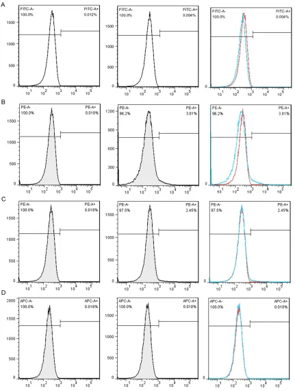
mesenchy-mal stem cell property of the BMSCs was char-acterized by the presence of surface pheno-types by flow cytometry. Figure 3 showed the characterization of cultured BMSCs with mono-clonal antibodies CD45, CD34, CD 90and CD105. Primary cultures of BMSCs were expanded from passage 0 to passage 4. As shown, the percent of CD45, CD34, CD90 and CD105 was 0.004%, 3.81%, 2.45%, 0.018% respectively. The flow cytometery data showed that our cultured BMSCs expressed standard surface markers of BMSCs and therefore used for experiments described below.
Multi-lineage differentiation potential of BM-SCs
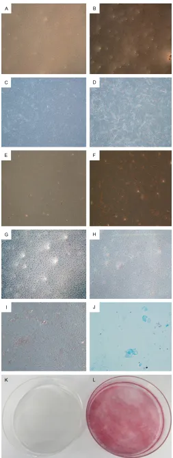
The effect of ICSII on fat droplets formation dur-ing adipogenic differentiation of BMSCs was studied and the results were shown in Figure 4A-F. As shown, lipid droplets formed when BMSCs were cultured in adipogenic medium; while in complete medium, there was no lipid droplets formed. It indicated ICSII could inhibit-ed the adipogenic differentiation of BMSCs. Mineralization is an important functional index of osteogenic differentiation in vitro and bone
regeneration in vivo. Alizarin Red S (ARS) staining was per-formed to study the effect of ICSII on mineralization in osteogenic differentiation of BMSCs and the results were shown in Figure 4G-L. As shown, at day 25, a large amount of calcium nodules were formed when BMSCs were cultured in osteogenic medium (Figure 4H, 4I, 4L); while in complete medium, there was no calcium nodules formed (Figure 4G, 4K).
ICSII enhanced mineraliza-tion in osteogenic differentia-tion of BMSCs
Mineralization is the mature sign of osteogenic differentia-tion of BMSCs. We analyzed the calcium nodule formation situation in ICSII, ICSII+OM, OM, CM group by Alizarin red staining. Results were shown in Figure 5. As shown in Figure Figure 1. Chemical structure of icariin and icariside II. A: Icariin B: Icariside II.
The common structure is 8-prenylkaempferol.
Figure 2. Morphology of BMSCs in passage 1 (A) and passage 3 (B). P0 cultures were maintained for 8 days, after which they were trypsinized and
replated as passage1cells. Magnification ×50 (A), ×100 (B).
5A and 5B, when BMSCs were treated in medi-um with 10-5 icariside II for12 days (Figure 5B), showed the typical morphology of osteoblasts-cobblestone compared with that cultured in complete medium (Figure 5A). As shown in Figure 5C and 5D, a large amount of calcium nodules were formed when BMSCs were cul-tured in medium with 10-5 Icariside II while there was no calcium nodules formed in com-plete medium. Moreover, the red staining in ICSII+OM group is more densely than that in ICSII group.
ICSII promoted cell proliferation of BMSCs
concentra-Figure 3. Characterization of cultured BMSCs with monoclonal antibodies. Primary cultures of BMSCs were ex-panded to passage 4. Populations of these cells were analyzed with monoclonal antibodies CD45, CD34, CD 90and
CD105 by flow cytometry. A: CD45; B: CD34; C: CD90; D: CD105. P2: Passage 2.
tions in D2 and D4 stage. Therefore, we
[image:5.612.91.522.71.641.2]ICSII promoted osteogenic differentia-tion of BMSCs
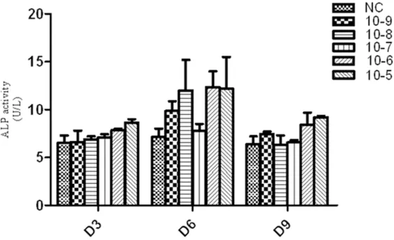
ALP is one of the early markers of osteogenic differentiation of BMSCs. To study the function of ICSII in osteo-genic differentiation of MSCs, the dose-dependent effects and optimal concentrations need to be determined. In this study, ALP activity with ICSII con-centration of 10-9, 10-8, 10-7, 10-6 and 10-5 M/L was determined, respectively. As Figure 7 showed, compared with the control, ALP activity in ICSII con-centration of 10-5 and 10-6 group was significantly higher (P<0.05). Although in other groups with ICSII concentra-tion of 10-9, 10-8 and 10-7 M/L, ALP activity were not significantly different from the control (P>0.05), it was slight-ly enhanced. Therefore, we concluded ICSII could enhance ALP activity and the concentration of 10-5 and 10-6 M/L was selected.
Discussion
The flavonoid compound icariside II is derived from the stems and leaves of Epimedium koreanum Nakai (Berbe- ridaceae) [27]. It was known to have antioxidant activity and inhibit melano-genesis and hypoxia inducible factor [28]. It has shown potential anti-cancer activity as indicated by its ability to suppress the proliferation and to induce apoptosis of various human cancer cells including osteosarcoma, breast carcinoma and prostate carci-Figure 4. Multi-lineage differentiation po-tential of BMSCs. A-F: Adipogenic differ-entiation of BMSCs. Lipid droplets formed only when BMSCs were cultured in ad-ipogenic medium (B, D, F) compared with complete medium (A, C, E), as shown by Oil
red O staining. Magnification ×50 (A, B),
×100 (C, D), ×200 (E, F). G-L: Mineraliza-tion in osteogenic differentiaMineraliza-tion of BMSCs. Calcium nodules formed only when BMSCs were cultured in osteogenic medium (H, I, L) compared with complete medium (G, K), as indicated by Alizarin red staining.
Mag-nification ×50 (G, H), ×100 (I), ×1 (K, L). K:
Chondrogenic differentiation of BMSC: cell pellet of BMSCs expressed proteoglycan as indicated by positive for Alcian blue
[image:6.612.88.341.67.735.2]noma [20, 29]. It has been used as a traditional medicine for neurasthenia, amnesia and impo-tence [30]. However, the effect of icariside II on osteogenic differentiation of BMSWs is poorly defined. Therefore, in this study Beagle canine BMSCs were selected to perform this research. BMSCs are also designated mesenchymal stem cells, which are the most commonly used
[image:7.612.89.377.69.539.2]cell source for bone tissue engineering [31]. They nor-mally give rise to bone, carti-lage, and mesenchymal cells [32]. However, the percentage of putative stem cells in BMSCs in whole bone marrow is considered as less than 0.01% and indeed the majori-ty of the cells are not true stem cells [33]. The Me- senchymal and Tissue Stem Cell Committee of the In- ternational Society for Cellular Therapy proposes minimal cri-teria to define human MSC. They reported MSC must express CD105 CD73 and CD90, lack expression of CD45, CD34, CD14 or CD11b, CD79a or CD19 and HLA-DR surface molecules and MSC must differentiate to osteo-blasts, adipocytes and chon-droblasts in vitro [34]. How- ever, those markers are not sufficient to define a specific population for osteogenic stem/progenitor cells. There is no established, widely accepted marker for osteo-genic stem/progenitor cells in bone marrow [35]. In this study, density gradient cen-trifugation-isolated canine bone marrow mononuclear cells were cultured in vitro and BMSCs of the 4th pas-sages were chosen as target cells. First, the morphology, phenotype, and function of BMSCs were examined. The result showed the percent (%) of CD45, CD34, CD90 and CD105 were 0.004%, 3.81%, 2.45%, 0.018%, respectively. Figure 5. The calcium nodule formation situation in ICSII, ICSII+OM, OM, CM
group by Alizarin red staining. A, C, G: BMSCs cultured in complete medium; B, E: BMSCs cultured in osteogenic medium; D, H: 10-5 M/L Icariside II me-dium; F, I: Osteogenic medium containing 10-5 Mol/L Icariside II. Magnifica -tion ×50 (A-F), ×1 (G-I).
formed mineralized nodules, since their ability to differentiate into osteoblast-like cells can be easily diminished during culture and passage [37, 38]. In this study, the cells we isolated from bone marrow aspirated from the iliac crest of the Beagle dog exhibited the same phenotype as mentioned above.
Next, the effect of on fat droplets formation and mineralization during adipogenic differen-tiation of BMSCs was studied. The ability of BMSCs to undergo matrix mineralization can be detected by von kossa staining and a high level of matrix mineralization in ICSII with or without
osteogenic medium can detect mineralized nodules. When cultured in the pres-ence of ICSII, OM and OM+ICSII group, mineralized nodules were observed. Therefore, we concluded ICSII inhibited the fat droplets for-mation and promoted os- teoblast mineralization. ICSII is one metabolite of Icariin. Icaritin could promote osteo-genic but inhibit adipoosteo-genic differentiation of MSCs by regulating osteogenesis and adipogenesis related gene [39, 40]. The inhibition on adi- pogenic differentiation result-ed in the number of fat re- duced, which indirectly pro-moted the conversion of BM- SCs to osteoblast direction. When the occurrence of os- teogenic differentiation, BM- SCs become cuboid or polygo-nal in shape, resembling osteoblasts, and they cluster concentrically, forming miner-alised nodules [41]. Such nod-ules are considered reliable indicators of in vitro osteogen-ic differentiation [42].
[image:8.612.93.373.71.229.2]Then the effect of ICSII with different concentration on ALP activity and BMSCs prolif-eration was studied. Results showed ALP activity and OD value increased with ICSII concentration increased from 10-9 M/L to 10-5 M/L and an Figure 6. The effect of icariside II on BMSCs proliferation at a wide range of
doses measured by Cell Counting Kit-8. The cells were incubated with icari-side II (10-9 M to 10-5 M) for 2, 4, 6 and 8 days. The complete medium served as control. All experiments were carried out in 5 replicates and the data were expressed as means ± SD.
Figure 7. Icariside II induced alkaline phosphatase (ALP) activity during os-teogenic differentiation of BMSCs. BMSCs treated with Icariside II (10-9 M to 10-5 M) in absence of OM or in presence of OM for 3, 6 and 9 days respec-tively, then the cells were lysed and ALP activity assay was performed.
[image:8.612.93.374.313.484.2]In conclusion, ICSII could inhibit adipogenic dif-ferentiation, promote osteoblast mineraliza-tion, BMSCs proliferation and enhance ALP activity. Therefore, we concluded ICSII could promote osteogenic differentiation of BMSCs. The optimal ICSII concentration was 10-5 in our study.
Acknowledgements
Current research was financially supported by Shanghai Pudong New Area Science and Technology Development Fund Innovation Fund Project (Number: PKJ2013-Y12).
Disclosure of conflict of interest
None.
Address correspondence to: Dr. Yuanliang Huang, Department of Stomatology, Shanghai East Hospital
Affiliated with Tongji University, 150 Jimo Road,
Shanghai 200120, P. R. China. Tel: +086133- 01921076; Fax: +086-21-58765119; E-mail: 13301921076@163.com
References
[1] Bianco P, Riminucci M, Gronthos S, Robey PG. Bone marrow stromal stem cells: nature, biol-ogy, and potential applications. Stem Cell 2001; 19: 180-92.
[2] Deng W, Obrocka M, Fischer I, Prockop DJ. In vitro differentiation of human marrow stromal cells into early progenitors of neural cells by conditions that increase intracellular cyclic AMP. Biochem Biophys Res Commun 2001; 282: 148-52.
[3] Woodbury D, Schwarz EJ, Prockop DJ, Black IB. Adult rat and human bone marrow stromal cells differentiate into neurons. J Neuro Res 2000; 61: 364-70.
[4] Dezawa M, Kanno H, Hoshino M, Cho H, Mat-sumoto N, Itokazu Y, Tajima N, Yamada H, Sawada H, Ishikawa H, Mimura T, Kitada M, Suzuki Y, Ide C. Specific induction of neuronal
cells from bone marrow stromal cells and ap-plication for autologous transplantation. J Clin-Invest 2004; 113: 1701-10
[5] Banfi A, Muraglia A, Dozin B, Mastrogiacomo M, Cancedda R, Quarto R. Proliferation kinet-ics and differentiation potential of ex vivo ex-panded human bone marrow stromal cells: implications for their use in cell therapy. Exp Hematol 2000; 28: 707-15.
[6] Colter DC, Class R, DiGirolamo CM, Prockop DJ. Rapid expansion of recycling stem cells in cul-tures of plastic-adherent cells from human
bone marrow. Proc Natl Acad Sci 2000; 97: 3213-8.
[7] Koc ON and Lazarus HM. Mesenchymal stem cells: heading into the clinic. Bone Marrow Transplant 2001; 27: 235-9.
[8] Huang KC. The pharmacology of Chinese herbs. CRC press; 2011.
[9] Meng FH, Li YB, Xiong ZL, Jiang ZM, Li FM. Os-teoblastic proliferative activity of Epimedium brevicornum Maxim. Phytomedicine 2005; 12: 189-93.
[10] Li L, Peng L, Miao J, Qiu Y, Zhou Y, Gao X, Xu Y, Shi Z, Shao D, Ma Z. Icariin induces the Expres-sion of Toll-like Receptor 9 in Ana-1 Murine Macrophages. Phytother Res 2011; 25: 1732-5.
[11] Liang Q, Wei G, Chen J, Wang Y, Huang H. Vari-ation of Medicinal Components in a Unique Geographical Accession of Horny Goat Weed Epimedium sagittatum Maxim. (Berberidace-ae). Molecules 2012; 17: 13345-56.
[12] Liu ZQ, Luo XY, Sun YX, Wu W, Liu CM, Liu ZQ, Liu SY. The antioxidative effect of icariin in hu-man erythrocytes against free-radical-induced haemolysis. J Pharm Pharmacol 2004; 56: 1557-62.
[13] Makarova MN, Pozharitskaya ON, Shikov AN, Tesakova SV, Makarov VG, Tikhonov VP. Effect of lipid-based suspension of Epimedium kore-anum Nakai extract on sexual behavior in rats. J Ethnopharmacol 2007; 114: 412-6.
[14] Zhao J, Ohba S, Shinkai M, Chung UI, Nagamune T. Icariin induces osteogenic differ-entiation in vitro in a BMP-and Runx2-depen-dent manner. BiochemBiophy Res Commun 2008; 369: 444-8.
[15] Liang W, Lin M, Li X, Li C, Gao B, Gan H, Yang Z, Lin X, Liao L, Yang M. Icariin promotes bone formation via the BMP-2/Smad4 signal trans-duction pathway in the hFOB 1.19 human os-teoblastic cell line. Int J Mol Med 2012; 30: 889-95.
[16] Zhao J, Ohba S, Komiyama Y, Shinkai M, Chung UI, Nagamune T. Icariin: a potential osteoin-ductive compound for bone tissue engineer-ing. Tissue Eng Part A 2009; 16: 233-43. [17] Li D, Yuan T, Zhang X, Xiao Y, Wang R, Fan Y,
Zhang X. Icariin: a potential promoting com-pound for cartilage tissue engineering. Osteo-arthritis Cartilage 2012; 20: 1647-56. [18] Fan JJ, Cao LG, Wu T, Wang DX, Jin D, Jiang S,
Zhang ZY, Bi L, Pei GX. The dose-effect of icari-in on the proliferation and osteogenic differen-tiation of human bone mesenchymal stem cells. Molecules 2011; 16: 10123-33.
com-pound formulation) by LC-MS/MS. J Chro-matogr B Analyt Technol Biomed Life Sci 2007; 860: 113-20.
[20] Choi HJ, Eun JS, Kim DK, Li RH, Shin TY, Park H, Cho NP, Soh Y. Icariside II from Epimedium
koreanum inhibits hypoxia-inducible factor-1α
in human osteosarcoma cells. Eur J Pharmacol 2008; 579: 58-65.
[21] Kang SH, Jeong SJ, Kim SH, Kim JH, Jung JH, Koh W, Kim JH, Kim DK, Chen CY, Kim SH. Ica-riside II induces apoptosis in U937 acute my-eloid leukemia cells: role of inactivation of STAT3-related signaling. PLoS One 2012; 7: e28706.
[22] Meng F. Osteoblastic proliferation stimulating activity of Epimedium koreanum. Nakai
ex-tracts and its flavonol glycosides. Pharmaceuti -cal Biology 2005; 43: 92-95.
[23] Huang J, Yuan L, Wang X, Zhang TL, Wang K. Icaritin and its glycosides enhance osteoblas-tic, but suppress osteoclasosteoblas-tic, differentiation and activity in vitro. Life Sci 2007; 81: 832-40. [24] Trzeciakiewicz A, Habauzit V and Horcajada
MN. When nutrition interacts with osteoblast function: molecular mechanisms of polyphe-nols. Nutr Res Rev 2009; 22: 68-81.
[25] Wang CG. LC determination of icariside II in rat plasma and tissues: application to a tissue dis-tribution study. Chromatographia 2011; 74: 251-258.
[26] Sun XJ, Xia LG, Chou LL, Zhong W, Zhang XL, Wang SY, Zhao J, Jiang XQ, Zhang ZY. Maxillary
sinus floor elevation using a tissue engineered bone complex with BMP-2 gene modified bM -SCs and a novel porous ceramic scaffold in rabbits. Arch Oral Biol 2010; 55: 195-202. [27] Kim SH, Ahn KS, Jeong SJ, Kwon TR, Jung JH,
Yun SM, Han I, Lee SG, Kim DK, Kang M, Chen CY, Lee JW, Kim SH. Janus activated kinase 2/ signal transducer and activator of transcrip-tion 3 pathway mediates icariside II-induced apoptosis in U266 multiple myeloma cells. Eur J Pharmacol 2011; 654: 10-6.
[28] Xia Q, Xu D, Huang Z, Liu J, Wang X, Wang X, Liu S. Preparation of icariside II from icariin by en-zymatic hydrolysis method. Fitoterapia 2010; 81: 437-42.
[29] Lee KS, Lee HJ, Ahn KS, Kim SH, Nam D, Kim DK, Choi DY, Ahn KS, Lu J, Kim SH. Cyclooxy-genase-2/prostaglandin E2 pathway mediates icariside II induced apoptosis in human PC-3 prostate cancer cells. Cancer Lett 2009; 280: 93-100.
[30] Wang YK ,Huang ZQ. Protective effects of icari -in on human umbilical ve-in endothelial cell - in-jury induced by H2O2 in vitro. Pharmacoll Res 2005; 52: 174-82.
[31] Chen J, Li Y, Wang L, Zhang Z, Lu D, Lu M, Chopp M. Therapeutic benefit of intravenous
administration of bone marrow stromal cells after cerebral ischemia in rats. Stroke 2001; 32: 1005-11.
[32] Sanchez-Ramos J, Song S, Cardozo-Pelaez F, Hazzi C, Stedeford T, Willing A, Freeman TB, Sa-porta S, Janssen W, Patel N, Cooper DR, San-berg PR. Adult bone marrow stromal cells dif-ferentiate into neural cells in vitro. Exp Neurol 2000; 164: 247-56.
[33] Toma C, Pittenger MF, Cahill KS, Byrne BJ, Kes-sler PD. Human mesenchymal stem cells dif-ferentiate to a cardiomyocyte phenotype in the adult murine heart. Circulation 2002; 105: 93-8.
[34] Dominici M, Le Blanc K, Mueller I, Slaper-Cor-tenbach I, Marini F, Krause D, Deans R, Keat-ing A, Prockop DJ, Horwitz E. Minimal criteria
for defining multipotent mesenchymal stromal
cells. The International Society for Cellular Therapy position statement. Cytotherapy 2006; 8: 315-7.
[35] Majumdar MK, Thiede MA, Mosca JD, Moor-man M, Gerson SL. Phenotypic and functional comparison of cultures of marrow-derived mesenchymal stem cells (MSCs) and stromal cells. J Cell Physiol 1998; 176: 57-66.
[36] Dissanayaka WL, Zhu X, Zhang C, Jin L. Charac-terization of dental pulp stem cells isolated from canine premolars. J Endod 2011; 37: 1074-80.
[37] Sugiura F, Kitoh H and Ishiguro N. Osteogenic potential of rat mesenchymal stem cells after several passages. Biochem Biophys Res Com-mun 2004; 316: 233-9.
[38] Agata H, Asahina I, Watanabe N, Ishii Y, Kubo N, Ohshima S, Yamazaki M, Tojo A, Kagami H. Characteristic change and loss of in vivo osteo-genic abilities of human bone marrow stromal cells during passage. Tissue Eng Part A 2010; 16: 663-73.
[39] Yao D, Xie XH, Wang XL, Wan C, Lee YW, Chen SH, Pei DQ, Wang YX, Li G, Qin L. Icaritin, an exogenous phytomolecule, enhances
osteo-genesis but not angioosteo-genesis-an in vitro effi -cacy study. PLoS One 2012; 7: e41264. [40] Wang XF and Wang J. Icaritin suppresses the
proliferation of human osteosarcoma cells in vitro by increasing apoptosis and decreasing MMP expression. Acta Pharmacologica Sinica 2014; 35: 531-9.
[41] Payushina O, Domaratskaya E and Starostin V. Mesenchymal stem cells: sources, phenotype, and differentiation potential. Biology Bulletin 2006; 33: 2-18.
[43] Hessle L, Johnson KA, Anderson HC, Narisawa S, Sali A, Goding JW, Terkeltaub R, Millan JL.
Tissue-nonspecific alkaline phosphatase and
plasma cell membrane glycoprotein-1 are cen-tral antagonistic regulators of bone mineraliza-tion. Proc Natl Acad Sci 2002; 99: 9445-9. [44] Anderson HC, Sipe JB, Hessle L, Dhanyamraju
R, Atti E, Camacho NP, Millán JL. Impaired
cal-cification around matrix vesicles of growth plate and bone in alkaline phosphatase-defi -cient mice. Am J Pathol 2004; 164: 841-7.