Original Article
Knockdown of lncRNA XIST suppresses osteosarcoma
progression by inactivating AKT/mTOR signaling
pathway by sponging miR-375-3p
Xin Sun, Bo Wei, Zhi-Heng Peng, Qing-Long Fu, Chao-Jun Wang, Jin-Chang Zheng, Jie-Cong Sun
Orthopaedic Center, Affiliated Hospital of Guangdong Medical University, Xiashan District, Zhanjiang, Guangdong Province, China
Received January 21, 2019; Accepted February 22, 2019; Epub May 1, 2019; Published May 15, 2019
Abstract: Background: Osteosarcoma (OS) is one of the most common bone tumors in adolescents and young adults. Emerging evidence suggested ncRNA (lncRNA and miRNA) are closely associated with cell progression, apoptosis and autophagy. However, the role of regulatory network between ncRNA and mRNA in OS has not been
fully verified. Methods: lncRNA XIST, miRNA expression were detected by qRT-PCR. The protein expression of LC3, p62, AKT, p-AKT, mTOR and p-mTOR was measured by western blot. MTT assay and flow cytometry were applied to
measure cell proliferation and apoptosis. Luciferase assay was used to ensure the relationship between lncRNA,
miRNA and mRNA. GFP-LC3 cells were observed using fluorescence microscope. Results: XIST expression was
up-regulated but miR-375-3p was down-up-regulated in OS tissues and cells. Luciferase assay results demonstrated that
miR-375-3p was a target of XIST and mTOR was a target mRNA of miR-375-3p. In addition, knockdown of XIST and mTOR inhibited OS cell proliferation and autophagy, but induced apoptosis. Knockdown of XIST could reverse the effect of 375-3p inhibitor on OS cells. The effects of si-mTOR of OS cells could be reversed by silencing miR-375-3p. Moreover, knockdown of XIST inhibited AKT/mTOR signaling pathway via sponging miR-miR-375-3p. Conclusion: Knockdown of XIST inhibited cell growth and autophagy but induced cell apoptosis by suppressing the AKT/mTOR
signaling pathway by sponging miR-375-3p.
Keywords: Osteosarcoma, XIST, miR-375-3p, AKT/mTOR
Introduction
Osteosarcoma (OS) is the most common pri-mary bone tumor in adolescents and young adults, exhibiting osteogenic differentiation and producing malignant bones [1, 2]. How- ever, research on the underlying mechanisms of OS is still lacking.
Long non-coding RNAs (lncRNAs) are a class of RNAs, which are ~200 nucleotides in length. Evidence determined that lncRNAs are widely involved in multiple biologic processes, includ-ing cell progression, inflammation and tumori -genesis [3-5]. LncRNA-XIST (X inactive-specific transcript) is an oncogenic lncRNA in various cancers, including pancreatic cancer, non-small cell lung cancer, hepatocellular carcino-ma, pancreatic cancer and osteosarcoma [6-9]. Some studies showed that LncRNA-XIST was up-regulated in OS and associated with cellular processes [9, 10].
MicroRNAs (miRNAs) are ~22 nucleotides small non-coding RNAs that repress gene expression at post-transcription by binding to mRNAs [11]. Additionally, miRNAs, as oncogenes or tumor suppressors, play an important role in cell pro-liferation, migration, apoptosis, and drug resis-tance of many cancers [12-15]. For example, miR-155, as an oncogene, is up-regulated in liposarcoma and promotes tumor cell growth through targeting casein kinase-1α [16]. miR-145, as a tumor suppressor, was down-regulat-ed in colon cancer and inhibits cell growth by regulating HDAC4 [17].
that XIST was up-regulated and miR-375-3p was down-regulated in OS, and bioinformatic analysis showed that miR-375-3p was a poten-tial target miRNA of XIST and mTOR was a potential target mRNA of miR-375-3p. In addi-tion, autophagy plays an important role in regu-lating cellular progression and tumor formation [23]. Therefore, we hypothesized XIST can regu -late mTOR expression by sponging miR-375-3p and that the regulatory network was associated with OS cell growth and autophagy.
Materials and methods
Patients and tissues specimens
20 pairs of tissues and adjacent tissues speci-mens were obtained from 20 OS patients with no chemotherapy or radiation therapy at Affi-liated Hospital of Guangdong Medical Univer- sity. Written informed consent was obtained from patients and their guardians. This study has been approved by Experiments committee of Affiliated Hospital of Guangdong Medical University.
Cell culture
The human OS cell lines MG-63, HOS, U2-OS and Saos-2, and the human normal cell line hFOB1.19 were purchased from the American Type Cultured Collection (Manassas, VA, USA). The cells were cultured in Dulbecco’s Modified Eagle Medium (DMEM) with 10% fetal bovine serum (FBS, Gemini Bio-Products, West Sacra- mento, CA), penicillin and streptomycin at 37°C with 5% CO2.
Cell transfection
miR-375-3p mimic, miR-375-3p inhibitor, sh-XIST, pc-XIST and their respective negative controls were obtained from GenePharma (Shanghai, China). si-mTOR, pc-mTOR and their respective negative controls were purchased form from Ribobio (Guangzhou, Guangdong, China). All the vectors and fragments were transfected into OS cell lines using Lipofecta- mine 2000 (Invitrogen, Carlsbad, CA) according to the manufacturer’s instruction.
Reverse transcription and quantitative real-time PCR
Total RNA was extracted from tissues and cells by TRIzol reagent (Invitrogen) according to the
manufacturer’s instructions. RNA concentra -tion was detected using the NanoDropND- 1000 spectrophotometer (NanoDrop Techno-logies, Wilmintgon, DE, USA). cDNA were re- verse transcribed using TaqMan MicroRNA Reverse Transcription Kit for microRNA (Applied Biosystems, Foster City, CA, USA) or M-MLV Reverse Transcriptase (Invitrogen) for mRNA. Then the SYBR® Green (Promega, Madison, WI, USA) was applied to detect the expression of XIST, miR-375-3p and mTOR, according to the manufacturer’s instruction. GAPDH and U6 were used as reference gene for lncRNA, mRNA and miRNA. Quantitative real-time PCR was performed by using an iQTM5 Multicolor Real-Time PCR Detection System (Bio-Rad, Hercules, CA, USA). The primer: U6: forward CTCGCTTCGGCAGCACA-3’ and reverse 5’-AACGCTTCACGAATTTGCGT-3’; miR-375-3p: for -ward 5’-ACACTCCAGCTGGGTTTGTTCGTTCGGC-TC-3’ and reverse 5’-CTCAACTGGTGTCGTGGA-GTCGGCAATTCAGTTGAGTCACGCGA-3’ GAPDH: forward 5’-GCACCGTCAAGGCTGAGAAC-3’ and reverse 5’-ATGGTGGTGAAGACGCCAGT-3’; XIST: forward 5’-ACGCTGCATGTGTCCTTAG-3’ and re-verse 5’-GAGCCTCTTATAAGCTGTTTG-3’; mTOR: forward 5’-ACATGCAGCTGTCCTGGTTC-3’ and reverse 5’-TGAGGCTTCTGCATCTCCTT-3’.
Western blot
Cell proliferation
2 × 10³ cells were seeded into the 96-well cell culture plates (Corning Inc., Corning, NY, USA). Cell proliferation was measured using 3-(4,5- Dimethyl-2-thiazolyl)-2,5-diphenyl-2Htetrazoli-um bromide (MTT) assay kit (Sigma-Aldrich, St. Louis, MO, USA) according to the manufactur-er’s protocol. 30 µL serum-free media with MTT solution were added into each well and incu-bated for 4 h at 37°C. Then 150 ul MTT solvent were added and incubated for 3 h at 37°C. The absorbance at 450 nm was measured using a spectrophotometric microplate reader (Beyo- time Institute of Biotechnology, Haimen, China).
Luciferase assay
XIST wild-type (WT) or mutant (MUT) and mTOR wild-type (WT) or mutant (MUT) fragment were amplified and inserted the downstream of the luciferase vector pGL3-control vector (Promega, Madison, WI, USA). Then XIST WT, XIST MUT, mTOR WT and mTOR MUT were co-transfected with miR-375-3p mimic into MG-63 and U2-OS cell lines using Lipofectamine 2000 (Invitro-
Statistical analysis
All statistical analyses were performed using GraphPad Prism (GraphPad Software, San Diego, CA, USA). All results were showed as mean ± SD (standard deviation) at least three repeated individual experiments. All compari-sons groups were analyzed using Student t-test. P-value < 0.5 was considered significant. Results
XIST expression was up-regulated and miR-375-3p was down-regulated in OS tissues and cells
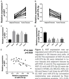
[image:3.612.89.376.72.400.2]In our experiments, we obtained tumor tissues and matched adjacent tissues from 20 pa- tients. As shown in Figure 1A and 1B, XIST was up-regulated and miR-375-3p was down-regulated in tumor tissues compared with adja-cent tissues. Then to test their role in OS cell lines, we chose hFOB1.19, MG-63, HOS, U2-OS and Saos-2 and measured XIST, miR-375-3p expression by qRT-PCR. The results showed that the expression of XIST was increased and the expression of miR-375-3p was decreased Figure 1. XIST expression was
up-regulated and miR-375-3p was down-regulated in OS tissues and cells. (A
and B) The expression of XIST (A) and
miR-375-3p (B) were detected in tu-mor tissues and adjacent tissues by
qRT-PCR. (C and D) The expression of XIST (C) and miR-375-3p (D) were de -tected in hFOB1.19, MG-63, HOS,
U2-OS and Saos-2 cell lines by qRT-PCR. (E) XIST and miR-375-3p correlation relationship was analyzed by Pear
-son’s correlation analysis. *P < 0.05.
gen). Cell collected at 48 h after transfection, and the luciferase activity was detect-ed using the Dual luciferase reporter assay system (Pro- mega).
Cell apoptosis
Cell apoptosis was detect- ed using flow cytometry and Annexin-V fluorescein isothio -cyanate and propidium iodide (FITC-Annexin V/PI) apoptosis detection kit (Life Technolo-gy, Waltham, MA, USA). Cells were stained by Annexin-V FITC/PI. Then each sample was measured using flow cy-tometry.
Microcopy
in MG-63, HOS, U2-OS, Saos-2 cell lines com-pared with hFOB1.19 cell line (Figure 1C and 1D). We selected MG-63 and U2-OS cell lines for following experiments. Meanwhile, miR-375-5p expression was negatively correlated with the expression of XIST in OS tissues (Fig- ure 1E).
Knockdown of XIST inhibited cell proliferation and autophagy, and induced apoptosis in OS cells
To further access the function of XIST, siRNA was conducted to knock down its expression (Figure 2A). Subsequently, MTT assay showed
[image:4.612.90.520.69.462.2]that knockdown of XIST inhibited cell prolifera -tion in MG-63 and U2-OS cell lines (Figure 2B and 2C). Apoptosis rate of sh-XIST group was significantly higher than control (Figure 2D and 2E). Autophagy plays a critical role in regulating the cell progression in cancers, so we detected the protein expression LC-3 and p62 in OS, which are the important markers of autophagy [24]. As shown in Figure 2F, GFP-LC3 positive cells was significantly lower in sh-XIST group compared with sh-NC groups. In addition, west-ern blot data verified that knockdown of XIST down-regulated the expression of LC3-II/I, and up-regulated p62 expression (Figure 2G). In conclusion, knockdown of XIST inhibited cell Figure 2. Knockdown of XIST inhibited cell proliferation and autophagy, but induced apoptosis in OS cells. (A) The expression of XIST was detected in sh-NC and sh-XIST groups of MG-63 and U2-OS cell lines via qRT-PCR. (B and C) Cell proliferation were measured in sh-NC and sh-XIST groups of MG-63 (B) and U2-OS (C) cell lines via MTT assay. (D and E) Cell apoptosis were detected in sh-NC and sh-XIST groups of MG-63 and U2-OS cell lines by flow cytom
proliferation and autophagy, but induced apop-tosis in OS cells.
miR-375-3p was a target of XIST
To further explore the relationship of XIST and miR-375-sp, we predicted that miR-375-3p was a candidate miRNA target of XIST in miRCode and miRBase database (Figure 3A). To verify this, we co-transfected the luciferase reporter plasmids XIST-WT or XIST-MUT with miR-NC or miR-375-3p into MG-63 and U2-OS cell lines respectively. The results showed that miR-317-3p decreased the luciferase activity by binding transfection of XIST WT, but not XIST MUT (Figure 3B). Furthermore, we verified that miR-375-3p expression was negatively regulated by XIST (Figure 3C and 3D). With sh-NC or sh-XIST transfected into MG-63 and U2-OS cell lines, we found that the miR-375-3p expression was increased by sh-XIST (Figure 3C). Transfection of miR-375-3p inhibitor decreased the expres-sion of miR-375-3p in MG-63 and U2-OS cell lines, while sh-XIST restored its expression (Figure 3D). Taken together, we proved that miR-375-3p was a target miRNA of XIST. Down-regulated XIST reversed the effect of low miR-375-3p expression on OS cells
To explore the function of miR-375-3p in OS cells, we obtained the MG-63 and U2-OS cell
3p increased the protein level of LC3-II/I, and reduced the protein level of p62. However, knockdown of XIST could reverse the effect of miR-375-3p inhibitor on the protein level of LC3-II/I and p62 in MG-63 and U2-OS cell lines (Figure 4E). Therefore, miR-375-3p inhibitor transfection increased OS cells proliferation and autophagy, but decreased apoptosis, which was recovered by XIST knockdown.
mTOR was a target of miR-375-3p
miRNA usually functions to bind with specific mRNA to mediate itsdegradation or transcrip-tion [25]. Bioinformatic analysis revealed that mTOR is a potential target gene of miR-375-3p, and miR-375-3p binds to mTOR 3’UTR (Figure 5A). Then mTOR WT or mTOR MUT were co-transfected with miR-NC or miR-375-3p into MG-63 and U2-OS cell lines. Luciferase report-er assay results showed that lucifreport-erase activity was significantly decreased in mTOR 375-3p group compared with mTOR WT+miR-NC group, while mTOR MUT+miR-375-3p gro-ups showed no difference with mTOR MUT+ mTOR MUT+miR-NC group, implying that mTOR was a target of miR-375-3p (Figure 5B). To further clarify our speculation, the protein level of mTOR was measured by western blot. We found that mTOR protein level was remarkably Figure 3. miR-375-3p is a target of XIST. A. miRCode and miRBase predic
-tion of miR-375-3p binding to XIST. B. Luciferase reporter assay was used to detected luciferase activity in XIST WT+miR-NC, XIST WT+miR-375-3p, XIST MUT+miR-NC and XIST MUT+miR-375-3p groups. C. The expression of miR-375-3p was detected in sh-NC and sh-XIST groups by qRT-PCR. D. The expression of 3p was detected in miR-NC inhibitor, miR-375-3p inhibitor, miR-375-miR-375-3p inhibitor+sh-NC and miR-375-miR-375-3p inhibitor+sh-XIST groups by qRT-PCR. *P < 0.05.
lines with transfectionof miR-375-3p inhibitor. As shown in Figure 4A and 4B, MTT assay results showed that cell prolif-eration were significantly pro -moted by miR-375-3p inhibi-tor, whereas knockdown of XIST can reverse the effect of miR-375-3p inhibitor (Figure 4C).
miR-375-increased by miR-375-3p inhibitor in MG-63 and U2-OS cell lines (Figure 5C). Then, when the si-mTOR and miR-NC inhibitor or si-mTOR and miR-375-3p inhibitor were co-transfected into OS cells, we found that down-regulated
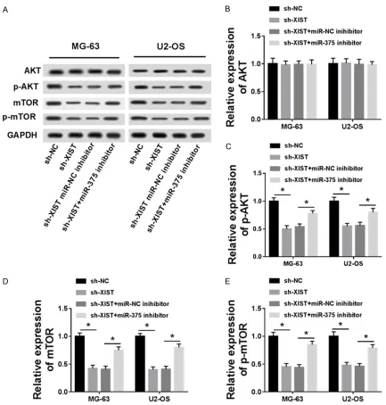
Knockdown of XIST inhibited AKT/mTOR sig-naling pathway through regulating miR-375-3p
[image:6.612.93.373.72.509.2]AKT, p-AKT, mTOR and p-mTOR are key proteins in AKT/mTOR signaling pathway, for which we Figure 4. Knockdown of XIST reversed the effect of miR-375 inhibitor on
cell proliferation, autophagy and apoptosis in OS cells. (A and B) Cell prolif-eration was measured in miR-NC inhibitor, miR-375-3p inhibitor, miR-375
inhibitor+sh-NC and miR-375-3p inhibitor+sh-XIST groups of MG-63 (A) and
U2-OS (B) cell lines. (C) Cell apoptosis was detected in NC inhibitor,
miR-375-3p inhibitor, miR-375 inhibitor+sh-NC and miR-miR-375-3p inhibitor+sh-XIST groups of MG-63 and U2-OS cell lines by flow cytometry. (D) GFP-LC3 posi -tive cells were calculated in miR-NC inhibitor, miR-375-3p inhibitor, miR-375
inhibitor+sh-NC and miR-375-3p inhibitor+sh-XIST groups of MG-63 and U2-OS cell lines. (E) The expressions of LC3-І, LC3-ІІ and p62 were measured in NC inhibitor, 375-3p inhibitor, 375 inhibitor+sh-NC and miR-375-3p inhibitor+sh-XIST groups of MG-63 and U2-OS cell lines by western blot. *P < 0.05.
miR-375-3p can recovered mTOR expression reduced by si-mTOR (Figure 5D). There-fore, mTOR protein level cou- ld be negatively regulated by miR-375-3p and mTOR was a target of miR375-3p.
mTOR knockdown inhibited OS cell proliferation and autophagy but induced cell apoptosis in vitro, which was reversed by down-regulating miR-375-3p
detected their expression in NC, XIST, sh-XIST+miR-NC inhibitor and sh-XIST+miR-375-3p inhibitor groups by western blot (Figure 7A). Our results demonstrated that down-regulated XIST suppressed the protein level of p-AKT, mTOR and p-mTOR, whereas had no significant effect on AKT. Therefore, the AKT/mTOR sig-naling pathway was inactivated by sh-XIST. Otherwise, miR-375-3p inhibitor could restore p-AKT, mTOR and p-mTOR expression inhibited by sh-XIST (Figure 7B-E). Therefore, down-regu -lated XIST inhibited the AKT/mTOR signaling pathway, which was reversed by miR-375-3p silencing.
Discussion
LncRNA plays an indispensable role in the occurrence and development of tumors. Emer- ging studies indicated that lncRNAs were wide- ly involved in cell progression, inflammation, immune response, drug resistance and progno-sis in cancers [3, 22, 26]. Of note, lncRNA XIST has been investigated as an oncogenic lncRNA to affect cell growth in several cancers, includ-ing OS [7-9]. Previous studies suggested that lncRNA-XIST was up-regulated in OS and XIST/ miR-320b/RAP2B axis and XIST/miR-21-5p/
PDCD axis affected OS cell proliferation, inva-sion and metastasis [9, 10]. As the regulatory network of lncRNA is very complex and one lncRNA may have multiple targeted miRNAs, the regulatory mechanism of lncRNA comple-ment still needs further research and explora-tion. In present study, lncRNA-XIST was up-reg -ulated in OS tissues and cells. Besides, inhi- bition of XIST can reduce cell proliferation and induce cell apoptosis. LC3, as an autophago-some marker, can be incorporated into autoph-agy protein, which is a critical protein in auto- phagy [27]. The signaling adaptor p62 is a multidomain protein, which is associated with autophagy and apoptosis in cancers [28]. Thus, we found that knockdown of XIST inhibited cell autophagy and LC3-І and LC3-ІІ protein level and induced p63 protein level in OS cells. miR-375-3p is processed from the precursor has-miR-375 and associated with cell growth and is prognostic in cancers [29-31]. For exam-ple, Ding et al. showed that miR-375 was down-regulated in gastric cancer and inhibited cell proliferation [29]. As a tumor suppressor, miR-375 is also implicated in OS diagnosis, progno-sis and chemosensitivity [32-34]. However, the underlying mechanism has been not fully illumi-Figure 5. mTOR is a target of miR-375-3p. A. miRCode and miRBase prediction of miR-375-3p binding sites in the 3’UTR of mTOR. B. Luciferase reporter assay was used to detect luciferase activity in mTOR WT+miR-NC, mTOR WT+miR-375-3p, mTOR MUT+miR-NC and mTOR MUT+miR-375-3p groups. C. The expression of mTOR was detect
[image:7.612.91.526.70.312.2]nated. In our study, miR-375-3p was down-reg-ulated in OS tissues and cells and negatively correlated with XIST. Luciferase reporter assay verified that miR-375-3p was a target miRNA of XIST, which was reported for the first time. Moreover, down-regulated miR-375-3p could promote OS cells growth and autophagy but reduce apoptosis which had the opposite effect compared to knockdown of XIST.
Rapamycin (mTOR) was a downstream of AKT, which is a key factor in AKT/mTOR signaling pathway [35, 36]. AKT/mTOR signaling pathway plays an important role in cell autophagy, cell cycle progression, and apoptosis in several cancers, such as prostate cancer, endometrial cancer, and OS [19, 37, 38]. Miwa et al. show- ed that caffeine contributed to apoptosis via inhibition of AKT/mTOR/S6K, NF-kB and MAPK
pathway [39]. In our study, we proved for the first time that mTOR was the target gene of miR-375-3p and verified the function of mTOR in OS. More than that, knockdown of mTOR suppressed OS cells growth and autophagy but promoted apoptosis.
Notably, in our study, miR-375-3p silencing can reverse the effect of XIST knockdown on OS cells. Meanwhile, si-mTOR transfection inhibit -ed OS cells proliferation and autophagy but induced cell apoptosis in vitro, which was reversed by down-regulation of miR-375-3p. Taken together, knockdown of XIST sponged miR-375-3p to inhibit cell growth and autopha-gy by down-regulating mTOR.
[image:8.612.89.519.70.389.2]AKT, p-AKT, mTOR and p-mTOR were key pro -teins in the AKT/mTOR signaling pathway. Also, to further realize whether lncRNA-XIST affect Figure 6. si-mTOR transfection inhibited OS cell proliferation and autophagy and induced cell apoptosis in vitro, which was reversed by down-regulating miR-375-3p. (A and B) Cell proliferation was measured in si-NC, si-mTOR, si-mTOR+miR-NC inhibitor and si-mTOR+miR-375-3p inhibitor groups of MG-63 (A) and U2-OS (B) cell lines. (C) Cell apoptosis was detected in si-NC, si-mTOR, si-mTOR+miR-NC inhibitor and si-mTOR+miR-375-3p inhibitor groups of MG-63 and U2-OS cell lines by flow cytometry. (D) GFP-LC3 positive cells were calculated in NC, mTOR, si-mTOR+miR-NC inhibitor and si-mTOR+miR-375-3p inhibitor groups of MG-63 and U2-OS cell lines. (E) The expres
AKT/mTOR signaling pathway, we measured the AKT, p-AKT, mTOR and p-mTOR protein level using western blot and found that down-regu-lated XIST inhibited p-AKT, mTOR and p-mTOR protein level, whereas it had no significant effect on AKT. In addition, down-regulated miR-375-3p restored p-AKT, mTOR and p-mTOR pro -tein level inhibited by sh-XIST. However, our study showed that the expression of p-AKT, which is the upstream protein of mTOR, was changed by sh-XIST and miR-375-3p. This may be caused by other interactions between
miR-NAs and their target genes, which remains unclear. Taken together, lncRNA-XIST inhibited AKT/mTOR signaling pathway to affect cell pro -gression by sponging miR-375-3p.
Conclusions
[image:9.612.91.524.69.523.2]In conclusion, we first elucidated lncRNA-XIST/ miR-375-3p/mTOR axis plays an important role in OS cells, and lncRNA XIST promoted OS progression by activating AKT/mTOR signaling pathway through sponging miR-375-3p.
Acknowledgements
This study was approved by the Science and Technology Planning Project of Guangdong Province (Grant No.: 2017ZC0311).
Disclosure of conflict of interest
None.
Address correspondence to: Xin Sun, Orthopaedic
Center, Affiliated Hospital of Guangdong Medical
University, No. 57, South Renmin Avenue, Xiashan District, Zhanjiang 524001, Guangdong Province,
China. Tel: +86-759-2387-291; E-mail: sunxingd-mu@126.com
References
[1] Cortini M, Avnet S, Baldini N. Mesenchymal stroma: role in osteosarcoma progression. Cancer Lett 2017; 405: 90-99.
[2] Ottaviani G, Jaffe N. The epidemiology of os -teosarcoma. Cancer Treat Res 2009; 152: 421-436.
[3] Li H, Yu B, Li J, Su L, Yan M, Zhu Z, Liu B. Over -expression of lncRNA H19 enhances carcino-genesis and metastasis of gastric cancer. On-cotarget 2014; 5: 2318-2329.
[4] Rapicavoli NA, Qu K, Zhang J, Megan M,
Remi-Martin L, Chang HY. A mammalian pseu
-dogene lncRNA at the interface of inflamma
-tion and anti-inflammatory therapeutics. Elife 2013; 2: e00762.
[5] Yang YR, Zang SZ, Zhong CL, Li YX, Zhao SS,
Feng XJ. Increased expression of the lncRNA
PVT1 promotes tumorigenesis in non-small
cell lung cancer. Int J Clin Exp Path 2014; 7: 6929-6935.
[6] Zhang YL, Li XB, Hou YX, Fang NZ, You JC, Zhou QH. The lncRNA XIST exhibits oncogenic prop -erties via regulation of miR-449a and Bcl-2 in
human non-small cell lung cancer. This article
has been corrected since advanced online publication, and an erratum is also printed in this issue. Acta Pharmacol Sin 2017; 38: 371-381.
[7] Kong Q, Zhang S, Liang C, Zhang Y, Kong Q, Chen S, Qin J, Jin Y. LncRNA XIST functions as
a molecular sponge of miR-194-5p to regulate MAPK1 expression in hepatocellular carcino-ma cell. J Cell Biochem 2017; 119: 4458-4468.
[8] Wei W, Liu Y, Lu Y, Yang B, Tang L. LncRNA XIST promotes pancreatic cancer
prolifera-tion through miR-133a/EGFR. J Cell Biochem 2017; 118: 3349-3358.
[9] Lv GY, Miao J, Zhang XL. Long non-coding RNA XIST promotes osteosarcoma progression by
targeting ras-related protein RAP2B via miR-320b. Oncol Res 2018; 26: 837-846. [10] Rui Z, Tian X. Long non-coding RNA XIST regu
-lates PDCD4 expression by interacting with miR-21-5p and inhibits osteosarcoma cell growth and metastasis. Int J Oncol 2017; 51: 1460-1470.
[11] Gurtan AM, Sharp PA. The role of miRNAs in
regulating gene expression networks. J Mol Biol 2013; 425: 3582-3600.
[12] Ping H, Jie X, Liu S. MiR-139-3p induces cell apoptosis and inhibits metastasis of cervical cancer by targeting NOB1. Biomed Pharmaco-ther 2016; 83: 850-856.
[13] Zhong S, Li W, Chen Z, Xu J, Zhao J. MiR-222 and miR-29a contribute to the drug-resistance of breast cancer cells. Gene 2013; 531: 8-14. [14] Tang H, Deng M, Tang Y, Xie X, Guo J, Kong Y, Ye
F, Su Q, Xie X. miR-200b and miR-200c as prognostic factors and mediators of gastric cancer cell progression. Clin Cancer Res 2013; 19: 5602-5612.
[15] Xu D, Takeshita F, Hino Y, Fukunaga S, Kudo Y, Tamaki A, Matsunaga J, Takahashi R, Takata T,
Shimamoto A. miR-22 represses cancer pro-gression by inducing cellular senescence. J Cell Biol 2011; 193: 409-424.
[16] Zhang P, Bill K, Liu J, Young E, Peng T, Bolsha-kov S, Hoffman A, Song Y, Demicco EG, Terrada
DL, Creighton CJ, Anderson ML, Lazar AJ, Calin GG, Pollock RE, Lev D. MiR-155 is a
liposarco-ma oncogene that targets casein kinase-1α and enhances β-catenin signaling. Cancer Res
2012; 72: 1751-1762.
[17] Wu G, Yu W, Zhang M, Yin R, Wu Y, Liu Q. Mi -croRNA-145-3p suppresses proliferation and promotes apotosis and autophagy of osteosar-coma cell by targeting HDAC4. Artif Cells Nano-med Biotechnol 2018; 1-8.
[18] Qu J, Li M, Zhong W, Hu C. Competing endoge-nous RNA in cancer: a new pattern of gene ex-pression regulation. Int J Clin Exp Med 2015; 8: 17110-17116.
[19] Li E, Zhao Z, Ma B, Zhang J. Long noncoding
RNA HOTAIR promotes the proliferation and
metastasis of osteosarcoma cells through the
AKT/mTOR signaling pathway. Exp Ther Med
2017; 14: 5321-5328.
[20] Ren YF, Zhang TH, Zhong S, Zhao YT, Lv YN.
miR144 suppresses proliferation and induces apoptosis of osteosarcoma cells via direct
reg-ulation of mTOR expression. Oncol Lett 2018;
15: 1163-1169.
[21] Huang G, Nishimoto K, Zhou Z, Hughes D, Kleinerman ES. 20a encoded by the miR-17-92 cluster increases the metastatic poten-tial of osteosarcoma cells by regulating Fas expression. Cancer Res 2012; 72: 908-916. [22] Zhang CL, Zhu KP, Ma XL. Antisense lncRNA
in osteosarcoma by increasing the expression of FOXC2. Cancer Lett 2017; 13: 1180-1191. [23] Koren I, Kimchi A. Promoting tumorigenesis by
suppressing autophagy. Science 2012; 338: 889-890.
[24] Shibuya K, Yamada T, Ichimura K. Autophagy
regulates progression of programmed cell death during petal senescence in Japanese morning glory. Autophagy 2009; 5: 546-547. [25] Bartel DP. MicroRNAs: genomics, biogenesis,
mechanism, and function. Cell 2004; 116: 281-297.
[26] Chen Z, Yu C, Zhan L, Pan Y, Chen L, Sun C.
LncRNA CRNDE promotes hepatic carcinoma cell proliferation, migration and invasion by suppressing miR-384. Am J Cancer Res 2016; 6: 2299-2309.
[27] Kuma A, Matsui M, Mizushima N. LC3, an au -tophagosome marker, can be incorporated into protein aggregates independent of au-tophagy: caution in the interpretation of LC3
localization. Autophagy 2007; 3: 323-328.
[28] Moscat J, Diazmeco MT. p62 at the
cross-roads of autophagy, apoptosis, and cancer. Cell 2009; 137: 1001-1004.
[29] Ding L, Xu Y, Zhang W, Deng Y, Si M, Du Y, Yao H, Liu X, Ke Y, Si J. MiR-375 frequently down -regulated in gastric cancer inhibits cell prolif-eration by targeting JAK2. Cell Res 2010; 20: 784-793.
[30] Wang F, Li Y, Zhou J, Xu J, Peng C, Ye F, Shen Y,
Lu W, Wan X, Xie X. miR-375 is down-regulated
in squamous cervical cancer and inhibits cell
migration and invasion via targeting transcrip-tion factor SP1. Am J Pathol 2011; 179: 2580-2588.
[31] Zhang X, Yan Z, Zhang J, Gong L, Li W, Cui J, Liu Y, Gao Z, Li J, Shen L. Combination of
hsa-miR-375 and hsa-miR-142-5p as a predictor for recurrence risk in gastric cancer patients following surgical resection. An Oncol 2011; 22: 2257-2266.
[32] Hu W, Xiao Z. Formononetin induces apoptosis of human osteosarcoma cell line U2OS by reg-ulating the expression of Bcl-2, Bax and MiR-375 in vitro and in vivo. Cell Physiol Biochem 2015; 37: 933-939.
[33] Liu W, Zhao X, Zhang YJ, Fang GW, Xue Y. Mi -croRNA-375 as a potential serum biomarker for the diagnosis, prognosis, and chemosensi-tivity prediction of osteosarcoma. J Int Med Res 2018; 46: 975-983.
[34] Shi ZC, Chu XR, Wu YG, Wu JH, Lu CW, Lü RX,
Ding MC, Mao NF. MicroRNA-375 functions as a tumor suppressor in osteosarcoma by
target-ing PIK3CA. Tumor Biol 2015; 36: 8579-8584.
[35] Guertin DA, Sabatini DM. Defining the role of mTOR in cancer. Cancer Cell 2007; 12: 9-22.
[36] Gao N, Zhang Z, Jiang BH, Shi X. Role of PI3K/
AKT/mTOR signaling in the cell cycle progres -sion of human prostate cancer. Biochem Bio-physical Res Commun 2003; 310: 1124-1132. [37] Kinkade CW, Castillo-Martin M, Puzio-Kuter A,
Yan J, Foster TH, Gao H, Sun Y, Ouyang X, Ger
-ald WL, Cordon-Cardo C. Targeting AKT/mTOR
and ERK MAPK signaling inhibits hormone-re-fractory prostate cancer in a preclinical mouse model. J Clin Invest 2008; 118: 3051-3064. [38] Slomovitz BM, Coleman RL. The PI3K/AKT/
mTOR pathway as a therapeutic target in endo -metrial cancer. Clin Cancer Res 2012; 18: 5856-64.
[39] Miwa S, Sugimoto N, Yamamoto N, Shirai T, Nishida H, Hayashi K, Kimura H, Takeuchi A, Igarashi K, Yachie A. Caffeine induces apopto