Original Article
miR-181a and miR-203 inhibit migration and invasion
of laryngeal carcinoma cells by interacting with ATF2
Yacuo Dai1,2, Yanzi Zang2, Jing Li2, Yangfan Liu2, Baoluo Wan2
1Department of Otolaryngology, Jinzhou Medical University, Jinzhou, China; 2Department of Otolaryngology, Henan
Provincial People’s Hospital, Zhengzhou, China
Received October 7, 2018; Accepted November 21, 2018; Epub January 1, 2019; Published January 15, 2019
Abstract: MicroRNAs (miRNAs) have been recognized to modulate the progression of tumorigenesis by serving as oncogenes or tumor suppressors. Despite the involvement of miR-181a and miR-203 in several cancers as has been substantiated, their roles in laryngeal carcinoma (LC) remain unclear. In this study, the abundances of miR-181a, miR-203 and activating transcription factor 2 (ATF2) mRNA in LC cell lines were detected by RT-qPCR. Western blot was performed to detect the protein levels of N-cadherin, E-cadherin and ATF2. Cell migration and invasion ability were assessed by Trans-well assay. The putative binding sites between miR-181a or miR-203 and ATF2 were predicted using Bioinformatics software and further validated by Dual-Luciferase reporter and RNA im-munoprecipitation (RIP) assays. Results showed reduced abundances of miR-181a and miR-203 in LC cell lines. Introduction of miR-181a or miR-203 reduced cell migration and invasion, which was further confirmed by the reduction of N-cadherin and increase of E-cadherin in LC cells. ATF2 was identified to be a potential target of miR-181a and miR-203. Absence of ATF2 overturned the stimulatory effects of anti-miR-miR-181a and anti-miR-203 on cell migration and invasion in LC cells. Our findings suggested that miR-181a and miR-203 attenuated cell migration and invasion ability by directly targeting ATF2 in LC, providing novel insight into the regulatory mechanisms of miR-181a and miR-203 in LC.
Keywords: Laryngeal carcinoma, miR-181a, miR-203, activating transcription factor 2, migration, invasion
Introduction
Laryngeal carcinoma (LC) is a prime leading cause of head and neck carcinoma worldwide with a fast-growing morbidity and mortality [1, 2]. In 2018, about 13150 new cases were diag-nosed in the United States, leading to an esti-mated 3710 deaths from LC [3]. Although great advances in surgery and radiotherapy have been achieved over the past years, the progno-sis for patients with advanced LC remains dispiriting [4]. Therefore, clarification of the detail molecular mechanisms underlying LC progression will help to develop novel targets for LC diagnosis and therapy.
MicroRNAs (miRNAs) are a class of conserva-tive, endogenous, non-protein coding RNAs ranging from 21-23 nucleotides in length [5]. Reliable evidence has confirmed the central regulatory roles of miRNAs in gene expression by base-pairing with the 3’-UTR of downstream
miR-203 in the progression of malignancies. For instances, lowered expression of miR-203 was responsible for cell proliferation, invasion, and migration in renal cancer [18]. Likewise, the function of miR-203 in pancreatic cancer was fully consistent with that in renal cancer [19]. In LC, miR-203 was obviously downregulated and re-expression of miR-203 inhibited tumorigen-esis by regulation of cell proliferation and apop-tosis [20]. However, the effects of miR-203 in LC cell invasion and migration are uncertain. Here, we investigated the roles of miR-181a and miR-203 in the development of LC. According to our results, notable losses of miR-181a and miR-203 were observed in LC cell lines. Moreover, activating transcription factor 2 (ATF2) was directly targeted by miR-181a and miR-203. Exogenous restoration of miR-181a or miR-203 reduced cell invasion and migration by suppressing ATF2.
Materials and methods
Cell culture
Normal Human Oral Keratinocyte NHOK cells were purchased from the Institute of Bio- chemistry and Cell Biology of the Chinese Academy of Sciences (Shanghai, China). Hu- man LC cell lines Hep-2, AMC-HN-8 and TU-177 were obtained from American Type Culture Collection (ATCC, Manassas, VA, USA) and TU-212 cells were purchased from Type Culture Collection of the Chinese Academy of Sciences (Shanghai, China). All LC cells were grown in RPMI-1640 medium (Gibco, Carlsbad, CA, USA) supplemented with 10% fetal bovine serum (Gibco) and 1% penicillin/streptomycin (Invi- trogen, Carlsbad, CA, USA) during this study. NHOK cells were grown in indicated keratino-cyte-serum free medium (KSFM, Thermo Fisher, Waltham, MA, USA) as a control.
RNA extraction and reverse transcription quan-titative PCR (RT-qPCR)
For the analyses of miR-181a, miR-203, and ATF2 mRNA expressions, total RNA in LC cells was extracted using Trizol reagent (Thermo Fisher) according to the manufacturer’s proto-cols, followed by the detection of RNA purity using a Spectrophotometer (NanoDrop, Wil- mington, DE, USA). Reverse transcription of miRNA or mRNA was performed using Taq-
Man™ Advanced miRNA cDNA Synthesis Kit (Thermo Fisher) or M-MLV Reverse Tran- scriptase (Thermo Fisher). Then, the relative expression of miRNA or mRNA was determined by TaqMan Advanced miRNA Assay reagents (Thermo Fisher) or SYBR™ Select Master Mix (Thermo Fisher), respectively. Results were cal-culated by 2-ΔΔCt method with U6 snRNA and
GAPDH as housekeeping genes to normalize miRNA or mRNA. All primers for miRNA and mRNA were obtained from GenePharma Co., Ltd (Shanghai, China).
Cell transfection
miR-181a mimics (miR-181a), miR-203 mimics (miR-203) and their matched control (miR-NC), miR-181a inhibitor (anti-miR-181a), miR-203 inhibitor (anti-miR-203) and negative control (anti-miR-NC), si-RNA targeting ATF2 (si-ATF2) and scrambled control (si-NC) were purchased from GenePharma Co., Ltd. Hep-2 cells were seeded into 6-well plates, followed by the transfection of above oligonucleotides using Lipofectamine 2000 reagent (Thermo Fisher) referring to the manufacturer’s instructions.
Trans-well assay
For migration assay, transfected cells resus-pended in serum-free RPMI-1640 medium were seeded into the upper chamber (Corning, New York, USA), and complete medium contain-ing 10% FBS was added into the bottom cham-ber. After incubation for 24 h, cells remaining on the upper membrane were carefully re- moved, and those migrating to the basal side of the membrane were fixed with 4% parafor-maldehyde and stained with crystal violet (Sigma, St. Louis, MO, USA) for 30 min. Finally, migrated cells in random three visual fields were photographed and counted under a micro-scope (Olympus, Tokyo, Japan). Besides, the chambers coated with matrigel (BD, San Jose, CA, USA) were employed for invasion assay as mentioned above.
Western blot assay
amounts of protein were divided by SDS-PAGE gel, and electro-transferred to the polyvinyli-dene difluoride (PVDF) membranes (Millipore, Billerica, MA, USA). After blocked with 5% non-fat milk, the membranes were hatched over-night at 4°C with primary antibodies against N-cadherin, E-cadherin, ATF2, and β-actin (Abcam, Cambridge, UK). Next, HRP-conjugated secondary antibodies (Abcam) were further added for another 1.5 h incubation. Protein bands were visualized by Pierce™ ECL Western Blotting Substrate (Thermo Fisher) and quanti-fied using Bio-Rad Image Lab software (Bio-Rad, Hercules, CA, USA).
Luciferase reporter assay
Partial sequences of ATF2 3’-untranslated re- gions (3’-UTR) containing the putative binding sites of miR-181a were cloned into psiCHECK-2 luciferase vector (Promega, Madison, WI, USA) to generate the wild-type ATF2 construct WT). After that, mutant ATF2 construct (ATF2-MUT) containing mutant miR-181a binding sites was generated by using KOD-plus-mutagenesis kit (Toyobo, Osaka, Japan). Wild-type wt) or mutant ATF2 reporter (ATF2-mut) containing the binding sites of wild-type or mutant miR-203 were cloned parallelly as above. 293T cells (2×10⁵) were seeded into 96-well plates and co-transfected with lucifer-ase reporter and miR-181a, miR-203 or corre-sponding controls using Lipofectamine 2000. At 48 h post-transfection, cells were harvested for luciferase activity assay using Dual-Luciferase Reporter Assay System (Promega).
RNA immunoprecipitation (RIP)
Ago2 immunoprecipitation was employed us- ing Magna RIPTM RNA-Binding Protein Immu-
ved with proteinase K buffer and total RNA was extracted for the detection of ATF2 by RT-qPCR assay.
Statistical analysis
All quantitative data are shown as mean ± stan-dard deviation (SD) with three independent experiments. Statistical analyses were carried out using SPSS 20.0 (SPSS Inc., Chicago, IL, USA) with Student’s t-test and one-way ANOVA to estimate significant differences between groups. P < 0.05 was considered significant.
Results
miR-181a and miR-203 are impaired in LC cells
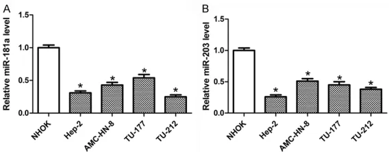
To clarify the roles of miR-181a and miR-203 in LC, RT-qPCR assay was initially performed to detect the two miRNAs’ expression. As present-ed in Figure 1A, a strong rpresent-eduction of miR-181a was observed in four LC cell lines (Hep-2, AMC-HN-8, TU-177, TU-212) compared to NHOK cells. Likewise, the expression of miR-203 was also lessened in LC cells, particularly in Hep-2 cells with severest loss (Figure 1B). Thus, Hep-2 cells were selected for the following functional studies.
miR-181a and miR-203 inhibited the migration and invasion of LC cells
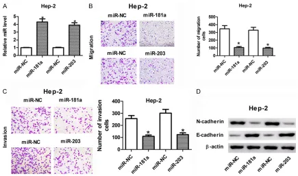
[image:3.612.90.374.71.182.2]In the present study, we observed enforced ab- undances of miR-181a and miR-203 in Hep-2 cells after transfected with 181a or 203 mimics (Figure 2A), indicating that miR-181a or miR-203 mimics could be used for the following gain-of-function experiments. Given that migration and invasion were required for
Figure 1. miR-181a and miR-203 were downregulated in LC cell lines. The expression of miR-181a (A) and miR-203 (B) in NHOK and four LC cells (Hep-2, AMC-HN-8, TU-177, and TU-212) was measured by RT-qPCR.
tumor progression, we addressed the roles of miR-181a and miR-203 in LC. As shown in Figure 2B, cell migration ability was markedly suppressed in Hep-2 cells after the transfec-tion of miR-181a or miR-203. Similarly, overex-pression of miR-181a or miR-203 also
attenu-ated cell invasion ability (Figure 2C). E- and N-cadherin, known as calcium-dependent cell adhesion molecules, are major landmarks of tumor metastasis [21]. Alterations of E- and N-cadherin are closely involved in the transfer of epithelial tumors from a non-invasive to
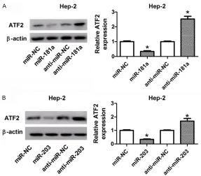
[image:4.612.88.523.69.326.2]inva-Figure 3. ATF2 was identified to be a direct target of 181a and 203. A. The putative binding sites of miR-181a or miR-203 within the 3’-UTR fragments of ATF2. B. 293T cells were co-transfected with miR-miR-181a, miR-203, or matched controls and luciferase reporter carrying the wild-type or mutated 3’-UTR of ATF2, followed by the detec-tion of luciferase activity by Dual-Luciferase reporter assay. C. Hep-2 cells were transfected with 181a, miR-203, or respective controls, then RIP analysis was used to evaluate the enrichment degree of ATF2 in IgG or Ago2 immunoprecipitation complex.
[image:4.612.93.521.403.558.2]sive status [22]. Next, the expression levels of E- and N-cadherin in miR-181a or miR-203-up-regulated LC cells were determined by western blot assay. Results revealed that addition of miR-181a and miR-203 resulted in the down-regulation of N-cadherin and updown-regulation of E-cadherin in Hep-2 cells (Figure 2D), denoting the inhibitory effects of miR-181a and miR-203 on cell migration and invasion.
ATF2 was a direct target of miR-181a and miR-203
Having confirmed that miR-181a and miR-203 were required for LC progression, we expected to explore a potential target gene. Bioinfor- matics software revealed the existence of puta-tive binding regions of miR-181a and miR-203 within the 3’UTR fragments of ATF2 (Figure 3A). To further demonstrate this prediction, Dual-Luciferase and RIP assays were carried out to probe the interplay between 181a or miR-203 and their target gene ATF2. As displayed in Figure 3B, elevated expression of miR-181a or miR-203 attenuated the luciferase activity of wile-type ATF2 reporters ATF2-WT and ATF2-wt in 293T cells, but no substantial alteration of mutant ATF2 reporters MUT and
ATF2-mut. Next, RIP assay revealed that ATF2 could be highly enriched by Ago2 antibody in miR-181a or miR-203-upregulated Hep-2 cells compared to miR-NC group, but IgG antibody failed to pull down ATF2 (Figure 3C). These findings suggested that ATF2 was a potential target of miR-181a and miR-203.
Knockdown of ATF2 inhibited migration and invasion of LC cells
ATF2 expression was first detected by RT-qPCR and results exhibited an elevated expression of ATF2 mRNA in all four LC cell lines compared to NHOK cells (Figure 4A), indicating that ATF2 might be required for LC progression. For the following loss-of-function experiments, si-ATF2 was transiently transfected into Hep-2 cells to interfere ATF2 (Figure 4B). Functionally, ATF2 depletion remarkably reduced cell migration and invasion ability, reflected by the decreased number of migrated (Figure 4C) and invaded cells (Figure 4D), as well as the lowered expres-sion of N-cadherin, and enhanced abundance of E-cadherin (Figure 4E). Altogether, these findings suggested that ATF2 deficiency sup-pressed cell migration and invasion in Hep-2 cells.
ATF2 was negatively modulated by miR-181a and miR-203
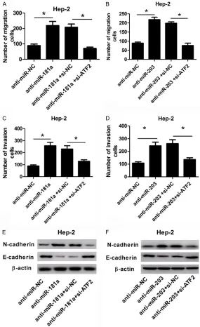
In this study, we probed the regulatory impacts of miR-181a and miR-203 on ATF2 expression in Hep-2 cells by western blot assay. Results displayed that overexpression of miR-181a or 203 suppressed, while reduction of miR-181a or miR-203 induced the expression of ATF2 protein in Hep-2 cells (Figure 5A, 5B).
miR-181a or miR-203 impeded cell migration and invasion by targeting ATF2 in LC
To further investigate the functional interaction between miR-181a or miR-203 and ATF2, res-cue experiments were performed in anti-miR-181a or anti-miR-203-transfected Hep-2 cells through silencing ATF2. Trans-well assay revealed that absence of miR-181a or miR-203 evidently stimulated cell migration (Figure 6A, 6B) and invasion (Figure 6C, 6D), which was reversed following ATF2 knockdown. Western blot analysis showed that downregulated ATF2 strikingly abrogated the stimulatory effect of anti-miR-181a or anti-miR-203 on Ncadherin expression, as well as the inhibitory effect on
nosis of patients. However, reduction of miR-149 or miR-101 foreboded a short overall sur-vival, metastasis, and an advance clinical stage in LC patients [28, 29]. These findings propose the underlying roles of miRNAs as diagnostic or therapeutic markers for LC.
[image:6.612.89.376.69.321.2]In this study, we provided the prior evidence that miR-181a and miR-203 expressions were obviously decreased in LC cells, suggesting the potential correlation between 181a or miR-203 and LC progression. Accumulated evi-dence has generally validated the central roles of miR-181a and miR-203 in tumorigenesis. Other than miR-203, miR-181a exhibits pro- or anti-tumor effects in diverse cancers. For example, miR-181a acted as an oncogene by inducing cell proliferation and repressing apop-tosis in cervical cancer [30]. Inversely, in NSCLC, lowered expression of miR-181a was significantly associated with severe histological grade and advanced TNM stage. The addition of miR-181a markedly impeded cell prolifera-tion, invasion, and colony formation through repressing CDK1 [31]. Additionally, elevated miR-181a expression evidently retarded the growth of oral squamous cell carcinoma (OSCC)
Figure 5. miR-181a and miR-203 suppressed ATF2 expression by direct complementary binding sites. A, B. Hep-2 cells were transfected with miR-181a, anti-miR-miR-181a, miR-203, anti-miR-203, and matched controls. At 48 h post-transfetion, ATF2 expression at protein level was detected by western blot assay.
E-cadherin expression (Figure 6E, 6F). These results indicat-ed that overexpression of miR-181a or miR-203 markedly reduced cell migration and invasion by directly targeting ATF2.
Discussion
prog-by directly targeting K-ras [11]. In line with previous reports, our study also demonstrated the anti-tumor role of miR-181a in LC progres-sion through regulation of cell migration and invasion.
miR-203, located on human chromosome 12, is generally recognized as a tumor suppressor.
identified as a target of miR-181a and miR-203. Restoration experiments further revealed that ATF2 knockdown abrogated anti-miR-181a or anti-miR-203-mediated migration and pro-invasion effects in LC cells.
In sum, our study indicated that miR-181a and miR-203 attenuated cell migration and
inva-Figure 6. The stimulatory effects of anti-miR-181a and anti-miR-203 on cell migration and invasion were abrogated following ATF2 knockdown. Hep-2 cells were transfected with anti-miR-181a, anti-miR-203, or matched con-trols or combined with si-ATF2 or si-NC. About 48 h post-transfection, cell migration (A and B), invasion (C and D), and protein levels of N-cadherin and E-cadherin (E and F) were measured by trans-well assay or western blot.
Dysregulation of this miRNA was observed in a range of human cancers. As reported by Zhang et al., exogenous restoration of miR-203 dra-matically delayed tumor gro- wth of esophageal cancer by downregulation of target gene Ran [32]. Xiang et al. indicated that the inhibitory effects of miR-203 on cell proliferation, adhesion, invasion, as well as tumor growth in prostate can-cer were partially reversed by Rap1A [33]. In addition, miR-203 was recognized to serve as a tumor suppressor by af- fecting cell proliferation, apop-tosis, invasion, and migration [20, 34]. These findings were fully consistent with our rese- arch that miR-203 inhibited migration and invasion of LC cells.
[image:7.612.89.374.72.538.2]sion by co-targeting ATF2 in LC. These data uncover that miR-181a and miR-203 may be a therapeutic target for LC.
Acknowledgements
This research was funded by the key scientific and technological project of Henan province (No. 132102310521).
Disclosure of conflict of interest
None.
Address correspondence to: Baoluo Wan, Depart- ment of Otolaryngology, Henan Provincial People’s Hospital, 7 Weiwu Road, Jinshui District, Zhengzhou 450003, China. Tel: +86-15290827301; E-mail: wanbaoluo2016@163.com
References
[1] Tamaki A, Miles BA, Lango M, Kowalski L and Zender CA. AHNS series: do you know your guidelines? Review of current knowledge on laryngeal cancer. Head Neck 2017; 40: 170-181
[2] Tomeh C and Holsinger FC. Laryngeal cancer. Curr Opin Otolaryngol Head Neck Surg 2014; 22: 147-153.
[3] Siegel RL, Miller KD and Jemal A. Cancer sta-tistics, 2018. Ca A Cancer J Clin 2018; 68: 11. [4] Mou S, Zhou Z, He Y, Liu F and Gong L. Cur-cumin inhibits cell proliferation and promotes apoptosis of laryngeal cancer cells through Bcl-2 and PI3K/Akt, and by upregulating miR-15a. Oncol Lett 2017; 14: 4937.
[5] Lagosquintana M, Rauhut R, Lendeckel W and Tuschl T. Identification of novel genes coding for small expressed RNAs. Science 2001; 294: 853-858.
[6] Flynt AS and Lai EC. Biological principles of microRNA-mediated regulation: shared the- mes amid diversity. Nat Rev Genet 2008; 9: 831.
[7] Esquelakerscher A and Slack FJ. Oncomirs-mi-croRNAs with a role in cancer. Nat Rev Cancer 2006; 6: 259-269.
[8] Chen X, Ba Y, Ma LJ, Cai X, Yin Y, Wang KH, Guo JG, Zhang YJ, Chen JN, Guo X, LI QB, Li XY, Wang WJ. Characterization of microRNAs in se-rum: a novel class of biomarkers for diagnosis of cancer and other diseases. Cell Res 2008; 18: 997-1006.
[9] Hui AB, Lenarduzzi M, Krushel T, Waldron L, Pintilie M, Shi W, Perez-Ordonez B, Jurisica I, O’Sullivan B and Waldron J. Comprehensive MicroRNA profiling for head and neck
squa-mous cell carcinomas. Clin Cancer Res 2010; 16: 1129-1139.
[10] Gao W, Yu Y, Cao HL, Shen H, Li XD, Pan SY and Shu YQ. Deregulated expression of miR-21, miR-143 and miR-181a in non small cell lung cancer is related to clinicopathologic charac-teristics or patient prognosis. Biomed Pharma-cother 2010; 64: 399-408.
[11] Shin KH, Bae SD, Hong HS, Kim RH, Kang MK and Park NH. miR-181a shows tumor suppres-sive effect against oral squamous cell carcino-ma cells by downregulating K-ras. Biochem Biophys Res Commun 2011; 404: 896-902. [12] Yao YH. Expression of miR-181a and miR-181b
in human gastric cancer cells and tissues. World Chinese Journal of Digestology 2015; 23: 30.
[13] Taylor MA, Sossey-Alaoui K, Thompson CL, Danielpour D, Schiemann WP. TGF-beta upreg-ulates miR-181a expression to promote breast cancer; metastasis. J Clin Invest 2013; 123: 150-163.
[14] Pichler M, Winter E, Ress AL, Bauernhofer T, Gerger A, Kiesslich T, Lax S, Samonigg H and Hoefler G. miR-181a is associated with poor clinical outcome in patients with colorectal cancer treated with EGFR inhibitor. J Clin Pathol 2014; 67: 198-203.
[15] Viticchiè G, Lena AM, Latina A, Formosa A, Gre-gersen LH, Lund AH, Bernardini S, Mauriello A, Miano R and Spagnoli LG. MiR-203 controls proliferation, migration and invasive potential of prostate cancer cell lines. Cell Cycle 2011; 10: 1121-1131.
[16] Shen J, Zhang J, Xiao M, Yang J and Zhang N. MiR-203 suppresses bladder cancer cell growth and targets the Twist1. Oncol Res 2018; 26: 11-1165.
[17] Zhou Y, Liang H, Liao Z, Wang Y, Hu X, Chen X, Xu L and Hu Z. miR-203 enhances let-7 bio-genesis by targeting LIN28B to suppress tu-mor growth in lung cancer. Sci Rep 2017; 7: 42680.
[18] Xu M, Gu M, Zhang K, Zhou J, Wang Z and Da J. miR-203 inhibition of renal cancer cell prolif-eration, migration and invasion by targeting of FGF2. Diagn Pathol 2015; 10: 24.
[19] Miao L, Xiong X, Lin Y, Cheng Y, Jiong LU, Zhang J and Cheng N. miR-203 inhibits tumor cell mi-gration and invasion via caveolin-1 in pancre-atic cancer cells. Oncol Lett 2014; 7: 658-662. [20] Tian L, Li M, Ge J, Guo Y, Sun Y, Liu M and Xiao
H. MiR-203 is downregulated in laryngeal squamous cell carcinoma and can suppress proliferation and induce apoptosis of tumours. Tumour Biol 2014; 35: 5953-5963.
pro-motes metastasis via multiple downstream transcriptional pathways. Cancer Res 2008; 68: 3645-3654.
[22] Hugo H, Ackland ML, Blick T, Lawrence MG, Clements JA, Williams ED and Thompson EW. Epithelial-mesenchymal and mesenchymal-epithelial transitions in carcinoma progres-sion. J Cell Physiol 2007; 213: 374-383. [23] Koskinen WJ, Brøndbo K, Dahlstrand HM,
Luostarinen T, Hakulinen T, Leivo I, Molijn A, Quint WG, Røysland T and Munck-Wikland E. Alcohol, smoking and human papillomavirus in laryngeal carcinoma: a nordic prospective mul-ticenter study. J Cancer Res Clin Oncol 2007; 133: 673-678.
[24] Vlachos IS, Paraskevopoulou MD, Karagkouni D, Georgakilas G, Vergoulis T, Kanellos I, Anas-tasopoulos IL, Maniou S, Karathanou K and Kalfakakou D. DIANA-TarBase v7.0: indexing more than half a million experimentally sup-ported miRNA:mRNA interactions. Nucleic Ac-ids Res 2015; 43: 153-159.
[25] Di LG and Croce CM. Roles of small RNAs in tumor formation. Trends Mol Med 2010; 16: 257-267.
[26] Wu S, Jia S and Xu P. MicroRNA-9 as a novel prognostic biomarker in human laryngeal squamous cell carcinoma. Int J Clin Exp Med 2014; 7: 5523-5528.
[27] Zhang XW, Liu N, Chen S, Wang Y, Zhang ZX, Sun YY, Qiu GB and Fu WN. High microRNA-23a expression in laryngeal squamous cell carcinoma is associated with poor patient prognosis. Diagn Pathol 2015; 10: 22.
[28] Xu Y, Lin YP, Yang D, Zhang G and Zhou HF. Clinical significance of miR-149 in the survival of patients with laryngeal squamous cell carci-noma. Biomed Res Int 2016; 2016: 8561251. [29] Li MH, Tian LL, Ren H, Chen XX, Wang Y, Ge JC,
Wu SL, Sun YN, Liu M and Xiao H. MicroR-NA-101 is a potential prognostic indicator of laryngeal squamous cell carcinoma and modu-lates CDK8. J Transl Med 2015; 13: 271.
[30] Xu H, Zhu J, Hu C, Song H and Li Y. Inhibition of microRNA-181a may suppress proliferation and invasion and promote apoptosis of cervi-cal cancer cells through the PTEN/Akt/FOXO1 pathway. J Physiol Biochem 2016; 72: 1-12. [31] Shi Q, Zhou Z, Ye N, Chen Q, Zheng X and Fang
M. MiR-181a inhibits non-small cell lung can-cer cell proliferation by targeting CDK1. Cancan-cer Biomark 2017; 20: 1-8.
[32] Zhang F, Yang Z, Cao M, Xu Y, Li J, Chen X, Gao Z, Xin J, Zhou S and Zhou Z. MiR-203 suppress-es tumor growth and invasion and down-regu-lates MiR-21 expression through repressing ran in esophageal cancer. Cancer Lett 2014; 342: 121-129.
[33] Xiang J, Bian C, Hao W, Huang S and Wu D. MiR-203 down-regulates Rap1A and suppress-es cell proliferation, adhsuppress-esion and invasion in prostate cancer. J Exp Clin Cancer Res 2015; 34: 1-10.
[34] Xu L, Shen B, Chen T and Dong P. miR-203 is involved in the laryngeal carcinoma pathogen-esis via targeting VEGFA and cox-2. Onco Tar-gets Ther 2016; 9: 4629-4637.
[35] Rodríguez M, Domingo E, Alonso S, Frade JG, Eiros J, Crespo MS and Fernández N. The un-folded protein response and the phosphoryla-tions of activating transcription factor 2 in the trans-activation of il23a promoter produced by β-glucans. J Biol Chem 2014; 289: 22942-22957.
[36] Wu D, Chen C, Wu Z, Liu B, Gao L, Yang Q, Chen W, Chen J, Bao Y and Qu L. ATF2 predicts poor prognosis and promotes malignant pheno-types in renal cell carcinoma. J Exp Clin Cancer Res 2016; 35: 108.