Original Article
Kidney and lung tissue modifications after BDL-induced
liver injury in mice are associated with increased
expression of IGFBPrP1 and activation of
the NF-κB inflammation pathway
Zhi-Hui Hu1, Yang-Yang Kong1, Jun-Jie Ren1, Ting-Juan Huang1, Yan-Qin Wang1, Li-Xin Liu1,2,3
1Department of Gastroenterology and Hepatology, 2Experimental Center of Science and Research, The First Hos-pital of Shanxi Medical University, Taiyuan 030001, China; 3Key Laboratory of Cell Physiology, Department of The Ministry of Education, Shanxi Medical University, Taiyuan 030001, China
Received November 25, 2019; Accepted January 23, 2020; Epub February 1, 2020; Published February 15, 2020 Abstract: Background: Hepatorenal and hepatopulmonary syndrome are common clinical diseases; however, their mechanisms have not been fully elucidated. Our aim was to determine whether liver injury by bile duct ligation (BDL) causes modifications in kidney and lung tissue in mice, and to explore the possible mechanism of these changes. Methods: BDL in mice was used as a research model. Pathologic changes of liver, kidney, and lung tissue were ob-served by hematoxylin-eosin (H&E) staining. The expression of IGFBPrP1, NF-κB, TNF-α, and IL-6 were investigated in liver, kidney, and lung tissue by immunohistochemical staining and western blot. The correlation between IGFBPrP1 and NF-κB, TNF-α, and IL-6 protein expression in liver, kidney, and lung tissues of each group was analyzed by the Pearson method. Results: H&E staining showed, after BDL administration in mice, different degrees of inflammatory change in liver, kidney, and lung tissues of mice in each group. The results of immunohistochemical staining and western blot analysis showed increased expressions of IGFBPrP1, NF-κB, TNF-α, and IL-6 after BDL. Pearson correla-tion analysis showed that IGFBPrP1 positively correlated with the expressions of NF-κB, TNF-α, and IL-6. Conclusion: Liver injury caused by bile duct ligation can lead to kidney and lung tissue injury in mice. The mechanism of injury may be related to the high expression of liver injury factor IGFBPrP1, transcription factor NF-κB, proinflammatory cytokine TNF-α, and IL-6 in kidney and lung tissue. Moreover, an increased expression level of IGFBPrP1 may be accompanied by the activation of the NF-κB inflammatory pathway.
Keywords: Bile duct ligation, insulin-like growth factor binding protein related protein 1, nuclear factor-κB, mouse, kidney, lung
Introduction
Cholestasis caused by stones, inflammation, and tumors of the biliary tract system are a common clinical problem. When cholestasis occurs, a large amount of bile accumulates in the liver and destroys the structure of the bili-ary tract. The bile enters the blood, causing serious damage to the body. The liver becomes the first damaged organ. Inflammatory cell infil-tration, fibroblast proliferation, and hepatocyte degeneration and necrosis in liver tissue lead to cholestasis of hepatic fibrosis and cirrhosis, and may further develop into hepatopulmonary syndrome, hepatorenal syndrome, and multiple organ failure [1, 2]. The multiple organ (liver,
kidney and lung) dysfunction caused by cho-lestasis, has a high incidence rate, high mortal-ity, is expensive to treat, and threatens lives [3, 4]. Therefore, it is of clinical significance to study the mechanism of multiple organ damage caused by cholestasis.
193 Int J Clin Exp Pathol 2020;13(2):192-202 immune regulation, and participates in fever
and inflammation. IL-6 is a cytokine that is a sensitive index reflecting the severity of inflam-mation and tissue injury. Its elevation can exac-erbate oxidative stress and lead to the release of toxic metabolites, which causes tissue injury [5, 6]. Moreover, IL-6 can be induced by TNF-α. Nuclear factor (NF)-κB is a transcription factor that plays a key role in the expression of pro-inflammatory cytokine genes [7]. At rest, NF-κB is a heterodimer composed of p65 and p50 (p65/p50) and its endogenous inhibitor of NF-κB (I-κB) binds to the cytoplasm and is regu-lated by cytokines. When stimuregu-lated by various damage factors such as lipopolysaccharides, I-κB degrades, releasing the NF-κB subunit that enters the nucleus to bind to the corresponding target gene, enhancing transcriptional activity, and regulating pro-inflammatory cytokines TNF-α, and IL-6. Other inflammatory mediators are also produced and in large quantities, trigger-ing an inflammatory response. Therefore, detecting the expression of the p65 subunit in the nucleus can indirectly reflect the activation of NF-κB and the degree of inflammatory response.
Insulin-like growth factor binding protein relat-ed protein 1 (IGFBPrP1) is a secretrelat-ed protein that can independently participate in a variety of biologic functions. Our previous study found that IGFBPrP1 can promote the activation of hepatic stellate cells (HSCs) in mice with bile duct ligation [8], produce excessive extracellu-lar matrices, and promote the development of liver fibrosis [9]. Moreover, it could enhance the DNA binding activity of NF-κB p65 in the HSC [10]. In addition, adenovirus carrying IGFBPrP1 can induce kidney and lung tissue injury in Sprague Dawley (SD) rats to varying degrees. The presence or absence of kidney and lung tis-sue damage, after hepatic injury induced by bile duct ligation in mice is yet to be deter-mined. If this injury does exist, its relation to the pro-inflammatory cytokine TNF-α, to IL-6 which responds to tissue damage, and to the regulation of transcriptional activation of NF-κB, and the liver injury factor IGFBPrP1 has not yet been reported.
A large number of studies have shown that cho-lestatic cirrhosis can be simulated by perform-ing bile duct ligation (BDL) surgery on mice. This model is ideal for studying the pathogenesis of cholestasis, since it closely mimics human
cho-lestasis liver fibrosis. It is also similar to the his-tologic changes of human small nodular cirrho-sis. Consequently, as a research model, we will use bile duct ligation in mice to determine if there is damage to kidney and lung tissues after liver injury. We will also explore whether this damage is related to pro-inflammatory cytokine TNF-α, or IL-6 which responds to tis-sue damage, the regulation of transcriptional activation of NF-κB and liver injury factor IGFBPrP1; and the relationship between the four. This study aims to provide new ideas for elucidating the pathogenesis of hepatorenal and hepatopulmonary syndrome.
Materials and methods
Animal models
A total of 78 clean grade C57BL/6 wild type male mice, approximately 3-4 weeks old with a body weight of 18±2 g, were used in this study. These mice were provided by the Institute of Experimental Animals, Academy of Military Medical Sciences (Beijing, China), with animal license number SCXK (Beijing) 2014-0013. The animal study protocol was in compliance with the guidelines of China for animal care, and confor to the internationally accepted princi-ples for care and use of experimental animals. All animals were kept in the animal room of the Department of Pharmacology, Shanxi Medical University. The indoor temperature was 23±2°C, with a 12 h light-dark cycle, relative humidity of 50±10%. Mice were free to eat specific-pathogen-free (SPF) large mouse growth and breeding feed (Beijing Keao Xieli Feed Co., Ltd.) and had access to drinking water. Experimental work was performed on all animals after one week of adaptive feeding. Animal grouping and disposal methods
The 78 mice were randomly divided as follows: 24 in the normal control group (normal group), 24 in the sham operation group (sham group) and 30 in the BDL group. The bile duct was iso-lated and ligated in the BDL group, and isoiso-lated but not ligated in the sham group, while the nor-mal group did not undergo any treatment. Experimental methods
the end of 2, 4 and 6 weeks, eight animals in each group were anesthetized by intraperito-neal injection of 4% chloral hydrate. Liver, kid-ney and lung tissues were removed by laparoto-my. Some were fixed with 10% formaldehyde solution, embedded in paraffin, and sectioned for H&E and immunohistochemical staining. The rest of liver, kidney and lung tissues were frozen in liquid nitrogen for 2 h and subse-quently frozen at -80°C for western blot analysis.
Hematoxylin-eosin (H&E) staining
The liver, kidney and lung tissue sections of mice were routinely dewaxed, hydrated, stained with hematoxylin-eosin and dried with neutral gum. Thereafter, the pathological changes of each tissue were observed under light microscopy.
Immunohistochemical staining
The distribution and expression of IGFBPrP1, NF-κB, TNF-α, and IL-6 in liver, kidney and lung tissues of each group were observed by immu-nohistochemical staining. Liver, kidney, and lung tissue sections were routinely dewaxed, hydrated, and incubated with 3% H2O2 for 15 min to inactivate endogenous peroxidase. After high-pressure heat repair with citrate buffer, the corresponding primary antibodies, IGFB- PrP1 (Abcam), TNF-α (Abcam), IL-6 (Abcam), and NF-κB (Protein-tech), were added and incu-bated for 1 h at 37°C. Thereafter, a biotinylated secondary antibody was added and incubated for 20 min at 37°C. Finally, staining was detect-ed using DAB (Beyotime). Each stain was replaced with phosphate buffered saline (PBS) as a negative control instead of the primary antibody. Brown or yellow granules were posi-tively expressed in each tissue. The images were semi-quantitatively analyzed using the Image-Pro Plus 6.0 automatic medical image color analysis system. Five fields were random-ly selected for each slice to anarandom-lyze the integral optical density (IOD) value of the brown and tan positive expression sites in the tissues. The higher the IOD value, the stronger the positive expression.
Western blot analysis
The liver, kidney, and lung tissues were stored in a biofreezer at -80°C. Protein from tissue was extracted according to the operation
instructions of the KGI whole protein extraction kit. The protein concentration was determined by the BCA method. Sodium dodecyl sulfate-polyacrylamide gel electrophoresis (SDS-PAGE) was performed and the gels were transferred to polyvinylidene fluoride (PVDF) membranes. The membranes were blocked with tris-buffered saline containing Tween 20 (TBST) and 5% skim milk powder at 23°C. Thereafter, the mem-branes were incubated with primary antibodies for IGFBPrP1 (Abcam), TNF-α (Abcam), IL-6 (Abcam), NF-κB (Protein-tech) and β-actin (Abcam) at 4°C overnight. Horseradish peroxi-dase (HRP)-labeled secondary antibody was added thereafter. The ECL chemiluminescence method was used to image through the Bio-Rad gel imaging system. The quantitative value of the target protein and the corresponding inter-nal reference protein (β-actin) was determined by Quantity One analysis software, and the ratio was the relative expression of the target protein.
Statistical analysis
Experimental data were expressed as means ± standard deviation (SD), and all calculations were performed using SPSS 22.0 statistical software. Statistical significance was evaluated using the factorial design analysis of variance. In addition, the Pearson correlation coefficients (r values) were calculated. Results were consid-ered significant at P<0.05.
Results
General condition and body weight of mice in each group
The normal group and the sham group had a good diet and mental state. Their responses were rapid and their body weight increased steadily. After the bile duct ligation, the mice showed a loss in appetite, poor cognitive func-tion, and a gradual decrease in sensitivity. Stimulation by sound and light was weakened, body hair was dull and scattered, and skin was yellow and discolored. Mice also suffered hair loss, yellow urine and feces, and gradual weight loss.
Histopathologic changes of the liver, kidney, and lung in mice
195 Int J Clin Exp Pathol 2020;13(2):192-202
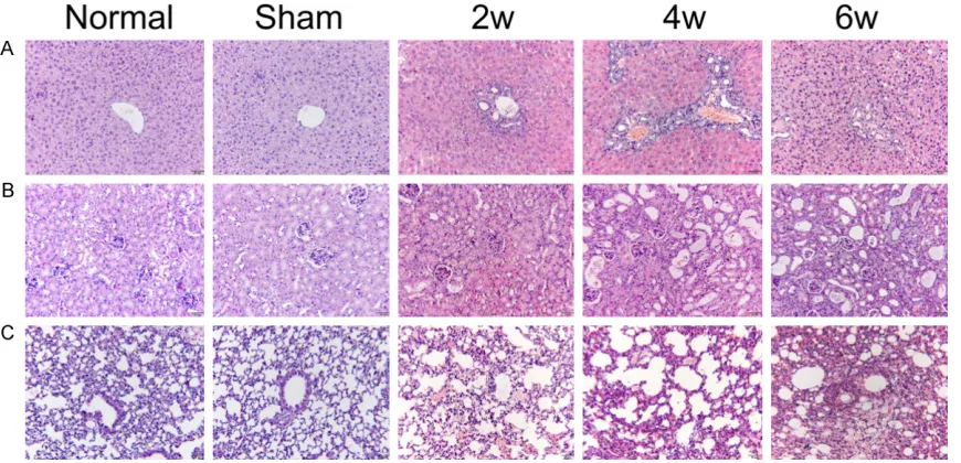
Figure 1. Histologic changes of liver, kidney and lung tissues at different time points were observed by H&E staining (original magnification ×200). A. Liver histology changes; B. Kidney histology changes; C. Lung histology changes.
hepatic cell cords were arranged neatly and radially around the central vein. The H&E stain-ing of liver tissue in the BDL group showed that the portal area was enlarged at 2 weeks, the bile duct was dilated, and the inflammatory cells infiltrated the portal area and bile duct; at 4 weeks, the portal area gradually increased, the bile duct expanded, and a large number of hyperplastic small bile ducts and inflammatory cells infiltrated the portal area; at 6 weeks of hepatocyte arrangement disorder, marked inflammatory cell infiltration occurred, together with some hepatocyte deformation and necro-sis(Figure 1A).
In the normal group and the sham group, epi-thelial cells of glomeruli and tubules were neat-ly arranged. There were no obvious pathologic changes in the kidneys, but only in a few con-nective tissues. At 2 weeks, swelling of renal cells and infiltration of inflammatory cells in the renal interstitium were observed in the BDL group; at 4 weeks, the renal tubular epithelial cells were irregularly arranged, and infiltration of the inflammatory cells in the renal intersti-tium increased; at 6 weeks, the renal tubular epithelial cells were unsystematically arranged. Infiltration of inflammatory cells in the intersti-tium, together with a marked increase of con-nective tissue were observed (Figure 1B). In the normal group and the sham group, alveo-lar structure was intact, and the alveoalveo-lar cavity
was clear. At 2 weeks of the BDL group, some alveolar spaces and interstitial inflammation were seen, and thickening of the alveolar sep-tum was noticed; at 4 weeks, some alveolar col-lapse and atelectasis were observed, accom-panied by pulmonary interstitial hemorrhage and a large amount of inflammatory cell infiltra-tion; at 6 weeks, extensive hyperemia and pul-monary vascular macrophages were observed in the lung tissue, and inflammatory cells increased(Figure 1C).
Distribution and expression of IGFBPrP1,
NF-κB, TNF-α, and IL-6 protein in liver, kidney, and
lung tissues of each group
Immunohistochemical staining showed positive staining from brown-yellow to tan particles. At different time points in liver, kidney, and lung tissues, only a small amount of IGFBPrP1, NF-κB, TNF-α, and IL-6 protein were expressed in the normal group and sham group (Figures 2A, 2D and 3A).
Dynamic changes of IGFBPrP1, NF-κB, TNF-α, and IL-6 protein expression levels in liver tis -sues of each group
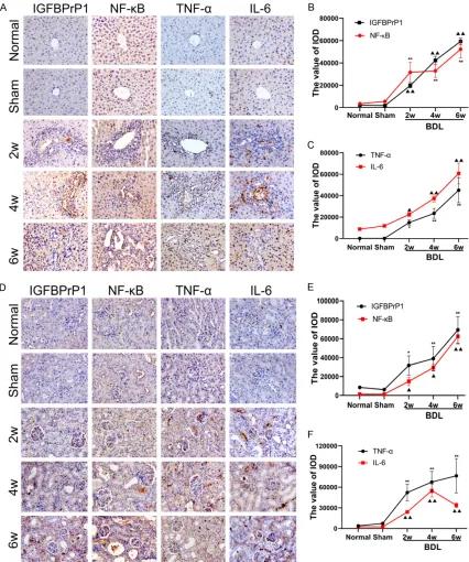
[image:4.612.93.528.74.284.2]Figure 2. The expression of insulin-like growth factor binding protein-related protein 1 (IGFBPrP1), NF-κB, TNF-α, and IL-6 in liver and kidney of mice.A. Immunohistochemical staining of IGFBPrP1, NF-κB, TNF-α and IL-6 in liver tissue of mice (original magnification ×400); B and C. Dynamic changes of expression of IGFBPrP1, NF-κB, TNF-α, and IL-6 in liver tissue; D. Immunohistochemical staining of IGFBPrP1, NF-κB, TNF-α and IL-6 in kidney tissue of mice (original magnification ×400); E and F. Dynamic changes of expression of IGFBPrP1, NF-κB, TNF-α and IL-6 in kidney tissue. PBS used as a negative control. Data are presented as the mean ± SD (n=8 per group). Compared with the normal group and the sham group, ▲: P<0.05, ▲▲: P<0.01; *: P<0.05, **: P<0.01.
increase in a time-dependent manner. As the disease progressed, the expression of the above proteins increased to all hepatic lobules
[image:5.612.93.519.73.583.2]197 Int J Clin Exp Pathol 2020;13(2):192-202
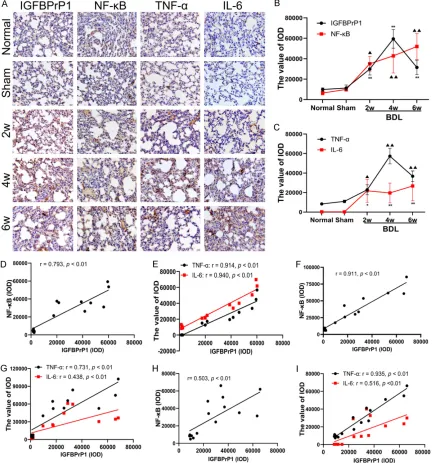
Figure 3. The expression of insulin-like growth factor binding protein-related protein 1 (IGFBPrP1), NF-κB, TNF-α, and IL-6 in lung of mice; the correlation between IGFBPrP1 and the expression of NF-κB, TNF-α and IL-6 in liver, kidney and lung tissues.A. Immunohistochemical staining of IGFBPrP1, NF-κB, TNF-α, and IL-6 in lung tissue of mice (original magnification ×400); B and C. Dynamic changes of expression of IGFBPrP1, NF-κB, TNF-α, and IL-6 in lung tissue; D and E. The correlation between IGFBPrP1 and the expression of NF-κB, TNF-α, and IL-6 in liver tissues of mice; F and G. The correlation between IGFBPrP1 and the expression of NF-κB, TNF-α and IL-6 in kidney tissues of mice; H and I. The correlation between IGFBPrP1 and the expression of NF-κB, TNF-α and IL-6 in lung tissues of mice. PBS was used as a negative control. Data are presented as the mean ± SD (n=8 per group). Compared with the normal group and the sham group, ▲: P<0.05, ▲▲: P<0.01; *: P<0.05, **: P<0.01.
Dynamic changes of IGFBPrP1, NF-κB, TNF-α, and IL-6 protein expression levels in renal tis -sues
After BDL was performed on mice, the four pro-teins were mainly expressed in renal tubular
[image:6.612.91.525.74.537.2]the normal group and sham group, positive expression of these proteins increased with the progression of the disease.
Dynamic changes of expression levels of
IGFB-PrP1, NF-κB, TNF-α, and IL-6 in lung tissue
After BDL was performed on mice, the four pro-teins were mainly expressed in alveolar mesen-chymal cells and alveolar vascular endothelial cells (Figure 3A). The expression of NF-κB and IL-6 in lung tissue increased at 2 weeks in a time-dependent manner. The IGFBPrP1 and TNF-α began to increase at 2 weeks, peaked at 4 weeks and decreased slightly at 6 weeks (Figure 3B, 3C). When compared with the nor-mal group and the sham group, the positive expression of the above-mentioned proteins increased with the progression of the disease.
Correlation analysis of IGFBPrP1 with NF-κB, TNF-α, and IL-6 in liver, kidney and lung tissues
In liver tissues of mice that underwent BDL, the correlation coefficients of IGFBPrP1 with NF-κB, TNF-α, and IL-6 were r=0.793, 0.914 and 0.940, respectively (P<0.01); and displayed a positive correlation(Figure 3D, 3E). In renal tis-sues, the correlation coefficients of IGFBPrP1 with NF-κB, TNF-α, and IL-6 were r=0.911, 0.731 and 0.438, respectively (P<0.01); and displayed a positive correlation(Figure 3F, 3G). In lung tissue, the correlation coefficients of IGFBPrP1 with NF-κB, TNF-α, and IL-6 were r=0.503, 0.935 and 0.516, respectively (P<0.01); and displayed a positive correlation (Figure 3H, 3I).
Protein expression levels of IGFBPrP1, NF-κB, TNF-α, and IL-6 in liver, kidney, and lung tis -sues
Western blot showed that the expression of IGFBPrP1, NF-κB, TNF-α, and IL-6 in the normal group and sham group were less at different time points in liver, kidney and lung tissues (Figure 4A-I).
After BDL was performed on mice, the expres-sion of IGFBPrP1, NF-κB, TNF-α, and IL-6 pro-tein in liver tissue increased with the progres-sion of the disease at 2 weeks (Figure 4A-C). The expression of IGFBPrP1, NF-κB, and TNF-α protein increased at 2 weeks and continued to gradually increase with time in renal tissues; the expression of IL-6 protein increased at 2
weeks, peaked at 4 weeks, and slightly declined at 6 weeks (Figure 4D-F). Additionally, the expression of NF-κB and IL-6 protein in lung tis-sue increased at 2 weeks in a time-dependent manner; the expression of IGFBPrP1 and TNF-α protein increased at 2 weeks, peaked at 4 weeks, and decreased at 6 weeks (Figure 4G-I). Discussion
199 Int J Clin Exp Pathol 2020;13(2):192-202
can induce IL-6 overexpression, and increase local endothelial cell permeability and local microcirculatory disorder, leading to an inflam-mation cascade, promotion of cytokine net-work formation, and involvement in hepatore-nal syndrome [13] and hepatopulmonary syndrome [14]pathogenesis. The results show that the expression of TNF-α in liver tissue gradually increased after BDL in mice. Immunohistochemistry and western blot analy-sis showed that TNF-α expression in renal tis-sues increased at 2 weeks and continued to increase in a time-dependent manner. Studies have shown that endotoxin-induced overex-pression of the cytokine TNF-α is an important mediator of high-power cycles associated with hepatorenal syndrome [15]. This also suggests a role for TNF-α in kidney damage. We specu-late that after BDL in mice, local inflammation occurs in the liver tissues, and inflammatory cytokines such as TNF-α reach the kidney through blood circulation, resulting in corre-sponding changes in kidney tissues after liver injury. In lung tissue of mice, the expression of TNF-α in the BDL group was significantly higher than that in the normal group (P<0.05) and reached a peak at 4 weeks. In addition, Zhang et al. [16] found that inhibition of TNF-α reduced mononuclear/macrophage accumulation and angiogenesis in the pulmonary arteries. This shows that the pro-inflammatory cytokine TNF-α plays an important role in lung tissue damage of BDL mice.
Cytokine IL-6 is the best marker of tissue inflammation and injury. In cholestasis, it is mainly produced by monocytes and macro-phages induced by TNF-α. The interaction of IL-6 and TNF-α can enhance T cell proliferation and activation of polymorphonuclear leuko-cytes, which aggravate the severity of tissue and organ damage and increase mortality [17]. Many studies have shown that early infection, body injury, and inflammation can lead to abnormal increase of IL-6 expression, and to organ metabolic disorders and functional damage [18]. In our study, the expression of IL-6 in liver tissues of mice after BDL showed an increasing trend that was consistent with its pathologic changes, suggesting that IL-6 may reflect the degree of liver injury. Immuno- histochemistry and western blot analysis showed that the expression of IL-6 in kidney tis-sue of BDL group was significantly higher (P<0.05) than that of the normal group at each
time point, reached a peak at 4 weeks and slightly decreased at 6 weeks, suggesting that the inflammation injury in kidney tissue of mice after BDL may be most severe at 4 weeks. This may later inhibit the activity of interleukins due to the accumulation of chemokines such as prostaglandin El, thereby reducing the expres-sion level of IL-6 [19]. The expresexpres-sion of IL-6 in lung tissue increased with time and was signifi-cantly higher than that in the normal group (P<0.05), suggesting that the degree of inflam-matory injury in lung tissue increased with time. As a transcription factor, NF-κB plays a major role in inflammation and immune response, because many inflammation-related genes are regulated by NF-κB [20]. Over-activation of NF-κB can induce the overexpression of pro-inflammatory cytokines TNF-α and IL-6, thus accelerating the toxicity of multi-organ cells [21]. This study has confirmed the pro-inflam-matory cytokine TNF-α and IL-6 as major target genes of NF-κB regulation and are expressed in kidney and lung tissues of cholestasis mice. This study also showed by western blot analy-sis, in the liver tissues of mice in the BDL group that the expression of NF-κB protein began to increase at 2 weeks and continued to increase in a time-dependent manner. The immunohisto-chemical staining results showed that the nucleus was markedly stained, i.e., there was activation and nuclear translocation of NF-κB, which was consistent with the results of Moczydlowska et al. [22]. The expression of NF-κB in renal and lung tissues is similar to that of liver tissue. It suggests that after BDL, when stimulated by endotoxin, cytokines, and reac-tive oxygen species (ROS), NF-κB can be acti-vated in the cytoplasm of hepatocytes and Kupffer cells, resulting in the release of pro-inflammatory cytokines TNF-α and IL-6. This causes liver damage and systemic reactions, further leading to kidney and lung tissue damage.
path-201 Int J Clin Exp Pathol 2020;13(2):192-202 way, leading to infiltration of pro-inflammatory
cytokines and activation of hepatic stellate cells to promote liver injury [28]. In this study, IGFBPrP1 protein expression in the liver tissue of BDL mice began to increase in a time-depen-dent manner in 2 weeks, which was consistent with our previous study [8]. We also found that the expression of IGFBPrP1 in kidney and lung tissues was basically the same as that in liver tissues, suggesting that IGFBPrP1 may aggra-vate the injury of kidney and lung tissues by promoting the transcription and expression of NF-κB. Furthermore, Pearson correlation analy-sis showed that IGFBPrP1 was positively corre-lated with the expression of transcription factor NF-κB, proinflammatory cytokine TNF-α, and IL-6 which responds to tissue damage in liver, kidney and lung tissues of BDL mice. The results showed that the high expression of IGFBPrP1 in multiple organs of BDL mice was accompanied by activation of NF-κB and pro-duction of pro-inflammatory cytokines.
Conclusion
This study found that liver injury induced by bile duct ligation in mice can cause kidney and lung injury, and preliminarily found that the mecha-nism of injury may be related to the high expres-sion of liver injury factor IGFBPrP1, transcrip-tion factor NF-κB, proinflammatory cytokine TNF-α, and IL-6 which responds to tissue dam-age in kidney and lung tissues. In addition, increased expression of IGFBPrP1 may be associated with activation of the NF-κB inflam-matory pathway. This provides a new strategy for the study of pathogenesis of hepatorenal syndrome and hepatopulmonary syndrome caused by cholestasis.
Acknowledgements
This study was supported by grants from National Natural Science Foundation of China (81670559); sponsored by the Fund for Shanxi Key Subjects Construction (FSKSC), sponsored by the Fund for Shanxi “1331 Project” Key Subjects Construction (1331KSC); Key Rese- arch and Development Project of Shanxi Province (201603D421023). Graduate Student Education Innovation Project of Shanxi
(2019SY242; 2019BY075).
Disclosure of conflict of interest
None.
Address correspondence to: Dr. Li-Xin Liu, Experi- mental Center of Science and Research, The First Hospital of Shanxi Medical University, 85 Jiefang South Road, Yingze District, Taiyuan 030001, Shanxi, China. Tel: 351-4639039; Fax: +86-351-4639039; E-mail: [email protected]
References
[1] Zhang XP, Jiang J, Yu YP, Cheng QH and Chen B. Effect of Danshen on apoptosis and NF-kap-paB protein expression of the intestinal muco-sa of rats with severe acute pancreatitis or obstructive jaundice. Hepatobiliary Pancreat Dis Int 2010; 9: 537-546.
[2] Xiping Z, Chuyang L, Jie Z, Yuefang R and Meili M. Protection of Salvia miltiorrhizae to the spleen and thymus of rats with severe acute pancreatitis or obstructive jaundice. Mediators Inflamm 2009; 2009: 186136.
[3] Garcia-Tsao G, Parikh CR and Viola A. Acute kidney injury in cirrhosis. Hepatology 2008; 48: 2064-2077.
[4] Bittencourt PL, Farias AQ and Terra C. Renal failure in cirrhosis: emerging concepts. World J Hepatol 2015; 7: 2336-2343.
[5] Liu L, Mu Q, Li W, Xing W, Zhang H, Fan T, Yao H and He L. Isofraxidin protects mice from LPS challenge by inhibiting pro-inflammatory cyto-kines and alleviating histopathological chang-es. Immunobiology 2015; 220: 406-413. [6] Mei X, Xu D, Xu S, Zheng Y and Xu S. Novel role
of Zn(II)-curcumin in enhancing cell prolifera-tion and adjusting proinflammatory cytokine-mediated oxidative damage of ethanol-in-duced acute gastric ulcers. Chem Biol Interact 2012; 197: 31-39.
[7] Lappas M, Permezel M, Georgiou HM and Rice GE. Nuclear factor kappa B regulation of proin-flammatory cytokines in human gestational tis-sues in vitro. Biol Reprod 2002; 67: 668-673. [8] Huang TJ, Ren JJ, Zhang QQ, Kong YY, Zhang
HY, Guo XH, Fan HQ and Liu LX. IGFBPrP1 ac-celerates autophagy and activation of hepatic stellate cells via mutual regulation between H19 and PI3K/AKT/mTOR pathway. Biomed Pharmacother 2019; 116: 109034.
[9] Guo X, Zhang H, Zhang Q, Li X and Liu L. Screening for and validation of a hepatic fibro-sis-related pathway induced by insulin-like growth factor-binding protein-related protein 1. Eur J Gastroenterol Hepatol 2016; 28: 762-772.
[11] Belcher JM, Sanyal AJ, Peixoto AJ, Perazella MA, Lim J, Thiessen-Philbrook H, Ansari N, Coca SG, Garcia-Tsao G and Parikh CR; TRIBE-AKI Consortium. Kidney biomarkers and differ-ential diagnosis of patients with cirrhosis and acute kidney injury. Hepatology 2014; 60: 622-632.
[12] Kawut SM, Ellenberg SS, Krowka MJ, Goldberg D, Vargas H, Koch D, Sharkoski T, Al-Naamani N, Fox A, Brown R, Levitsky J, Oh JK, Lin G, Song N, Mottram C, Doyle MF, Kaplan DE, Gup-ta S and Fallon MB. Sorafenib in hepatopulmo-nary syndrome: a randomized, double-blind, placebo-controlled trial. Liver Transpl 2019; 25: 1155-1164.
[13] Liu C, Cai J, Cheng Z, Dai X, Tao L, Zhang J and Xue D. Xiayuxue decoction reduces renal injury by promoting macrophage apoptosis in hepatic cirrhotic rats. Genet Mol Res 2015; 14: 10760-10773.
[14] Liu L, Liu N, Zhao Z, Liu J, Feng Y, Jiang H and Han D. TNF-alpha neutralization improves ex-perimental hepatopulmonary syndrome in rats. Liver Int 2012; 32: 1018-1026.
[15] Wen Y, Wang JY and Liu P. Tumor necrosis fac-tor-alpha enhances the effect of endothelin on renal vasoconstriction in isolated perfused rat kidney. Zhonghua Gan Zang Bing Za Zhi 2003; 11: 583-585.
[16] Zhang J, Luo B, Tang L, Wang Y, Stockard CR, Kadish I, Van Groen T, Grizzle WE, Ponnazha-gan S and Fallon MB. Pulmonary angiogenesis in a rat model of hepatopulmonary syndrome. Gastroenterology 2009; 136: 1070-1080. [17] Li W, Wang X, Niu X, Zhang H, He Z, Wang Y, Zhi
W and Liu F. Protective effects of nobiletin against endotoxic shock in mice through inhib-iting TNF-alpha, IL-6, and HMGB1 and regulat-ing NF-kappaB pathway. Inflammation 2016; 39: 786-797.
[18] Lu Y, Yang Y, He X, Dong S, Wang W, Wang D and Zhang P. Esmolol reduces apoptosis and inflammation in early sepsis rats with abdomi-nal infection. Am J Emerg Med 2017; 35: 1480-1484.
[19] Matsuura Y, Nishi S, Kariya N, Shimadzu K and Asada A. The effects of norepinephrine and prostaglandin E1 on pharmacokinetics of lido-caine in isolated perfused rat liver. Life Sci 2001; 68: 2123-2129.
[20] Chen F, Castranova V, Shi X and Demers LM. New insights into the role of nuclear factor-kappaB, a ubiquitous transcription factor in the initiation of diseases. Clin Chem 1999; 45: 7-17.
[21] Padillo FJ, Cruz A, Segura-Jiménez I, Ruiz-Rabe-lo J, Vázquez-Ezquerra MR, Perea-Alvarez MD, Peña J, Briceño J and Muntané J. Anti-TNF-al-pha treatment and bile duct drainage restore cellular immunity and prevent tissue injury in experimental obstructive jaundice. Int J Immu-nopathol Pharmacol 2007; 20: 855-860. [22] Moczydlowska J, Miltyk W, Hermanowicz A,
Lebensztejn DM, Palka JA and Debek W. HIF-1 alpha as a key factor in bile duct ligation-in-duced liver fibrosis in rats. J Invest Surg 2017; 30: 41-46.
[23] Degeorges A, Wang F, Frierson HF Jr, Seth A and Sikes RA. Distribution of IGFBP-rP1 in nor-mal human tissues. J Histochem Cytochem 2000; 48: 747-754.
[24] van Breevoort D, van Agtmaal EL, Dragt BS, Gebbinck JK, Dienava-Verdoold I, Kragt A, Bier-ings R, Horrevoets AJ, Valentijn KM, Eiken-boom JC, Fernandez-Borja M, Meijer AB and Voorberg J. Proteomic screen identifies IGFBP7 as a novel component of endothelial cell-spe-cific Weibel-Palade bodies. J Proteome Res 2012; 11: 2925-2936.
[25] Rao C, Lin SL, Ruan WJ, Wen H, Wu DJ and Deng H. High expression of IGFBP7 in fibro-blasts induced by colorectal cancer cells is co-regulated by TGF-beta and Wnt signaling in a Smad2/3-Dvl2/3-dependent manner. PLoS One 2014; 9: e85340.
[26] Pen A, Durocher Y, Slinn J, Rukhlova M, Char-lebois C, Stanimirovic DB and Moreno MJ. In-sulin-like growth factor binding protein 7 exhib-its tumor suppressive and vessel stabilization properties in U87MG and T98G glioblastoma cell lines. Cancer Biol Ther 2011; 12: 634-646.
[27] Tomimaru Y, Eguchi H, Wada H, Kobayashi S, Marubashi S, Tanemura M, Umeshita K, Kim T, Wakasa K, Doki Y, Mori M and Nagano H. IGFBP7 downregulation is associated with tu-mor progression and clinical outcome in hepa-tocellular carcinoma. Int J Cancer 2012; 130: 319-327.