Original article
Abatacept in combination with
methotrexate in Japanese biologic- naive
patients with active rheumatoid
arthritis: a randomised placebo-
controlled phase IV study
Tsukasa Matsubara,1 Hiroshi Inoue,2 Toshihiro Nakajima,3,4 Kazuhide Tanimura,5 Akira Sagawa,6 Yukio Sato,7,8 Kei Osano,9 Shuji Nagano,10 Yukitaka Ueki,11 Tadamasa Hanyu,12 Koichi Hashizume,13 Norihito Amano,13 Yoshiya Tanaka,14 Tsutomu Takeuchi15
To cite: Matsubara t, inoue H, nakajima t, et al. abatacept in combination with methotrexate in Japanese biologic- naive patients with active rheumatoid arthritis: a randomised placebo- controlled phase iV study. RMD Open 2018;4:e000813. doi:10.1136/ rmdopen-2018-000813
►Prepublication history and additional material for this paper are available online. to view these files, please visit the journal online (http:// dx. doi. org/ 10. 1136/ rmdopen- 2018- 000813).
received 31 august 2018 revised 2 november 2018 accepted 4 november 2018
For numbered affiliations see end of article.
Correspondence to Dr tsukasa Matsubara; mats@ mayflower- hp. jp © author(s) (or their employer(s)) 2018. re- use permitted under cc BY- nc. no commercial re- use. See rights and permissions. Published by BMJ.
Key messages
What is already known about this subject?
► Previous studies have demonstrated that a high
pro-portion of Japanese patients with rheumatoid arthri-tis (ra) are anticitrullinated protein antibody (acPa) positive.
► abatacept, a co- stimulatory signal inhibitor, was
approved in Japan in 2010 for the treatment of pa-tients with ra who had an inadequate response to previous conventional synthetic disease- modifying antirheumatic drugs.
What does this study add?
► this randomised, double- blind, placebo- controlled
phase iV study demonstrated that abatacept with methotrexate (MtX) improved clinical symptoms and inhibited structural damage progression in acPa- positive Japanese patients with early erosive ra and an inadequate response to MtX alone.
How might this impact on clinical practice?
► this study suggests that abatacept with MtX
could be considered a suitable treatment option for biologic- naive Japanese patients with ra.
AbstrAct
Objectives to evaluate efficacy and safety of
abatacept+methotrexate (MtX) in biologic- naive, anticitrullinated protein antibody (acPa)- positive Japanese patients with active rheumatoid arthritis (ra) and early erosion versus placebo+MtX.
Methods in this phase iV, multicentre, double- blind study
(nct01758198), patients were randomised (1:1) to receive intravenous abatacept (~10 mg/kg) or placebo, plus MtX (≥6 mg/week). Primary efficacy objectives were to compare american college of rheumatology 20 (acr20) response rates at week 16 and mean change from baseline in van der Heijde- modified total Sharp score (vdH- mtSS) at week 24 between abatacept+MtX and placebo+MtX groups.
Results Overall, 203 and 202 patients received
abatacept+MtX and placebo+MtX, respectively. at week 16, acr20 response rates were higher in the abatacept (75.4%) versus placebo group (27.7%; p<0.001). Mean change from baseline in vdH- mtSS at week 24 was 0.84 in the abatacept and 1.26 in the placebo group (p=0.017). radiographic non- progression rates (change in vdH- mtSS≤smallest detectable change (1.9)) were 88.1% and 75.4% in abatacept and placebo groups, respectively. adjusted mean change from baseline in Disease activity Score 28 (c- reactive protein) (DaS28 (crP)) at week 16 demonstrated a numerically greater reduction in the abatacept versus placebo group. Proportions of patients with DaS28 (crP), Simplified Disease activity index and clinical Disease activity index remission up to week 52 were higher in the abatacept versus placebo group. the abatacept safety profile was consistent with previous observations.
Conclusions compared with MtX alone, abatacept+MtX
improved clinical symptoms and inhibited structural damage progression in acPa- positive, Japanese patients with ra, early erosion and inadequate response to MtX.
InTROduCTIOn
Rheumatoid arthritis (RA) is a chronic, inflammatory autoimmune disease
characterised by synovitis leading to progres-sive joint damage and subsequent impaired physical function and which requires long- term management.1 Poor prognostic factors (PPFs) for RA include the presence of rheu-matoid factor (RF) and/or anticitrullinated protein antibody (ACPA) or early erosions.1
The addition of biologic disease- modifying antirheumatic drugs (bDMARDs) or targeted synthetic DMARDs (tsDMARDs) to treat-ment with conventional synthetic DMARDs
on May 7, 2020 by guest. Protected by copyright.
http://rmdopen.bmj.com/
on May 7, 2020 by guest. Protected by copyright.
http://rmdopen.bmj.com/
on May 7, 2020 by guest. Protected by copyright.
(csDMARDs) is recommended for patients with PPFs who responded inadequately to previous csDMARDs.2
Abatacept is a human fusion protein that selectively modulates the CD28–CD80/CD86 co- stimulatory signal required for full T- cell activation.3 In Japan, abatacept was
approved in 2010 for the treatment of patients with RA who had an inadequate response to previous csDMARDs. To investigate safety and clinical efficacy, phase II and III studies of abatacept in Japanese patients with RA were conducted.4–6 However, none of these studies investigated
efficacy on joint protection or focused on those patients with PPFs. Previous studies have demonstrated that a high proportion of Japanese patients with RA are ACPA positive (79.2%–86.6%)7–10; thus, efficacy and safety of
bDMARDs or tsDMARDs with concomitant use of metho-trexate (MTX) are of interest in this patient population.
In this study, the efficacy and safety of abatacept with concomitant use of MTX were investigated in a randomised, double- blind and placebo- controlled phase IV trial in biologic- naive, ACPA- positive patients with active RA and early erosion.
MeTHOds study population
Biologic- naive and ACPA- positive patients aged ≥20 years with active RA (defined by American College of Rheu-matology (ACR) 198711/European League Against
Rheu-matism (EULAR) 2010 criteria12) for ≥16 weeks prior to
screening were eligible for inclusion. In addition, eligible patients were <5 years from diagnosis and had received MTX (≥6 mg/week) for ≥3 months. Eligible patients also had ≥6 swollen joints among 66 screened, ≥6 tender joints among 68 screened, investigator- diagnosed erosion and C- reactive protein (CRP) ≥2.0 mg/dL or erythrocyte sedimentation rate (ESR) ≥28 mm/hour. All patients provided written informed consent prior to participation in this study.
Patients were excluded if they met the diagnostic criteria for any other rheumatic disease such as systemic lupus erythematosus (those with Sjögren’s syndrome, osteoarthritis, gout and osteoporosis were eligible). Further exclusion criteria are detailed in the online supplementary material.
study design and treatment
This phase IV, randomised, double- blind, placebo- controlled, 52- week observation, postmarketing study ( ClinicalTrials. gov identifier, NCT01758198) was conducted between 24 April 2013 and 26 December 2016 at 71 sites in Japan.
After screening, eligible patients were randomised (1:1; without stratification) to receive intravenous abata-cept plus MTX (≥6 mg/week) or matched intravenous placebo plus MTX (≥6 mg/week). During the study period, patients could receive MTX continuously at the study initiation dose. Abatacept was administered by intravenous infusion at the dose approved in Japan in
accordance with the patients’ body weight (<60 kg: 500 mg; ≥60 to ≤100 kg: 750 mg; >100 kg: 1000 mg) on day 1, at weeks 2 and 4 and every 4 weeks thereafter for up to 48 weeks. Both investigator and patient remained blinded throughout the study. Randomisation and allowed concomitant medications are detailed in the online supplementary material.
Patients who did not achieve a ≥20% improvement from baseline in swollen and tender joint counts after the evaluation at week 16 were offered open- label intra-venous abatacept (~10 mg/kg) every 4 weeks as rescue therapy, regardless of the original randomisation arm allocation and without breaking the randomisation blind. However, the final decision regarding use of abata-cept rescue therapy was determined by the investigator.
disease activity and physical function assessments
Disease activity was evaluated using ACR response rates,13 Disease Activity Score 28 (CRP) (DAS28
(CRP)),14 15 Simplified Disease Activity Index (SDAI) and
Clinical Disease Activity Index (CDAI).16 The proportion
of patients achieving ACR20 response at week 16 was one of two co- primary efficacy endpoints. ACR50 and ACR70 response rates and the change from baseline in DAS28 (CRP) at week 16 were secondary efficacy endpoints. Tertiary efficacy endpoints included the proportion of patients with remission defined in this study by DAS28 (CRP) <2.6, SDAI≤3.3 or CDAI ≤2.816 at weeks 16, 24
and 52. Physical function was assessed using the Japa-nese version17 of the Health Assessment Questionnaire-
Disability Index (HAQ- DI).18 HAQ- DI response rates,
defined as a ≥0.3 reduction from baseline, were evaluated at week 24 and 52 as tertiary efficacy endpoints.
Radiographic assessment
Joint damage progression was evaluated by van der Heijde- modified total Sharp score (vdH- mTSS) method for scoring radiographs of hands and feet in RA including 16 areas in each hand for erosions and 15 for joint- space narrowing (JSN).19 The change from baseline
in vdH- mTSS at week 24 was the second co- primary effi-cacy endpoint. The rate of radiographic non- progression at week 24, defined as the proportion of patients with a change from baseline vdH- mTSS ≤1.9 (smallest detect-able change (SDC)),20 was a secondary endpoint. Tertiary
efficacy endpoints are detailed in the online supplemen-tary material.
safety assessment
Safety was assessed throughout the study by recording the incidence of all adverse events (AEs) or serious AEs (SAEs), discontinuations due to AEs and AEs of special interest including those associated with immunomodu-latory drug use, infections, prespecified autoimmune disorders, malignancies and infusional reactions (defi-nitions detailed in the online supplementary material). Further details on AE classification are presented in the online supplementary material.
on May 7, 2020 by guest. Protected by copyright.
Enrolled (N=547)
Randomised (n=406)
Treated (n=405)
Discontinued (n=3) Discontinued (n=9) Abatacept + MTX (n=203) Placebo + MTX (n=202)
Discontinued (n=2) Discontinued (n=3) Completed 16-week period (n=200, 98.5%) Completed 16-week period (n=193, 95.5%)
Discontinued (n=6) Discontinued (n=6) Completed 24-week period (n=198, 97.5%)
(Rescue therapy [n=25, 12.3%])
Completed 24-week period (n=190, 94.1%)
(Rescue therapy [n=116, 57.4%])
Completed 52-week period (n=192, 94.6%)
(Rescue therapy [n=37, 18.2%])
Completed 52-week period (n=184, 91.1%)
(Rescue therapy [n=138, 68.3%])
Disqualification (n=141)*
[image:3.595.115.481.405.697.2]Not treated (n=1)
Figure 1 Patient disposition over the 52- week study period. *Patients did not meet study inclusion criteria (patient may
have more than one reason), including 62 with CRP <2.0 mg/dL or ESR <28 mm/hour ESR, 26 ACPA- negative and 20 with ALT >2×ULN. ACPA, anti- citrullinated protein antibody; ALT, alanine transaminase; CRP, C- reactive protein; ESR, erythrocyte sedimentation rate; MTX, methotrexate; ULN, upper limit of normal.
statistical analyses
The study sample size of 400 patients was determined based on the following considerations. Assuming a placebo response rate of 20%, a difference of 45% in the ACR20 response rate can be detected with at least 99% power at a significance level of 5% (two- sided). Based on the hierarchical testing procedure used for the co- primary endpoints, the difference of 1.9 (assuming SD 5.5) in the change from baseline in mTSS using the vdH- S method can be detected with >90% power at a significance level of 5% (two- sided), using a Wilcoxon rank sum test. Patients who received rescue abatacept treatment or withdrew for any reason were considered non- responders from that time point onwards. Efficacy analyses were performed in the intent- to- treat popu-lation which included all randomised patients who received at least one dose of study medication. The differences in ACR20 response rates between treat-ment groups at week 16 were analysed by χ2 test with
continuity correction. Change from baseline in vdH- mTSS at week 24 was compared using a rank- based non- parametric analysis of covariance model, with the rank score for change from baseline in vdH- mTSS as the dependent variable, treatment group as a main effect and the rank score for baseline in vdH- mTSS as a covariate. Further statistical analyses are detailed in the online supplementary material.
ResulTs
Patient disposition and baseline characteristics
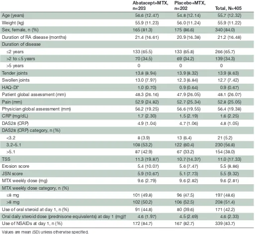
Patient disposition is shown in figure 1. Of 547 patients enrolled, 406 were randomised to treatment and 405 received at least one dose of the study drug; abatacept plus MTX group (n=203) and placebo plus MTX group (n=202). Baseline demographics, disease characteris-tics, treatment history and concurrent medication were comparable between the two treatment groups (table 1). Over the study period, a total of 29 patients discon-tinued; 12 (three abatacept plus MTX; nine placebo plus MTX) before week 16; five (two abatacept plus MTX; three placebo plus MTX) between weeks 16 and 24 and 12 (six abatacept plus MTX; six placebo plus MTX) between weeks 24 and 52. These discontinuations were mostly due to AEs (seven (3.4%) abatacept plus MTX; six (3.0%) placebo plus MTX) and lack of efficacy (two (1.0%) abatacept plus MTX; eight (4.0%) placebo plus MTX). AEs that led to discontinuation in the abatacept plus MTX group included interstitial lung disease (n=2), lymphoma, erythema nodosum, viral infection, colon cancer and abnormal liver function test. In the placebo plus MTX group, AEs that lead to discontinuation included suicide, Pneumocystis jirovecii pneumonia, drug hypersensitivity, anaphylactic shock, tendon rupture and drug- induced liver injury. Abatacept rescue therapy was received by 25 (12.3%) patients in the abatacept plus
on May 7, 2020 by guest. Protected by copyright.
Table 1 Patient demographics and baseline characteristics
Abatacept+MTX, n=203
Placebo+MTX,
n=202 Total, N=405
Age (years) 56.6 (12.47) 54.8 (12.14) 55.7 (12.32)
Weight (kg) 55.9 (11.23) 56.0 (11.24) 55.9 (11.22)
Sex, female, n (%) 165 (81.3) 175 (86.6) 340 (84.0)
Duration of RA disease (months) 21.4 (16.61) 20.9 (16.38) 21.2 (16.48)
Duration of disease
≤2 years 133 (65.5) 133 (65.8) 266 (65.7)
>2 to ≤5 years 70 (34.5) 69 (34.2) 139 (34.3)
>5 years 0 0 0
Tender joints 13.8 (8.94) 13.9 (8.32) 13.9 (8.63)
Swollen joints 13.0 (7.97) 12.3 (6.84) 12.7 (7.42)
HAQ- DI* 1.0 (0.70) 0.9 (0.64) 0.9 (0.67)
Patient global assessment (mm) 48.3 (26.16) 47.9 (26.05) 48.1 (26.07)
Pain (mm) 52.9 (24.82) 52.7 (25.34) 52.8 (25.05)
Physician global assessment (mm) 56.2 (19.25) 56.6 (19.55) 56.4 (19.38)
CRP (mg/dL) 1.7 (2.30) 1.5 (2.19) 1.6 (2.25)
DAS28 (CRP) 4.9 (1.04) 4.7 (1.06) 4.8 (1.05)
DAS28 (CRP) category, n (%)
<3.2 8 (3.9) 13 (6.4) 21 (5.2)
3.2–5.1 108 (53.2) 122 (60.4) 230 (56.8)
>5.1 87 (42.9) 67 (33.2) 154 (38.0)
TSS 11.3 (19.87) 10.7 (14.37) 11.0 (17.33)
Erosion score 5.4 (10.07) 5.6 (7.47) 5.5 (8.86)
JSN score 5.9 (10.67) 5.1 (7.73) 5.5 (9.32)
MTX weekly dose (mg) 9.6 (2.79) 9.6 (2.82) 9.6 (2.81)
MTX weekly dose category, n (%)
≤8 mg 101 (49.8) 96 (47.5) 197 (48.6)
>8 mg 102 (50.2) 106 (52.5) 208 (51.4)
Use of oral steroid at day 1, n (%) 91 (44.8) 80 (39.6) 171 (42.2)
Oral daily steroid dose (prednisone equivalents) at day 1 (mg)† 4.6 (1.97) 4.5 (2.69) 4.6 (2.33)
Use of NSAIDs at day 1, n (%) 172 (84.7) 167 (82.7) 339 (83.7)
Values are mean (SD) unless otherwise specified.
*Total (n=405), abatacept+MTX (n=203), placebo+MTX (n=202). †Total (n=171), abatacept+MTX (n=91), placebo+MTX (n=80).
CRP, C- reactive protein; DAS28, Disease Activity Score 28; HAQ- DI, Health Assessment Questionnaire- Disability Index; JSN, joint- space narrowing; MTX, methotrexate; NSAID, non- steroidal anti- inflammatory drug; RA, rheumatoid arthritis; TSS, total Sharp score.
MTX group between week 16 and 24 and by 12 patients (5.9%) between week 25 and 52. In contrast, 116 (57.4%) patients in the placebo plus MTX group received rescue therapy between week 16 and 24 and 22 (10.9%) between week 25 and 52 (figure 1).
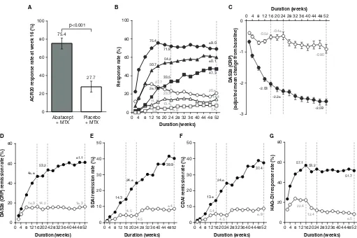
disease activity and physical function
A significantly greater proportion of patients achieved ACR20 response at week 16 with abatacept plus MTX (153/203 patients (75.4%), 95% CI 69.4 to 81.3) compared with placebo plus MTX (56/202 patients (27.7%), 95% CI 21.6 to 33.9). The estimated difference
between the groups was 47.6% (95% CI 38.6 to 56.7; p<0.001) (figure 2A). ACR20, 50 and 70 response rates over time from baseline to week 52 in the abatacept plus MTX and placebo plus MTX groups are shown in
figure 2B. In the abatacept plus MTX group, ACR20 response was detectable at week 2, rapidly increased thereafter, and plateaued from week 16 to week 52. In contrast, ACR20 response in the placebo plus MTX group increased from week 2, reached a maximum at week 8 and gradually decreased thereafter up to week 52. Similarly, ACR50 and 70 responses in the abatacept plus
on May 7, 2020 by guest. Protected by copyright.
0 20 40 60 80
0 4 8 1216202428323640444852
DAS28 (CRP) r
emission rate (%)
Duration (weeks) 16.3 53.2 14.2 61.1 16.3 46.8 D 0 10 20 30 40 50
0 4 8 12 16 20 24 28 32 36 40 44 48 52
SDAI r
emission rate (%)
Duration (weeks) 14.3 24.6 39.9 4.5 4.5 8.4 E 0 10 20 30 40 50
0 4 8 1216202428323640444852
CDAI r
emission rate (%)
Duration (weeks) 13.8 5.4 24.6 4.5 8.9 37.4 F G 0 20 40 60 80
0 4 8 1216202428323640444852
HAQ-DI
re
sponse rate (%)
Duration (weeks) 53.2 12.4 51.7 8.5 57.1 22.4 75.4 50.7 26.1 27.7 11.4 5.0 71.9 54.2 33.5 23.3 13.4 5.0 69.0 60.1 47.3 20.8 14.4 7.4 0 20 40 60 80 100
0 4 8 12 16 20 24 28 32 36 40 44 48 52
Response rate (%)
Duration (weeks) B 0 20 40 60 80 100 Abatacept
+ MTX Placebo+ MTX
ACR20 r
esponse rate at week 16 (%)
75.4 27.7 p<0.001 A -3 -2 -1
00 4 8 12 16 20 24 28 32 36 40 44 48 52
DAS28 (CRP)
(adjusted mean change fr
[image:5.595.42.553.42.382.2]om baseline) Duration (weeks) -2.03 -0.52 -2.26 -0.48 -2.59 -0.90 C
Figure 2 Disease activities evaluated as a comparison between abatacept plus MTX and placebo plus MTX groups. (A)
ACR20 response rate (co- primary endpoint) assessed at week 16. P value was based on the continuity corrected χ2 test. (B)
ACR20, 50 and 70 response rates over time up to 52 weeks. The patients who received rescue abatacept treatment, or who withdrew for any reason, were considered as non- responders from that time point forward. Dotted lines indicate time points at week 16 (secondary endpoints for ACR50 and 70 response rates), week 24 and week 52 (tertiary endpoints for ACR20, 50 and 70). The shapes indicate ACR20 (circles), ACR50 (triangles) and ACR70 (squares) response rates in the abatacept plus MTX (filled) and placebo plus MTX (open) groups. (C) Adjusted mean change from baseline in DAS28 (CRP) over time up to 52 weeks. Dotted lines indicate time points at week 16 (secondary endpoint), week 24 and week 52 (tertiary endpoints). Data after the initiation of rescue abatacept treatment were excluded. Error bars indicate the SE. (D–F) Remission rates with DAS28 (CRP) (D), SDAI (E) and CDAI (F) over time up to 52 weeks. Dotted lines indicate time points at week 16, week 24 and week 52 (tertiary endpoints). The circles show remission rates for the abatacept plus MTX (filled) and placebo plus MTX (open) groups. The remission was defined in this study as DAS28 (CRP) <2.6, SDAI ≤3.3 and CDAI ≤2.8. Patients with missing data due to early discontinuation or response after the initiation of rescue abatacept treatment were treated as non- responders. (G) Proportion of patients with HAQ- DI response over time up to 52 weeks. Dotted lines indicate time points at week 16, week 24 and week 52 (tertiary endpoints). The circles show response rates for the abatacept plus MTX (filled) and placebo plus MTX (open) groups. The HAQ- DI response was defined as a score reduction of ≥0.3 from baseline. ABA, abatacept; ACR20/50/70, American College of Rheumatology 20/50/70; CDAI, Clinical Disease Activity Index; CRP, C- reactive protein; DAS28, Disease Activity Score 28; HAQ- DI, Health Assessment Questionnaire- Disability Index; MTX, methotrexate; PBO, placebo; SDAI, Simplified Disease Activity Index.
MTX group improved rapidly from week 4 to week 8; were 50.7% and 26.1% at week 16 and 60.1% and 47.3% at week 52, respectively. Whereas, only marginal improve-ment was detectable for both parameters in the placebo plus MTX group. The adjusted mean change from base-line in DAS28 (CRP) rapidly decreased (week 4; greater than −1.0) with abatacept plus MTX, further improving thereafter (week 52; −2.59) (figure 2C), whereas it only decreased marginally with placebo plus MTX (week 52: −0.90). The score at week 16 was numerically greater in the abatacept plus MTX group versus placebo plus
MTX (−2.03 vs –0.52, respectively; adjusted mean differ-ence –1.50 (95% CI −1.73 to –1.28)). A higher DAS28 (CRP) remission rate (figure 2D) was also observed at week 16 with abatacept plus MTX (46.8%) compared with placebo plus MTX (16.3%) (estimated difference: 30.5, 95% CI 21.4 to 39.5), further increasing only with abatacept plus MTX up to week 52 (61.1% vs 16.3%; esti-mated difference: 44.7, 95% CI 35.8 to 53.7). SDAI remis-sion rates (figure 2E) at weeks 16 and 52 with abatacept plus MTX (14.3% and 39.9%) were numerically higher than those with placebo plus MTX (4.5% and 8.4%).
on May 7, 2020 by guest. Protected by copyright.
D E
0 2 4 6 8
10 Abatacept + MTX
(n=201) Placebo + MTX (n=199)
vdH-mTSS (mean change fr
om baseline)
p=0.017
24 weeks 52 weeks
A
0 2
1 3 4 5 6
Abatacept + MTX (n=201) Placebo + MTX (n=199)
Er
osion (mean change fr
om baseline)
24 weeks 52 weeks
B
0 2
1 3 4 5 8
7
6
Abatacept + MTX (n=201) Placebo + MTX (n=199)
JSN scor
e (mean change fr
om baseline)
24 weeks 52 weeks
C
Abatacept + MTX Placebo + MTX
45 40 35 30 25 20
Change from baseline in vdH-mTSS
at day 169
10 15
5 0
-10 -5
-15 -20
0 20 40 60
Cumulative probability (%)
80 100
Abatacept + MTX Placebo + MTX
90 80 70 60 50 40
Change from baseline in vdH-mTSS
at day 365 20
30
0 10
-20 -10
-30
0 20 40 60
Cumulative probability (%)
[image:6.595.42.548.43.477.2]80 100
Figure 3 Joint damage progression evaluated as a comparison between abatacept plus MTX (grey bar) and placebo plus
MTX groups (open bar). (A) The change from baseline in vdH- mTSS at week 24 (co- primary endpoint) and week 52. P value is based on a rank- based non- parametric analysis of covariance method. (B) The change from baseline in erosion score at week 24 and week 52 (tertiary endpoints). (C) The change from baseline in JSN score at week 24 and week 52 (tertiary endpoints). Data are shown as mean (SD). The number of patients with both baseline and post- baseline measurement are indicated in A–C. (D–E) Cumulative distribution function plot for the change from baseline in vdH- mTSS at week 24 (D) and week 52 (E). Missing data with patients who received rescue abatacept treatment, or who withdrew for any reason, were imputed with linear extrapolation. JSN, joint- space narrowing; MTX, methotrexate; vdH- mTSS, van der Heijde- modified total Sharp score.
Similarly, CDAI remission rates (figure 2F) at weeks 16 and 52 were numerically higher with abatacept plus MTX (13.8% and 37.4%) than placebo plus MTX (5.4% and 8.9%). Changes in HAQ- DI responses for the abatacept plus MTX group were detectable from week 2 and rapidly improved thereafter, plateauing from week 16 (57.1%) to week 52 (51.7%) (figure 2G). In contrast, HAQ- DI responses in the placebo plus MTX group exhibited only marginal improvement within the first 16 weeks (22.4%) and gradually declined up to week 52 (8.5%).
Radiographic assessment
The mean change from baseline vdH- mTSS at week 24 was significantly lower with abatacept plus MTX than
placebo plus MTX (0.84 vs 1.26; p=0.017; figure 3A). The mean change from baseline in vdH- mTSS at week 52 was numerically lower with abatacept plus MTX versus placebo plus MTX: 1.52 versus 2.62 (figure 3B). The numerical differences observed in mean change from baseline of erosion and JSN scores at weeks 24 and 52 are shown in figure 3B and C. Consistent with the vdH- mTSS, the mean change in erosion was numerically lower in the abatacept plus MTX group than in the placebo plus MTX group (week 24: 0.38 vs 0.74; week 52: 0.60 vs 1.52) (figure 3B). The change in JSN scores was rela-tively low in the abatacept plus MTX group compared with the placebo plus MTX group at week 24 (0.46 versus
on May 7, 2020 by guest. Protected by copyright.
Table 2 Summary of AEs reported in the 52- week study period
Week 16 Week 52
Abatacept+MTX (n=203, PY 62.7)
Placebo+MTX (n=202, PY 61.6)
Abatacept+MTX (n=203, PY 212.9)
Placebo+MTX (n=202, PY 108.7)
n IR n IR n IR n IR
All AEs 219 349.2 217 352.5 587 275.7 320 294.4
Related AEs 86 137.1 58 94.2 192 90.2 77 70.8
SAEs 4 6.4 6 9.7 17 8.0 9 8.3
Related SAEs 4 6.4 1 1.6 9 4.2 1 0.9
AEs of special interest
Infections 59 94.1 65 105.6 167 78.4 104 95.7
Malignancies 1 1.6 0 0 2 0.9 0 0
Autoimmune disorders 0 0 0 0 1 0.5 0 0
Peri- infusional AEs 3 4.8 7 11.4 4 1.9 8 7.4
Acute peri- infusional AEs 1 1.6 0 0 1 0.5 0 0
Other AEs within 24 hours of study drug 35 55.8 45 73.1 99 46.5 57 52.4 IR per 100 PY. Includes events with an onset date up to 113 days post first dose or 56 days post last dose. AEs occurring after the initiation of rescue abatacept are excluded.
AE, adverse event; IR, incidence rate; MTX, methotrexate; PY, person- years; SAE, serious adverse event. 0.52) and at week 52 (0.93 versus 1.10) (figure 3C). The
sensitivity analyses demonstrated that mean (SD) change from baseline in vdH- mTSS at week 24 was 0.76 (2.86) in the abatacept plus MTX group and 1.20 (2.72) in the placebo plus MTX group by the analysis as observed, and 0.56 (1.53) in the abatacept plus MTX group and 1.00 (1.69) in the placebo plus MTX group by the analysis with multiple imputation.
The cumulative distribution function plots of change from baseline in vdH- mTSS of the abatacept plus MTX group and the placebo plus MTX group at weeks 24 and 52 are shown in figure 3D and E. The radiographic non- progressor rates in the abatacept plus MTX group and the placebo plus MTX group were evaluated by the proportion of patients with change from baseline in vdH- mTSS below SDC (1.9), 0.5 or 0 at week 24 and week 52 (see online supplementary table S1). The non- progressor rates below SDC in the abatacept plus MTX and the placebo plus MTX groups were 88.1% and 75.4% (esti-mated difference 12.7%, 95% CI 4.7 to 20.7) at week 24 and were 83.6% and 68.3% (estimated difference 15.2%, 95% CI 6.5 to 24.0) at week 52. Additionally, the non- progressor rates by the cut- off levels ≤0.5 or ≤0 at week 24 and week 52 were numerically higher in the abatacept plus MTX group than placebo plus MTX group.
subgroup analysis
Subgroup analyses for ACR20 response rates at week 16 and for non- progression in change of mTSS at the cut- off level of ≤0 at week 24 are detailed in the online supple-mentary material and online supplesupple-mentary figures S1A and B. Higher ACR20 response rates and non- progressor rates were observed in the abatacept plus MTX group in all subgroups examined (except RF- negative; details in the online supplementary material).
safety
AEs and SAEs in the abatacept plus MTX and placebo plus MTX groups at weeks 16 and 52 are summarised in table 2. As a significant number of patients both in the abatacept plus MTX and placebo plus MTX groups received abatacept rescue therapy after week 16, the exposure- adjusted incidence rate (IR) per 100 person- years (PY) of exposure was calculated. Actual exposure of patients in the abatacept plus MTX and placebo plus MTX groups before entering abatacept rescue therapy up to week 52 were 212.9 and 108.7 PY, respectively. The exposure- adjusted IRs for SAEs throughout the study periods in the abatacept plus MTX and placebo plus MTX groups were 8.0 and 8.3 events/100 PY, respectively. In terms of AEs of special interests, the exposure- adjusted IR for infections in the abatacept plus MTX and placebo plus MTX groups were 78.4 and 95.7 events/100 PY, respectively. Notable clinical or laboratory test findings were not found in either group.
dIsCussIOn
To our knowledge, this is the first prospective, randomised, double- blind, placebo- controlled, multicentre study of abatacept in biologic- naive Japanese patients with RA who exhibited poor baseline prognostic factors. The co- primary endpoints demonstrated that abatacept in combination with MTX was superior to MTX alone, as evidenced by significantly higher ACR20 response rates at week 16 and lower mean change from baseline in vdH- mTSS at week 24. Furthermore, abatacept in combina-tion with MTX was highly effective for the duracombina-tion of the study, as compared with MTX alone, improving disease activity, ACR20/50/70 response rates, the mean change from baseline in DAS28 (CRP), remission rates (DAS28
on May 7, 2020 by guest. Protected by copyright.
(CRP)/SDAI/CDAI) as well as protection of joint damage destruction. Importantly, these positive clinical and radi-ographic outcomes with abatacept in combination with MTX were also consistently associated with improved patient- reported HAQ- DI response rates.
The achievement of clinical objectives for individual patients with RA, ‘treat- to- target strategy’ has been advo-cated by the EULAR recommendations2 21 and the ACR
guidelines22 with adjustment/change of treatment if the
target is not attained by 6 months. A few recent clinical studies for bDMARDs have focused on patients with ≤2 years of RA.23–31 In the present study, approximately 65%
of patients had early RA with average disease duration of 1.8 years. Additionally, mean baseline mTSS levels, erosion and JSN were 11.0, 5.5 and 5.5, respectively, indi-cating the consistency of this study with the current clin-ical setting.32
The mean dose of MTX in this study was 9.6 mg/week, which is similar to the typical dose range used in Japan.33–35
In addition, recent clinical studies with a tumour necrosis factor- alpha (TNF-α) inhibitor have reported concomi-tant use of MTX at similar dose ranges.36 37 The present
study reported similar ACR20, 50 and 70 response rates at week 24 with abatacept in combination with MTX to those for certolizumab and golimumab plus MTX studies.36 37
The impact of abatacept plus MTX treatment on joint damage progression, as observed by the mean change from baseline in vdH- mTSS at week 24 (0.84 vs 1.26 placebo plus MTX), differed from that reported for certolizumab plus MTX (0.2 vs 2.8 placebo plus MTX) and golimumab plus MTX (0.33 vs 2.51 placebo plus MTX).36 37 However, it should be noted that the mean
change from baseline in vdH- mTSS in the placebo plus MTX group in the present study was lower compared with the other studies, implicating differences in patient populations. For example, the mean disease durations and vdH- mTSS among these studies differed by 1.7 years and 11.0 (present study), 5.8 years and 55.1 (Japan RA Prevention of Structural Damage (J- RAPID) study) and 8.7 years and 52.7 (GO- FORTH study), respectively, suggesting that the inhibition of joint destruction data should be carefully interpreted.36 37
In the subgroup analyses, abatacept plus MTX tended to be more efficacious than MTX alone for all subgroups examined, except in prevention of joint destruction in RF- negative patients. Wider estimated difference between abatacept plus MTX and placebo plus MTX in ACR20 achievement at week 16 was observed for patients with lower baseline DAS28 (CRP), mTSS or MTX dose. While differences in non- progression rates in mTSS at the cut- off level of ≤0 on week 24 tend to be shown for those with longer disease duration or RF positivity, the RF- negative subgroup also showed higher non- progressor rate than the RF- positive subgroup in both treatment arms.
In this study, the incidence of AEs and SAEs were similar in both treatment arms. Additionally, there were no significant increases in incidence of AEs and SAEs
with abatacept plus MTX compared with placebo plus MTX over the dosing period. The safety profile of intra-venous abatacept in this study was consistent with that observed in previous Japanese4 6 38 and international39–41
clinical trials.
The data presented here should be interpreted within the context of the study limitations.
In this study, abatacept improved disease activities and reduced progression of joint damage in ACPA- positive patients with RA who had moderate to severe disease activity and low radiographic joint damage.
Author affiliations
1Department of Orthopedics, Matsubara Mayflower Hospital, Hyogo, Japan 2Department of Orthopaedic Surgery, inoue Hospital, gunma, Japan 3Department of rheumatology, Bay Side Misato Medical center, Kochi, Japan 4Department of locomotor Science, institute of Medical Science, tokyo Medical
University, tokyo, Japan
5Department of rheumatology, Hokkaido Medical center for rheumatic Diseases,
Sapporo, Japan
6akira Sagawa rheumatology clinic, Sapporo, Japan 7Sendai taihaku Hospital, Sendai, Japan
8Sendai Medical imaging clinic, Sendai, Japan
9Department of Orthopaedics, Fukuoka Mirai Hospital, Fukuoka, Japan
10Department of rheumatology, center for rheumatology, iizuka Hospital, Fukuoka,
Japan
11rheumatic and collagen Disease center, Sasebo chuo Hospital, nagasaki, Japan 12Department of rheumatology, nagaoka red cross Hospital, niigata, Japan 13Bristol- Myers Squibb K.K, tokyo, Japan
14First Department of internal Medicine, University of Occupational and
environmental Health, Kitakyushu, Japan
15Division of rheumatology, Department of internal Medicine, Keio University
School of Medicine, tokyo, Japan
Acknowledgements Professional medical writing and editorial assistance was provided by Sharon gladwin, PhD, and was funded by Bristol- Myers Squibb. Contributors tM, KH, na Yt and tt contributed to study conception and design and interpreting data. tM, Hi, tn, Kt, aS, YS, KO, Sn, YU, tH, Yt and tt contributed to collecting data. KH performed the statistical analysis. na managed the study. all authors contributed to critically reviewing and approving the manuscript. Funding this study was sponsored by Bristol- Myers Squibb K.K. and Ono Pharmaceutical co., ltd.
Competing interests tM received speaker honoraria from Pfizer Japan, nichi- iko, astellas, Meiji Seika, Bristol- Myers Squibb, abbVie gK, Janssen, chugai, eisai, aYUMi. Hi, aS, YS, KO, Sn and YU have nothing to disclose. tn received grants from Pfizer Japan, abbVie gK, eisai, aYUMi. Kt participated in speakers bureau for abbVie, astellas, Bristol- Myers Squibb, eisai, Mitsubishi tanabe. tH received grants from eli lilly, Bristol- Myers Squibb, abbVie. KH and na are employees of Bristol- Myers Squibb. Yt received speaking and/or honoraria from Daiichi- Sankyo, astellas, eli lilly, chugai, Sanofi, abbVie, Yl Biologics, Bristol- Myers Squibb, glaxoSmithKline, UcB, Mitsubishi tanabe, novartis, eisai, takeda, Janssen, asahi Kasei and has received research grants from Mitsubishi tanabe, Bristol- Myers Squibb, eisai, chugai, takeda, abbVie, astellas, Daiichi- Sankyo, Ono, MSD, taisho- toyama. tt received grants from astellas, chugai, Daiichi- Sankyo, takeda, abbVie gK, asahi Kasei, Mitsubishi tanabe, Pfizer Japan, eisai, aYUMi, nippon Kayaku, novartis K.K.; speaking fees from abbVie gK, Bristol- Myers Squibb K.K., chugai, Mitsubishi tanabe, Pfizer Japan, astellas, Daiichi- Sankyo, eisai, Sanofi K.K., teijin, takeda, novartis K.K. and consultant fees from astraZeneca K.K., eli lilly Japan K.K., novartis K.K., Mitsubishi tanabe, abbVie gK, nippon Kayaku, Janssen K.K., astellas, taiho, chugai, taisho toyama, glaxoSmithKline K.K., UcB Japan.
Patient consent not required.
ethics approval the study was conducted in accordance with the Declaration of Helsinki,42 international conference on Harmonisation good clinical Practice guidelines,43 the Pharmaceutical affairs law, good clinical Practice and good Post- marketing Study Practice as defined by the Ministerial Ordinance concerning the Standards for the implementation of clinical Studies on Pharmaceutical Products and concerning notifications in Japan.44 the protocol, any amendments and the
on May 7, 2020 by guest. Protected by copyright.
patient informed consent received local institutional review Board/independent ethics committee approval prior to initiation of this study.
Provenance and peer review not commissioned; externally peer reviewed. data statement Bristol- Myers Squibb policy on data sharing may be found at https://www. bms. com/ researchers- and- partners/ clinical- trials- and- research/ disclosure- commitment. html.
Open access this is an open access article distributed in accordance with the creative commons attribution non commercial (cc BY- nc 4.0) license, which permits others to distribute, remix, adapt, build upon this work non- commercially, and license their derivative works on different terms, provided the original work is properly cited, appropriate credit is given, any changes made indicated, and the use is non- commercial. See: http:// creativecommons. org/ licenses/ by- nc/ 4.0
RefeRences
1. Smolen JS, Aletaha D, Barton A, et al. Rheumatoid arthritis. Nat Rev Dis Primers 2018;4:18001.
2. Smolen JS, Landewé R, Bijlsma J, et al. EULAR recommendations for the management of rheumatoid arthritis with synthetic and biological disease- modifying antirheumatic drugs: 2016 update. Ann Rheum Dis 2017;76:960–77.
3. Cutolo M, Nadler SG. Advances in CTLA-4- Ig- mediated modulation of inflammatory cell and immune response activation in rheumatoid arthritis. Autoimmun Rev 2013;12:758–67.
4. Takeuchi T, Matsubara T, Nitobe T, et al. Phase II dose- response study of abatacept in Japanese patients with active rheumatoid arthritis with an inadequate response to methotrexate. Mod Rheumatol 2013;23:226–35.
5. Iwahashi M, Inoue H, Matsubara T, et al. Efficacy, safety, pharmacokinetics and immunogenicity of abatacept administered subcutaneously or intravenously in Japanese patients with rheumatoid arthritis and inadequate response to methotrexate: a Phase II/III, randomized study. Mod Rheumatol 2014;24:885–91. 6. Takeuchi T, Matsubara T, Urata Y, et al. Phase III, multicenter,
open- label, long- term study of the safety of abatacept in Japanese patients with rheumatoid arthritis and an inadequate response to conventional or biologic disease- modifying antirheumatic drugs.
Mod Rheumatol 2014;24:744–53.
7. Nakamura Y. The BioBank Japan project. Clin Adv Hematol Oncol
2007;5:696–7.
8. Kochi Y, Okada Y, Suzuki A, et al. A regulatory variant in CCR6 is associated with rheumatoid arthritis susceptibility. Nat Genet
2010;42:515–9.
9. Terao C, Yamada R, Ohmura K, et al. The human AIRE gene at chromosome 21q22 is a genetic determinant for the predisposition to rheumatoid arthritis in Japanese population. Hum Mol Genet
2011;20:2680–5.
10. Okada Y, Terao C, Ikari K, et al. Meta- analysis identifies nine new loci associated with rheumatoid arthritis in the Japanese population. Nat Genet 2012;44:511–6.
11. Arnett FC, Edworthy SM, Bloch DA, et al. The American Rheumatism Association 1987 revised criteria for the classification of rheumatoid arthritis. Arthritis Rheum 1988;31:315–24.
12. Aletaha D, Neogi T, Silman AJ, et al. Rheumatoid arthritis classification criteria: an American College of Rheumatology/ European League Against Rheumatism collaborative initiative. Arthritis Rheum 2010;2010:2569–81.
13. Felson DT, Anderson JJ, Boers M, et al. The American College of Rheumatology preliminary core set of disease activity measures for rheumatoid arthritis clinical trials. The Committee on Outcome Measures in Rheumatoid Arthritis Clinical Trials. Arthritis Rheum
1993;36:729–40.
14. van der Heijde DM, van 't Hof MA, van Riel PL, et al. Judging disease activity in clinical practice in rheumatoid arthritis: first step in the development of a disease activity score. Ann Rheum Dis
1990;49:916–20.
15. van der Heijde DM, van 't Hof M, van Riel PL, et al. Validity of single variables and indices to measure disease activity in rheumatoid arthritis. J Rheumatol 1993;20:538–41.
16. Aletaha D, Smolen JS. The Simplified Disease Activity Index (SDAI) and Clinical Disease Activity Index (CDAI) to monitor patients in standard clinical care. Best Pract Res Clin Rheumatol
2007;21:663–75.
17. Matsuda Y, Singh G, Yamanaka H, et al. Validation of a Japanese version of the Stanford Health Assessment Questionnaire in 3,763 patients with rheumatoid arthritis. Arthritis Rheum 2003;49:784–8. 18. Fries JF, Spitz P, Kraines RG, et al. Measurement of patient outcome
in arthritis. Arthritis Rheum 1980;23:137–45.
19. van der Heijde D. How to read radiographs according to the Sharp/ van der Heijde method. J Rheumatol 2000;27:261–3.
20. Bruynesteyn K, Boers M, Kostense P, et al. Deciding on progression of joint damage in paired films of individual patients: smallest detectable difference or change. Ann Rheum Dis 2005;64:179–82. 21. Smolen JS, Landewé R, Breedveld FC, et al. EULAR
recommendations for the management of rheumatoid arthritis with synthetic and biological disease- modifying antirheumatic drugs: 2013 update. Ann Rheum Dis 2014;73:492–509.
22. Singh JA, Saag KG, Bridges SL, et al. 2015 American College of Rheumatology guideline for the treatment of rheumatoid arthritis.
Arthritis Rheumatol 2016;68:1–26.
23. Conaghan PG, Peterfy C, Olech E, et al. The effects of tocilizumab on osteitis, synovitis and erosion progression in rheumatoid arthritis: results from the ACT- RAY MRI substudy. Ann Rheum Dis
2014;73:810–6.
24. Emery P, Burmester GR, Bykerk VP, et al. Evaluating drug- free remission with abatacept in early rheumatoid arthritis: results from the phase 3b, multicentre, randomised, active- controlled AVERT study of 24 months, with a 12- month, double- blind treatment period.
Ann Rheum Dis 2015;74:19–26.
25. Scott IC, Ibrahim F, Simpson G, et al. A randomised trial evaluating anakinra in early active rheumatoid arthritis. Clin Exp Rheumatol
2016;34:88–93.
26. Burmester GR, Kivitz AJ, Kupper H, et al. Efficacy and safety of ascending methotrexate dose in combination with adalimumab: the randomised CONCERTO trial. Ann Rheum Dis 2015;74:1037–44. 27. Smolen JS, Wollenhaupt J, Gomez- Reino JJ, et al. Attainment
and characteristics of clinical remission according to the new ACR- EULAR criteria in abatacept- treated patients with early rheumatoid arthritis: new analyses from the Abatacept study to Gauge Remission and joint damage progression in methotrexate (MTX)- naive patients with Early Erosive rheumatoid arthritis (AGREE).
Arthritis Res Ther 2015;17:157.
28. Westhovens R, van Vollenhoven RF, Boumpas DT, et al. The early clinical course of infliximab treatment in rheumatoid arthritis: results from the REMARK observational study. Clin Exp Rheumatol
2014;32:315–23.
29. Schiff M, Takeuchi T, Fleischmann R, et al. Patient- reported outcomes of baricitinib in patients with rheumatoid arthritis and no or limited prior disease- modifying antirheumatic drug treatment.
Arthritis Res Ther 2017;19:208.
30. Burmester GR, Rigby WF, van Vollenhoven RF, et al. Tocilizumab combination therapy or monotherapy or methotrexate
monotherapy in methotrexate- naive patients with early rheumatoid arthritis: 2- year clinical and radiographic results from the
randomised, placebo- controlled FUNCTION trial. Ann Rheum Dis
2017;76:1279–84.
31. Emery P, Bingham CO, Burmester GR, et al. Certolizumab pegol in combination with dose- optimised methotrexate in DMARD- naïve patients with early, active rheumatoid arthritis with poor prognostic factors: 1- year results from C- EARLY, a randomised, double- blind, placebo- controlled phase III study. Ann Rheum Dis 2017;76:96–104. 32. Nawata M, Saito K, Fukuyo S, et al. Clinically relevant radiographic
progression in joint destruction in RA patients with abnormal MMP-3 or high levels of CRP despite 1- year treatment with infliximab. Mod Rheumatol 2016;26:807–12.
33. Harigai M, Ishiguro N, Inokuma S, et al. Postmarketing surveillance of the safety and effectiveness of abatacept in Japanese patients with rheumatoid arthritis. Mod Rheumatol 2016;26:491–8. 34. Takeuchi T, Nakajima R, Komatsu S, et al. Impact of adalimumab
on work productivity and activity impairment in Japanese patients with rheumatoid arthritis: large- scale, prospective, single- cohort ANOUVEAU study. Adv Ther 2017;34:686–702.
35. Kanbori M, Suzuka H, Yajima T, et al. Postmarketing surveillance evaluating the safety and effectiveness of golimumab in Japanese patients with rheumatoid arthritis. Mod Rheumatol 2018;28:66–75. 36. Yamamoto K, Takeuchi T, Yamanaka H, et al. Efficacy and safety
of certolizumab pegol plus methotrexate in Japanese rheumatoid arthritis patients with an inadequate response to methotrexate: the J- RAPID randomized, placebo- controlled trial. Mod Rheumatol
2014;24:715–24.
37. Tanaka Y, Harigai M, Takeuchi T, et al. Golimumab in combination with methotrexate in Japanese patients with active rheumatoid arthritis: results of the GO- FORTH study. Ann Rheum Dis
2012;71:817–24.
38. Matsubara T, Yamana S, Tohma S, et al. Tolerability and efficacy of abatacept in Japanese patients with rheumatoid arthritis: a phase I study. Mod Rheumatol 2013;23:634–45.
39. Kremer JM, Dougados M, Emery P, et al. Treatment of rheumatoid arthritis with the selective costimulation modulator abatacept:
on May 7, 2020 by guest. Protected by copyright.
twelve- month results of a phase iib, double- blind, randomized, placebo- controlled trial. Arthritis Rheum 2005;52:2263–71. 40. Kremer JM, Genant HK, Moreland LW, et al. Effects of abatacept in
patients with methotrexate- resistant active rheumatoid arthritis: a randomized trial. Ann Intern Med 2006;144:865–76.
41. Schiff M, Keiserman M, Codding C, et al. Efficacy and safety of abatacept or infliximab vs placebo in ATTEST: a phase III, multi- centre, randomised, double- blind, placebo- controlled study in patients with rheumatoid arthritis and an inadequate response to methotrexate. Ann Rheum Dis 2008;67:1096–103.
41. . World Medical Association declaration of Helsinki.
Recommendations guiding physicians in biomedical research involving human subjects. JAMA 1997;277:925–6.
43. ICH harmonised tripartite guideline: guideline for good clinical practice. J Postgrad Med 2001;47:199–203.
44. Ministerial Ordinance, 2004. Ministerial ordinance on good post- marketing study practice for drugs. No.171. Available from: https://www. pmda. go. jp/ english/ index. html [Accessed 29 Jun 2018].
on May 7, 2020 by guest. Protected by copyright.
Correction: Abatacept in combination with methotrexate in
Japanese biologic- naive patients with active rheumatoid
arthritis: a randomised placebo- controlled phase IV study
Matsubara T, Inoue H, Nakajima T, et al. Abatacept in combination with methotrexate in Japanese biologic- naive patients with active rheumatoid arthritis: a randomised placebo- controlled phase IV study. RMD Open 2018;4:e000813.
The article has been corrected since it was published online.
In the Results section of the abstract (page 1), sentence 4 should read “Radio-graphic non- progression rates (change in vdH- mTSS≤smallest detectable change (1.9)) were 88.1% and 75.4% in abatacept and placebo groups, respectively.”
In the “Radiographic assessment” paragraph of the Methods section (page 2), sentence three should read “The rate of radiographic non- progression at week 24, defined as the proportion of patients with a change from baseline vdH- mTSS ≤1.9 (smallest detectable change (SDC)),20 was a secondary endpoint.”
In the “Radiographic assessment” section of the Results (page 7, paragraph 2), sentence two should read “The radiographic non- progressor rates in the abatacept plus MTX group and the placebo plus MTX group were evaluated by the proportion of patients with change from baseline in vdH- mTSS below SDC (1.9), 0.5 or 0 at week 24 and week 52 (see online supplementary table S1).”
Open access This is an open access article distributed in accordance with the Creative Commons Attribution Non Commercial (CC BY- NC 4.0) license, which permits others to distribute, remix, adapt, build upon this work non- commercially, and license their derivative works on different terms, provided the original work is properly cited, appropriate credit is given, any changes made indicated, and the use is non- commercial. See: http:// creativecommons. org/ licenses/ by- nc/ 4.0
© Author(s) (or their employer(s)) 2018. Re- use permitted under CC BY- NC. No commercial re- use. See rights and permissions. Published by BMJ.