Case Report
Secondary pyogenic ventriculitis following
erector spinae muscle abscess caused
by
Streptococcus intermedius
Chenjuan Tao1*, Yong Chen2, Xiaofei Zhao3, Xiaodong Lu1, Jun Cheng4, Ke Ma5*
Departments of 1Neurology, 2Radiology, 3Laboratory, 4Neurosurgery, Affiliated Hospital of Hangzhou Normal
Uni-versity, Hangzhou 310015, P. R. China; 5College of Traditional Chinese Medicine, Shandong University of
Tradi-tional Chinese Medicine, Jinan 250355, Shandong, P. R. China. *Equal contributors. Received July 4, 2017; Accepted April 2, 2018; Epub May 15, 2018; Published May 30, 2018
Abstract: Background: Pyogenic ventriculitis is a rare and severe cerebral infection. It may result from the rupture of brain abscess, neurosurgical procedures, hematogenous spread and so on. Contiguously diffusion through spinal dura from erector spinae muscle abscess is rare. Moreover, pyogenic ventriculitis caused by Streptococcus interme-dius is rare.We reported a patient with pyogenic ventriculitis secondary to erector spinae muscle abscess owing to
Streptococcus intermedius infection. Case presentation: A 61-year-old male with diabetes mellitus had been initially suffered from backache, then presented with fatigue and postural tremor when standing. On the second day of ad-mission, he suffered from retention of urine and disturbance of consciousness. Magnetic resonance imaging of the brain showed pyogenic ventriculitis. And enhanced computed tomography of the lumbar vertebra revealed abscess in the right erector spinae muscle, which helped us identified the pathogen as Streptococcus intermedius through the pus drainaged from the lesions. After antibiotic treatment for two and a half months, association with drainage of the abscess for one month, the cerebrospinal fluid tended to normal. Ventriculo-peritoneal shunt operation was performed at last as the complication of hydrocephalus. The outcome of the patient was relatively good. Conclusion:
S. intermedius has an affinity for invading the brain. Poor glycemic control with sustained hyperglycemia is the risk factor for abscess formation. Pyogenic ventriculitis can be caused by contiguously diffusion through spinal dura from the erector spinae muscle abscess.
Keywords: Pyogenic ventriculitis, streptococcus intermedius, abscess, erector spinae muscle, magnetic reso-nance imaging
Introduction
Pyogenic ventriculitis (also called pyocephalus or ventricular empyema) is a rare and serious intracranial infection characterized by the pres-ence of suppurative fluid in the cerebral ventri-cles. The clinical manifestation is usually char-acterized by headache, vomiting, impaired con- sciousness with fever and meningeal signs. Although a life threatening condition requiring the earliest diagnosis, symptoms and signs of pyogenic ventriculitis may sometimes be subtle and aspecific [1]. The possible mechanisms of infection of the ventricular system include intra-ventricular rupture of cerebral abscess, hema-togenous spread to the subependyma of the choroids plexus, complication of meningitis and direct implantation of pathogens following head
injury and neurosurgical procedures, e.g, ven-tricular drain insertion [2, 3]. The exact inci-dence of pyogenic ventriculitis is unknown. We here in present an unusual case with pyo-genic ventriculitis and erector spinae muscle abscess due to Streptococcus intermedius in- fection.
Case presentation
sought treatment at the urinary outpatient clin-ic. Urinary ultrasound and a computed tomog-raphy (CT) scan of the lumbar vertebra and lower abdomen were performed. CT scan sh- owed lumbar intervertebral disc bulging sli- ghtly at level of L4-L5 and L5-S1 (Figure 1A). He was diagnosed as prolapse of lumbar inter-vertebral disc and treated with nonsteroidal anti-inflammatory drugs, and was required to rest at home. After ten days, he showed no improvement but suffered from fatigue and mild postural tremor when standing. He sought for treatment again. As the elevated blood glu-cose was 25 mmol/L, he was admitted to the department of endocrinology for the purposes of further examination and treatment. The patient had lost approximately six kilogram dur-ing the past one month. He didn’t have a con-tact history of tuberculosis.
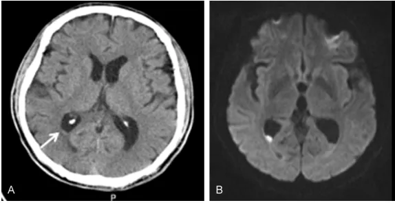
[image:2.612.88.379.73.167.2]from retention of urine and then became confu-sion. He could no longer eat a meal, change his clothes, or talk with others. His level of con-sciousness sharply deteriorated thereafter. An urgent CT scan of the brain was performed on hospital day 3, and showed no obvious ab- normalities (Figure 2A). So in consultation with the Department of Neurology, according to stiffness of his neck and a positive Kernig’s sign, he was diagnosed as suspected encepha-litis or and meningitis. Then he was transferred to our Neurology unit. Empirical ceftriaxone and acyclovir were commenced for suspected encephalitis and meningitis. Lumbar puncture was performed on the fourth day of admission, but failed. Magnetic resonance (MR) imaging of the brain was performed at the same day, and showed hyperintense lesion on diffusion-weighted image (DWI) with isointense on T2- Figure 1. Plain CT and contrast-enhanced CT of lumbar vertebra before
ad-mission and on day 5. A: Lumbar vertebra plain CT scan before adad-mission showed the right erector spinae muscle swelling, muscle gap blurred. B: Lumbar vertebra contrast enhancement CT scan on hospital day 5 showed hypodense lesions in the right erector spinae muscle with ring-like enhance-ment and bony destruction of right vertebral facet joint.
Figure 2. Plain CT and magnetic resonance imaging of the brain on days 3 and 4. A: Brain plain CT scan on hospital day 3 showed less obvious intraven-tricular isodense lesions (arrow) in bilateral occipital horns which had been lost. B: The diffusion-weighted imaging (b value of 800) clearly indicated hyperintense signals in right occipital horn of lateral ventricles and slightly hyperintensity in the left.
Physical examination showed a fleshless man and respon-sive slowly. Temperature was 36.5°C, and blood pressure, pulse and respiratory rate, as well pulse oximetry, were normal. The lips were dry and the limbs were slightly stre- ngthless. The rest of the ph- ysical examination was nor-mal. Blood tests revealed an elevated white blood cell count (WBC) of 16590/μl with a differential count of 93.5% neutrophils. The C-reactive protein (CRP) was high at 148.38 mg/L. Blood culture was negative. Other laborato-ry examination showed decr- eased sodium (127.3 mmol/ L) and albumin (29.7 g/L), increased glycosylated hemo-globin (13.1%). Test of purified protein derivative of tubercu-lin and T cell detection of tuberculosis infection were negative.
[image:2.612.89.376.257.405.2]weighted images (T2WI) and hypointense on apparent diffusion coefficient (ADC) at bilateral occipital horns of the lateral ventricles, and periventricular hyperintensity on fluid-attenuat-ed inversion recovery (Flair), which strongly supported infection (Figure 3A). Come back to read the cranial CT again, less obvious intra-ventricular isodense lesions were found in bi- lateral occipital horns compared with MR imag-ing (Figure 2B). Because the failure of lumbar puncture, a CT scan of the lumbar vertebra was performed again on hospital day 5, and showed a surprising finding. There were hypodense lesions in the right erector spinae muscle at lev-els of L1 to S1 with ring-like enhancement, and bony destruction were found of right facet joint at level of L4 to L5 (Figure 1B). As the unbeliev-able fact, we checked the back of the patient
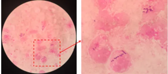
[image:3.612.94.524.71.434.2]ultrasound-guided aspiration of the muscle lesions was performed again and a drainage catheter was left in place. The material extract-ed from the abscess was smearextract-ed immextract-ediately and found Gram positive cocci in chain (Figure 5). Considering meropenem was not sufficient to control the inflammation, the antibiotic re- gimen was changed into linezolid associated with meropenem. Five days later, Streptococcus intermedius was isolated in the pus culture. It proved to be sensitive to penicillin, cefotaxime, levofloxacin and vancomycin. According to the sensitivity reports and the blood-brain barrier permeability of drugs, the antimicrobial therapy was later readjusted to ceftriaxone and linezol-id. On the twelfth day of drainage, cerebros- pinal fluid (CSF) like liquid was seen in the drainage pack. Microscopy showed pleocytosis (300 × 106/l, polymorphs). Protein content was 2550 mg/l. Three days later, the fluid decreas- ed gradually, and the drainage catheter was removed on day 26. At the same time, the patient was free from fever, conscious aware-ness, but cognitive function aggravated obvi-ously accompanied by delusion of persecution. Repeated MR of the brain showed gradually decreased dense material but increased hydro-cephalus (Figure 3C). Intravenous administra-tion of ceftriaxone was terminated on day 38. Two months later, repeated lumbar puncture showed decreased leukocytes count in the CSF to 40 × 106/l, and protein content was normal. The patient was treated by
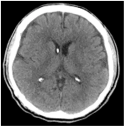
ventriculo-peritone-al shunt operation for hydrocephventriculo-peritone-alus. After the operation, oral linezolid was continued for half a month. The patient improved remarkably and can walk alone, with mild to moderate cognitive impairment. CT scan that performed one month after operation did not show any evidence of hydrocephalus (Figure 4).
Discussion
Pyogenic ventriculitis is a serious, life-threaten-ing infectious disease of the central nervous system. The clinical manifestations of pyogenic ventriculitis are often aspecific, since con-sciousness impairment may hide other symp-toms, and neurological focal deficits are hardly found when there are no abscesses or other lesions involving the brain [1]. Our patient main-ly characterized by motor symptoms, urinary retention and consciousness impairment, but with no evidence of fever and headache. The nonsteroidal anti-inflammatory drugs maybe was responsible for the absence of fever and pain. The most important feature of this case was the discovery of the abscess in the erector spinae muscle, which played an important role in the diagnosis. The pus aspirated from the abscess helped us to identify the pathogenic agent. We believed that the development of pyogenic ventriculitis owed to contiguously spread through the spinal dura, rather than hematogenous spread, because the blood cul-ture was negative. Moreover, the clinical fea-tures of this case pointed out that as to signs of paravertebral cold abscess, the diagnosis of bacteriogenic abscess should be considered too, although destruction of intervening inter-vertebral disc and a parainter-vertebral or psoas abscesst were the typical form of spinal tuber-culosis [4]. The cause of lumbar puncture fail-ure in our case maybe was due to destruction of lumbar structure or the turbid of cerebrospi-nal fluid.
[image:4.612.89.290.71.275.2]In the present case, neuroimaging played a critical role in the diagnosis especially magnet-ic resonance imaging. The DWI sequences cl- early indicated the presence of purulent mate-rial within the ventricles. On DWI, hyperintense signals were found in both lateral ventricles, with corresponding decreased signal intensi-ties in the apparent diffusion coefficient maps. Pus in the ventricles was a viscous fluid that consisted of inflammatory cells, cellular debris, microorganisms, and macromolecules. High vis- Figure 4. Brain plain CT scan performed one month
cosity, hypercellularity, and binding of water to macromolecules have been suggested to be responsible for restricted water diffusion[5]. Relative to cerebrospinal fluid, the debris was irregular and slightly hyperintense on T1-wei- ghted images, slightly hypointense on T2WI, and hyperintense on Flair images. Other find-ings may include hydrocephalus, periventricu-lar hyperintensity and ependymal enhance-ment [6, 7]. In our case, gradually enlargeenhance-ment of ventricular and periventricular hyperintensity were obvious too. Besides, another advantage of DWI was its high sensitivity for cortical and white matter cytotoxic edema, i.e., acute isch-emic infarction, likely due to septic emboli and/ or vasculitis. Lummel et al. [5] recently des- cribed the MR imaging features of 75 patients with acute bacterial meningitis, and revealed that signs of pyogenic ventriculitis were pres-ent in 41 cases (54.7%), with ischemic infarc-tions in 13 cases (17.3%). These MR imaging signs serve to distinguish intraventricular hem-orrhage from pyogenic ventriculitis and play a crucial role in the diagnosis of pyogenic ven-triculitis. However, in patients who are unstable to undergo MR scans, CT scans can still be informative. The irregular debris morphology is highly specific for purulent material. The ven-tricular debris sinks towards the occipital horns as the patient supine in the scanner. The sur-face of the viscous debris is often round or irregular, in contrast to the straight level of acute intraventricular blood. Moreover, we can see that CSF was isolated at the tip of the right occipital horn, by the overlying debris. Lo et al. [8] reported a case like this and described it as the ‘lodge sign’. Therefore, neuroimaging is a useful noninvasive method for diagnosis as well as follow-up of pyogenic ventriculitis.
other Gram-positive cocci, Enterobacter and
Klebsiella species [8]. In our case Streptococ- cus intermedius was cultured. They are part of normal flora of the oral cavity and gastrointesti-nal tract and possess the ability to cause abscesses, especially for the brain and liver. This characteristic can be explained by the viru-lence factors produced by S. intermedius. It produces the enzymes α-N-acetylneuramida- se (sialidase) and hyaluronidase, which destroy host tissues and, presumably, convert them into small nutrients to be utilized in bacterial growth. Moreover, S. intermedius specifically secretes a human-specific cytolysin, known as intermedilysin, which triggers the formation of deep-seated infection [10]. In the present case, poor glycemic control with sustained hypergly-cemia predisposed the patient to such severe infection.
As to the treatment, there are no guidelines for management of pyogenic ventriculitis. Woehrl
et al. [11] suggested that systemic intravenous antibiotic therapy seems sufficient to treat pneumococal meningitis-associated pyogenic ventriculitis. Marinelli et al. [1] reported a pa- tient underwent a number of surgical proce-dures and eventually developed pyogenic ven-triculitis. Intravenous vancomycin for 7 weeks and then replaced by oral linezolid were as the antimicrobial management which lead to a complete recovery. Akuzawa et al. [12] pre- sented a rare adult case of a left occipital lobe brain abscess that likely resulted from the hematogenous spread of an Escherichia coli
[image:5.612.88.377.73.201.2]right renal cyst infection followed by formation of pyogenic ventriculitis. The patient recovered after a 6-week course of meropenem. But Savardekar et al.[13] suggested that once the Figure 5. Light microscopy (1000 × magnification) of gram stained pus
sam-ple showed gram positive cocci in chain (arrow).
diagnosis of pyogenic ventriculitis secondary to brain abscess had been established, it was imperative to start simultaneous intrathecal and intravenous antibiotics, and frontal exter-nal ventricular drain should be placed and ven-tricular CSF drainage should be instituted. In conclusion, a neurosurgical intervention plus an antibiotic regimen is warranted for most patients. However, conservative management with antibiotics may be appropriate in patients too unwell for surgery and with significant co-morbidity. The treatment needs to be highly individualized in accordance with the patient’s clinical condition, the etiology of pyogenic ven-triculitis, the CSF reports, the brain imaging findings, the complications and the pathogens. The patient in this case, ultimately required two and a half months of antibiotic therapy, associ-ation with drainage of the erector spinae mus-cle abscess for one month. Ventriculo-peri- toneal shunt operation was performed as the complication of hydrocephalus. The outcome of the patient was relatively good with a sequela of mild to moderate cognitive impairment.
Conclusion
The clinical manifestations of pyogenic ventric-ulitis are often aspecific, with extremely unfa-vorable outcome. So early diagnose is impor-tant. Neuroimaging has a critical role in the diagnosis. Increased diffusion-weighted mag-netic resonance imaging and decreased appar-ent diffusion coefficiappar-ent maps are sensitive in pyogenic ventriculitis diagnosis, and are nonin-vasive methods rather than cerebrospinal fluid sampling. Fluid-attenuated inversion recovery sequences are specialized to detect periven-tricular signal abnormality. Moreover, S. inter-medius, found in normal intestinal flora, has an affinity for invading the brain [14-16]. This was the first case report of pyogenic ventriculitis secondary to erector spinae muscle abscess infected by S. intermedius. So as to patients with pyogenic ventriculitis and diabetes melli-tus, deep infection as a source should be con-sidered. Pyogenic ventriculitis can be caused by contiguously diffusion through spinal dura.
Disclosure of conflict of interest
None.
Address correspondence to: Dr. Chenjuan Tao, De- partment of Neurology, Affiliated Hospital of Hang-
zhou Normal University, 126 Wenzhou Road, Hang- zhou 310015, P. R. China. Tel: 86-571-2305984; Fax: 86-571-2305984; E-mail: taochenjuan@126. com; Dr. Ke Ma, College of Traditional Chinese Medicine, Shandong University of Traditional Chi- nese Medicine, 4655 University Road, Changqing, Jinan 250355, Shandong, P. R. China. Tel: 86-531-89628077; Fax: 86-531-86-531-89628077; E-mail: make- [email protected]
References
[1] Marinelli L, Trompetto C, Cocito L. Diffusion magnetic resonance imaging diagnostic rele-vance in pyogenic ventriculitis with an atypical presentation: a case report. BMC Res Notes 2014; 7: 149.
[2] Jorens PG, Voormolen MH, Robert D, Parizel PM. Imaging findings in pyogenic ventriculitis. Neurocrit Care 2009; 11: 403-5.
[3] Humphreys H, Jenks PJ. Surveillance and man-agement of ventriculitis following neurosur-gery. J Hosp Infect 2015; 89: 281-6.
[4] Momjian R, George M. Atypical imaging fea-tures of tuberculous spondylitis: case report with literature review. J Radiol Case Rep 2014; 8: 1-14.
[5] Lummel N, Koch M, Klein M, Pfister HW, Bruck-mann H, Linn J. Spectrum and prevalence of pathological intracranial magnetic resonance imaging findings in acute bacterial meningitis. Clin Neuroradiol 2016; 26: 159-67.
[6] Dandurand C, Letourneau L, Chaalala C, Magro E, Bojanowski MW. Pyogenic ventriculitis as clinical presentation of diverticulitis. Can J Neurol Sci 2016; 43: 576-7.
[7] Hatakeyama M, Kanazawa M, Ishihara A, Ta-nabe Y, Shimohata T, Nishizawa M. Pathogno-monic magnetic resonance imaging (MRI) find-ing of fluid-fluid level in pyogenic ventriculitis: two case reports. Rinsho Shinkeigaku 2014; 54: 732-7.
[8] Lo WB, Mitra R, Cadwgan A, Albanese E. Pyo-genic ventriculitis and the ‘lodge sign’. Acta Neurochir (Wien) 2016; 158: 1849-50.
[9] Hughes DC, Raghavan A, Mordekar SR, Griffiths PD, Connolly DJ. Role of imaging in the diagnosis of acute bacterial meningitis and its complications. Postgrad Med J 2010; 86: 478-85.
[10] Mishra AK, Fournier PE. The role of streptococ-cus intermedius in brain abscess. Eur J Clin Microbiol Infect Dis 2013; 32: 477-83. [11] Woehrl B, Linn J, Lummel N, Pfefferkorn T,
Ko-edel U, Pfister HW, Klein M. Pneumococcal meningitis-associated pyogenic ventriculitis. J Infect 2015; 70: 311-4.
abscess following simple renal cyst infection: a case report. BMC Neurol 2014; 14: 130. [13] Savardekar AR, Krishna R, Arivazhagan A.
Spontaneous intraventricular rupture of pyo-genic brain abscess: a short series of three cases and review of literature. Surg Neurol Int 2016; 7 Suppl 39: S947-S951.
[14] Gonzalez Mera L, Fernandez Gonzalez S, Mar-tinez Yelamos S, Fernandez Viladrich P. Brain abscess caused by Eikenella corrodens and Streptococcus intermedius associated with bowel carcinoid tumour. Med Clin (Barc) 2005; 125: 117-118.
[15] Baier B, Vogt T, Rohde F, Cuvenhaus H, Conrad J, Dieterich M. Deep brain stimulation of the nucleus ventralis intermedius: a thalamic site of graviceptive modulation. Brain Struct Funct 2017; 222: 645-650.