Original Article
New tissue processing protocol
for better long-fragment DNA preservation
Lixiong Ying, Ke Huang, Xiuming Zhang, Wei Ding
Department of Pathology, The First Affiliated Hospital, College of Medicine, Zhejiang University, Hangzhou, Zheji -ang Province, China
Received October 6, 2017; Accepted September 7, 2018; Epub November 15, 2018; Published November 30, 2018
Abstract: FFPE tissue blocks from pathology department are important resources for clinical diagnosis and scientific researches. Due to the chemical modification ability and fixation time of formalin, intact genetic materials such as the long-fragment DNA are difficult to achieve in the tissue blocks. How to properly process tissues especially during weekend and holidays remains complicated. Here we develop a new processing protocol in which the last step of paraffin immersion time is increased. Sections and DNA/RNA/protein from paired blocks (standard vs new protocol) were tested and compared for HE staining, IHC, FISH, RT-PCR, fragment analysis and Sanger sequencing. Our H&E, immunohistochemistry, FISH, RT-PCR and sequencing results suggested that tissues from the new method retained all the molecular features, and more importantly, fragment analysis showed the new method have better DNA frag-ment preservation. This new protocol can be easily applied during routine tissue processing including weekend and holidays to fulfill following elaborate molecular diagnostic requirements.
Keywords: Formalin-fixed, paraffin embedded (FFPE) tissue block, fragmentation, molecular diagnosis
Introduction
Formalin fixation and paraffin embedding has been the method of most diagnostic pathology archives for histological and pathological exam-ination. Formalin as a reagent can preserve tis-sue morphology and cytological features as well as the immunoreactivity of many antigens. However, FFPE application in many molecular diagnostic techniques like quantitative PCR, RT-PCR, Sanger sequencing, next-generation sequencing (NGS), copy number analysis and whole transcriptome/genome shotgun have its limitations. Formalin fixates tissues by robust crosslinks formation between proteins and nucleic acids [1]. Harsh conditions for breaking these crosslinks will have big effects on the genetic material retrieval [2-5]. Additionally, with fixation time increasing, the DNA and RNA long size fragments start to break into shorter sizes and it is recommended that tissues should not fixed in formalin over 48 hours [6, 7]. After fixation, tissues will proceed into dehy-dration process which formalin is washed out and finally tissues are embedded in the
paraf-fin. However, in many hospitals fixation longer than 48 hours cannot be avoided, for instance during the weekend and holidays. According to our knowledge, tissues during weekend and holidays are mainly kept in the formalin solu-tion. And these samples usually have problem in long fragment analysis [8]. How to handle samples during weekend and holidays to fulfill both pathological diagnosis as well as molecu-lar diagnosis? Here we develop a new tissue processing protocol which the tissues are immersed long-time in paraffin at the final step of dehydration process. In comparison with the standard protocol, our results revealed that this new protocol can preserve the pathological and molecular features of FFPE tissues.
Methods and materials
Tissue samples, fixation and processing
Table 1. Antibody clones, sources, dilutions and cellular distributions
Antibody Clones Sources Dilutions Cellular Distribution
ALK D5F3 Ventana Ready to use Lung adenocarcinoma (C)
Cadherin-E EP700Y Epitomics 1:100 Breast carcinoma (M; M+C)
CD2 AB75 DAKO 1:100 T cell lymphoma (M)
CD3 PS1 Novocastra 1:100 T celllymphoma (M; M+C)
CD4 4B12 Novocastra 1:50 T cell lymphoma (M)
CD7 CBC37 Novocastra 1:50 T cell lymphoma (M)
CD8 C8/114B Amsbio 1:300 T cell lymphoma (M)
CD20 L26 DAKO 1:200 B cell lymphoma (M)
CD79a JCB117 LabVision 1:200 B cell lymphoma (M)
CD117/C-kit Polyclonal Invitrogen 1:200 GIST (C; M+C)
C-erb-B2 4B5 Ventana Ready to use Breast cancer (M)
Chromogranin A Polyclonal LabVision 1:50 Pheochromocytoma (C)
Cytokeratin AE1/AE3 LabVision 1:100 Adenocarcinoma (C)
Cytokeratin 20 EP23 Epitomics 1:100 Colon adenocarcinoma (C)
DOG-1 SP31 Invitrogen 1:300 GIST (C; M+C)
Estrogen Receptor (ER) 1D5 DAKO 1:200 Breast cancer (N)
Ki-67 MIB-1 Invitrogen 1:500 Breast cancer (N)
Melan-A A103 Invitrogen 1:100 Melanoma (C)
Melanoma HMB45 DAKO 1:50 Melanoma (C)
Nestin 10C2 Invitrogen 1:200 Melanoma (C)
Progesterone Receptor (PR) PgR636 LabVision 1:200 Breast cancer (N)
P53 DO-7 DAKO 1:150 Breast carcinoma (N)
P120 Catenin EP66 Epitomics 1:200 Breast carcinoma (M; M+C)
Synaptophysin Polyclonal LabVision 1:100 Pheochromocytoma (C)
S-100 Polyclonal DAKO 1:5000 Melanoma (N+C)
TTF-1 8G7G3/1 LabVision 1:500 Lung adenocarcinoma (N)
Abbreviations: N = Nuclear; M = Membranous; C = Cytoplasmic.
node), non-Hodgkin T cell lymphoma (lymph node), breast invasive lobular carcinoma,
gas-trointestinal stromal tumor (GIST), melanoma and glioblastoma and 5 small tissues including lung biopsy, abdomen biopsy, breast biopsy, gastroscopic tissue and colonoscopic were selected from the Department of Pathology, the First Affiliated Hospital, College of Medicine, Zhejiang University. All above tissues were divided into 2 equal portions at <3 mm in thick-ness (for surgical samples) to have sections with similar sizes. Each portion was fixed in 4% neutral paraffin (Tongsheng Technology, Ning- bo) for 8 h and went through the following two dehydration protocols: protocol 1 (the standard weekend protocol in our department). 50 h in Formalin, 0.5 h in water, 2 h in 75% ethanol, 2 h in 85% ethanol, 1.5 h in 95% ethanol I, 2 h in 95% ethanol II, 1.5 h in 100% ethanol I, 2 h in 100% ethanol II, 1 h in xylene I, 1.5 h in xylene II, 1 h in paraffin I, 1.5 h in paraffin II and 1.5 h in paraffin III (Chaser, Fujian). Protocol 2 (the new developed paraffin weekend protocol), 2 h in Formalin, 0.5 h in water, 2 h in 75% ethanol,
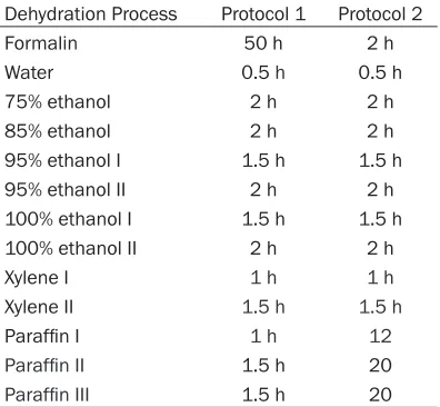
Table 2. Paired-FFPE tissue processing proto-col 1 and 2
Dehydration Process Protocol 1 Protocol 2
Formalin 50 h 2 h
Water 0.5 h 0.5 h
75% ethanol 2 h 2 h
85% ethanol 2 h 2 h
95% ethanol I 1.5 h 1.5 h
95% ethanol II 2 h 2 h
100% ethanol I 1.5 h 1.5 h 100% ethanol II 2 h 2 h
Xylene I 1 h 1 h
Xylene II 1.5 h 1.5 h
Paraffin I 1 h 12
Paraffin II 1.5 h 20
[image:2.612.91.289.501.684.2]xylene II, 12 h in paraffin I, 20 h in paraffin II and 20 h in paraffin III. The flowchart representation of two protocols was shown in Table 2. The study protocol was ap- proved by the Hospital Ethics Committee. 4 μm sections from paired For- malin-fixed, paraffin em- bedded (FFPE) tissue blo- cks were cut for hema-toxylin and eosin (H&E) staining and following analysis.
Immunohistochemistry
The slides were air-dried at room temperature (20°C) for 20 seconds. The details of 28 primary antibodies used are list-ed in Table 1. They are routinely used in our insti-tution to test specific anti-gen expression on tissue blocks. All IHC procedu- res were carried out in a fully-automatic IHC mac- hine (VENTANA BenchM- ark ULTRA, America; VE- NTANA BenchMark XT, America) by using a two-step En Vision system (DAKO, Denmark) with DAB colorization.
Fluorescent in situ hy
-bridization (FISH)
[image:3.612.91.396.69.607.2]FISH test was used to determine the HER-2/neu status. Sections were ba- ked overnight at 60°C, deparaffinized two times in the xylene for 10 min, transferred twice through 100% ethanol, one time in 85% ethanol, one time in 70% ethanol, each for Figure 1. Representative hematoxylin and eosin stained sections. Surgical lung
adenocarcinoma (A), lung biopsy (B), surgical colon adenocarcinoma (C), colono-scopic (D), surgical gastrointestinal stromal tumor (GIST) (E), abdomen biopsy (F) tissue samples treated with dehydrationprotocol 1 (A1-F1) and protocol 2 (A2-F2). The magnification for all images are ×200.
2 h in 85% ethanol, 1.5 h in 95% ethanol I, 2 h in 95% ethanol II, 1.5 h in 100% ethanol I, 2 h in 100% ethanol II, 1 h in xylene I, 1.5 h in
Figure 2. Immunohistochemical staining. Lung adenocarcinoma with ALK (A), B lymphoma with CD20 (B), breast cancer with CerbB2 (C), glioblastoma with P53 (D). (A1-D1) were tissues from dehydrationprotocol 1 and (A2-D2) were from pro-tocol 2. The magnification for all images are ×200.
After that, the slides were briefly washed in sodium saline citrate (SSC, pH 7.2) at room temperature, dehydrated through 70%, 85%, 100% ethanol and acetone. After drying in the open-air, 10 μl of probe (Zytovision, Germany) was applied onto each slide, cover slip was placed and sealed with rubber cement, and then the slides were transferred to the hybrid-ization oven (S500-24, Abbott molecular, USA). The procedure was as follows: denature at 83°C for 5 min, and hybridized overnight at 42°C. After that, the slides were washed in 46°C preheated post-hybridization buffer (2XSSC/0.1% sodium dodecyl sulfate) for 5 min and rinsed in 70% ethanol. After air-drying, the slides were counterstained with 15 μl DAPI and cover slip applied.
Thirty randomly selected invasive tumor nuclei in each of two separate, distinct microscopic
with 50 μl elution buffer. DNA/RNA concentra-tions were determined by Nanodrop. DNA frag-ment length was assessed using the DNA qual-ity control tube in Invivoscribe kit (Invivoscribe, USA), with amplicon lengths of 100, 200, 300, 400 and 600. The test was carried out in ABI3500Dx.
PCR and RT-PCR
Mutations of EGFRexon 18, 19, 20, 21, ROS1
and EML4-ALK fusion were tested in lung ade-nocarcinoma and lung biopsy blocks using ARMS Detection Kit (AmoyDx, Xiamen). Mu- tations of KRAS exon 2, 3 and 4, NRAS exon 2, 3 and 4, BRAF V600E mutations were also test-ed for colon cancer and gastroscopic tissue blocks using ARMS Detection Kit (AmoyDx, Xiamen).
areas were evaluated. Po- sitive for HER-2/neu is de- fined as HER-2/CEP 17 ra- tio ≥2.0 or HER-2/CEP17
ratio <2.0 with an average
HER-2 copy number ≥6.0. Equivocal for HER-2 is de- fined as HER-2/CEP17 ra- tio <2.0 with an average
HER-2 copy number ≥4.0 and <6.0 signals/cell. Ne- gative for HER-2 is defin- ed as HER-2/CEP17 ratio <2.0 with an average HE-R-2 copy number <4.0 sig-nals/cell.
Nucleic acid extraction
entical IHC staining pa- ttern and intensity in all paired samples. The representative selected sequential stains for ALK, CD20, CerbB2 and P53 were illustrated in
Figure 2.
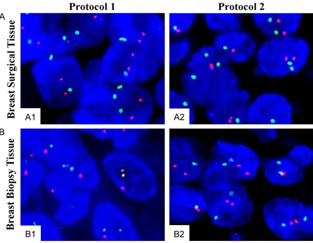
Fluorescent in situ hybridization (FISH)
Surgical breast invasive lobular carcinoma and breast biopsy tissues were chosen for FISH analysis. Two sets of paired samples went through same protocol and showed almost sa- me intensity of DAPI,
CEP17 and HER-2 stain-ing (Figure 3). The fluo-rescent signals of HER-2
[image:5.612.92.399.71.309.2]gene for both paired sa- mples were negative. Re- Figure 3. Fluorescent in situ hybridization (FISH) visualization of paired-breast
sam-ples.dehydration protocol 1 (A) and protocol 2 (B). Green dot: HER-2 gene. Red dot: CEP17 gene, blue background: DAPI staining. The magnification for both images are ×1000.
Sanger sequencing
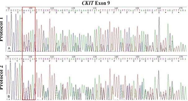
DNA from the paired GIST FFPE blocks were tested for KIT exon 9, 11, 13, 17 and PDGFRa
exon 12 and 18 mutations using Sanger Se- quencing Kit (Yuanqi, Shanghai). Sequencing was carried out in ABI3500Dx.
Results
Histological presentation
Representative H&E stained images (4 paired-tumor tissues) of dehydration protocol 1 and 2 are illustrated in Figure 1. Overall, the HE sta- ining of all sections from paired-FFPE tissue blocks with dehydration protocol 1 and 2 showed no significant differences in normal tissue architecture, cytological and nuclear details. We also noticed that sections of block-ing processblock-ing protocol 2 tended to have a slight better eosin stain.
Immunohistochemical examination
The amount of tissue from protocol 2 did not decrease obviously after IHC stains including heating antigen retrieval. The comparison be- tween protocol 1 and 2 showed the totally id-
sult proved that long time in the paraffin had no influence on the probe binding and fluorescent visualization.
DNA yields
DNA from total 30 FFPE blocks was ranging from 13.8-976.3 ng/ul. The variation of DNA yields between paired blocks was from 6.0 ng/ ulto 86.2 ng/ul. Moreover, the quality of all the blocks were very high (260/280 ratio ≥1.8 and 260/230 ration ≥1.6). There was no evidence that protocol 2 will influence the DNA yield.
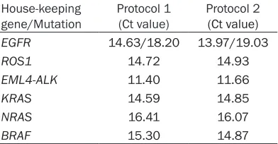
PCR results
In our paired lung adenocarcinoma FFPE blocks, we detected an EGFR 19-DEL mutation using ARMS PCR (Figure 4A). All the Ct values were listed in Table 3. The Ct value of house-keeping gene and EGFR 19-DEL between two blocks were only 0.21 and 0.18. No ROS1 or
EML4-ALK fusion were found in the lung adeno-carcinoma samples (Figure 4B and 4C). In ad- dition, KRAS exon 2, 3, 4, NRAS exon 2, 3, 4,
Table 3. Summary of Ct values House-keeping
gene/Mutation Protocol 1 (Ct value) Protocol 2 (Ct value) EGFR 14.63/18.20 13.97/19.03
ROS1 14.72 14.93
EML4-ALK 11.40 11.66
KRAS 14.59 14.85
NRAS 16.41 16.07
BRAF 15.30 14.87
(Figure 4D-F). T-test shown no significant differ-ence between the CT values. Overall there was no evidence of a difference in PCR success rates between protocol 1 and 2.
DNA length
DNA length was found highly correlated with time in formalin and paraffin. Most surgical samples from protocol 1 only retained frag-ment size within 300 bp. Moreover, the biopsy samples showed more fragmentation than the big tissue. In comparison, all sample blocks from protocol 2 had fragment sizes longer than 600 bp size (Figure 5B). This analysis revealed that paraffin had significantly better effect in preserving DNA from fragmentation.
RNA yields
RNA from total 30 FFPE blocks were ranging from 3.1-524.3 ng/ul. The variation of DNA yields between paired blocks were from 0.7 ng/ ul to 137.7 ng/ul. Moreover, the quality of all the blocks were high (260/280 ratio ≥2.0 and 260/230 ration ≥1.8). This rough RNA isolated test showed that protocol 2 also have a similar RNA preservation.
Sequencing results
We selected GIST, colon cancer, lung biopsy and gastroscopic tissue for Sanger sequencing. In both GIST samples, we observed same 502_503InsAY mutation in KIT exon 9 (Figure 6) and the sequencing signals from both meth-ods did not present obvious differences.
Discussion
Formalin fixation and paraffin embedded tissue samples are the major sources in the pathology department for routine histopathological diag-nose and research. Formalin can prevent
tis-sue autolysis, stabilize proteins and limit anti-gen and nucleic acids degradation thereby nicely preserve the tissue morphology. But for-malin introduce protein-protein and proteins-nucleic cross-linking, as well as chemical modi-fications of nucleic acids [9]. These reactions lead to significant degradation of nucleic acids in FFPE blocks. Further fragmentation of DNA and RNA can be caused by suboptimal fixation, prolonged storage of FFPE blocks, tissue pro-cessing, sectioning and staining procedures [10, 11]. With the development of molecular biology and genetics, requirements for nucleic acids preservation in clinical specimens be- come high. Besides, increased applications of minimal invasive surgery and biopsy produce much smaller pieces of pathology specimens. Therefore, finding better ways for pathological specimen processing and storage are of great necessity. Many groups from worldwide had published various papers aiming at nucleic preservation for molecular diagnosis. But these studies were mainly focus on testing different types of fixatives or DNA isolation methods [12-14]. Whereas different tissue processing proto-cols are not given enough attention.
suggest-Figure 5. Fragment analysis. Both surgical and biopsy samples were analyzed for fragmentation. Small tissues tended to have severe fragmentation than big samples at same tissue processing conditions. Tissues from protocol 2 showed nice long fragment preservation (>400 bp or 600 bp) whereas tissues from protocol 1 only had fragments smaller than 300 bp.
[image:9.792.97.703.127.453.2]ed lymphoma rearrangement should only be performed if the sample has the 300 bp frag-ment at the minimum [15]. This indicates that tissue treatment and duration of storage are highly related with assay success rates. Protocol 1 had significantly DNA degradation and contained short amplicons (<300 bp). This outcome will surely influence downstream molecular diagnostic assays. In contrast, proto-col 2 appeared to have better nucleic acid pres-ervation and yield long DNA fragment (>400 and 600 bp).
Overall, hydration protocol 1 and 2 showed no differences in H&E, IHC, FISH, DNA/RNA extrac-tion, quantitative PCR, RT-PCR and Sange sequencing but protocol 2 had been demon-strated to be a better method for long-fragment analysis from FFPE tissue specimens. The par-affin used in our department has a low melting point (54-56°C) and stable chemical properties and long-time immersion in paraffin showed no harm to DNA, RNA or proteins. Outcome of this tissue specimen treatment method have not been studied in previous articles.
In conclusion, we designed a new tissue pro-cessing protocol which the time of tissues in the paraffin, instead of in the formalin, is extended. All the data show that this method can retain all pathological features and more importantly, preserve large DNA fragment from fragmentation. This protocol can be widely applied in routine pathology departments with molecular diagnostic demands, especially dur-ing weekend and holidays.
Disclosure of conflict of interest
None.
Address correspondence to: Wei Ding, Department of Pathology, The First Affiliated Hospital, College of Medicine, Zhejiang University, No. 79 Qingchun Road, Hangzhou 310003, Zhejiang, China. Tel: +86-13958082289; Fax: +86-571-87236364; E-mail: [email protected]
References
[1] Masuda N, Ohnishi T, Kawamota S, Monden M, Okubu K. Analysis of chemical modification of RNA from formalin-fixed samples and optimiza-tion of molecular boology applicaoptimiza-tions for such
samples. Nucleic Acids Res 1999; 27: 4436-4443.
[2] Mcsherry EA, McGoldrick A, Kay EW, Hopkins AM, Gallagher WM, Dervan PA. Formalin-fixed paraffin-embedded clinical tissues show spuri-ous copy number changes in array-CGH pro-files. Clin Genet 2007; 72: 441-447.
[3] Tournier B, Chapusot C, Courcet E, Martin L, Lepage C, Faivre J, Piard F. Why do results con-flict regarding the prognostic value of the methylation status in colon cancers? The role of the preservation method. BMC Cancer 2012; 12: 12.
[4] Von Smolinski D, Leverkoehne I, von Samoson-Himmelstjerna G, Gruber AD. Impact of forma-lin-fixation and paraffin-embedding on the ra-tio between mRNA copy numbers of differently expressed genes. Histochem Cell Biol 2005; 124: 177-188.
[5] Williams C, Ponten F, Moberg C, Soderkvist P, Uhlen M, Ponten J, Sitbon G, Lundeberg J. A high frequency of sequence alterations is due to formalin fixation of archival specimens. Am J Pathol 1999; 155: 1467-1471.
[6] Bresters D, Schipper ME, Reesink HW, Boeser-Nunnink BD, Cuypers HT. The duration of fixa-tion influences the yield of HCV cDNA-PCR products from formalin-fixed, paraffin-embed-ded liver tissue. J Virol Method 1994; 48: 267-272.
[7] Macabeo-Ong M, Ginzinger DG, Dekker N, Mc-Millan A, Regezi JA, Wong DT, Jordan RC. Ef-fects of duration fixation on quantitative re-verse transcription polymerase chain reaction analyses. Mod Pathol 2002; 15: 979-987. [8] Greer CE, Peterson SL, Kiviat NB, Manos MM.
PCR amplification from paraffin-embedded tis-sues. Effects of fixative and fixation time. Am J Clin Pathol 1991; 95: 117-124.
[9] Werner M, Chott A, Fabiano A, Battifora H. Ef-fect of formalin tissue fixation and processing on immunohistochemistry. Am J Surg Pathol 2000; 24: 1016-1019.
[10] Cronin M, Pho M, Dutta D, Stephans JC, Shak S, Kiefer MC, Esteban JM, Baker JB. Measure-ment of gene expression in archival paraffin-embedded tissues: development and perfor-mance of a 92-gene reverse transcriptase- polymerase chain reaction assay. Am J Pathol 2004; 164: 35-42.
[11] Von Ahlfen S, Missel A, Bendrat K, Schlump-berger M. Determinants of RNA quality from FFPE samples. PLoS One 2007; 2: e1261. [12] Turashvili G, Yang W, McKinney S, Kalloger S,
blocks: defining optimal fixation, processing and DNA/RNA extraction techniques. Exp Mol Pathol 2012; 92: 33-43.
[13] Gilbert MT, Haselkorn T, Bunce M, Sanchez JJ, Lucas SB, Jewell LD, March EV, Worobey M. The isolation of nucleic acids from fixed, paraf-fin-embedded tissues-which methods are use-ful when? PLoS One 2007; 2: e537.
[14] Groelz D, Sobin L, Branton P, Compton C, Wyrich R, Rainen L. Non-formalin fixative ver-sus formalin-fixed tissue: a comparison of his-tology and RNA quality. Exp Mol Pathol 2013; 94: 188-194.