Original Article
Thrombin-activatable fibrinolysis inhibitor (TAFI) is
a potential biomarker for cerebral hemorrhage patients
Botao Wu1, Anyan Wen1, Xuebin Xu1, Cheng Zuo1, Baochang Shan1, Liang Li2
Departments of 1Neurosurgery, 2Orthopaedics, Dongying People’s Hospital, Dongying, China
Received September 18, 2017; Accepted January 10, 2018; Epub March 15, 2018; Published March 30, 2018
Abstract: Cerebral hemorrhage is the most common human cerebrovascular disease and frequently leads to pa-ralysis, a vegetative state, and even death. Previous studies have indicated that thrombin-activatable fibrinolysis inhibitor (TAFI) is associated with chronic thromboembolic pulmonary hypertension and cerebral hemorrhage. The purpose of this study was to investigate the correlation between serum levels of TAFI and inflammatory markers in cerebral hemorrhage patients. A total of 138 patients diagnosed with cerebral hemorrhage were enrolled in this clinical study, and 100 healthy volunteers were recruited as controls. Serum levels of IL-1β, IL-6, IL-17, and TNF-α were detected by ELISA. Serum levels of TAFI, procalcitonin (PCT), and C-reactive protein (CRP) were analyzed by col-loidal gold test strip. We showed that serum levels of inflammatory cytokines IL-1β, IL-6, IL-17, and TNF-α were mark-edly increased in cerebral hemorrhage patients compared to healthy volunteers (p < 0.01). Serum levels of TAFI, PCT, and CRP were also remarkably enhanced in cerebral hemorrhage patients compared to healthy volunteers (p < 0.01). Correlation analysis demonstrated that there was a significant correlation between serum TAFI concentration and illness degree of hemorrhage patients but not in healthy volunteers. We also found that serum TAFI concentra-tion was positively correlated with IL-1β, IL-6, IL-17, and TNF-α in the cerebral hemorrhage patients but not in healthy volunteers. In conclusion, these outcomes indicate that TAFI and inflammatory cytokines are up-regulated and cor-relate with illness degree in patients with cerebral hemorrhage, suggesting that TAFI may have potential ability to be considered as a biomarker for cerebral hemorrhage diagnosis or risk prediction.
Keywords: Cerebral hemorrhage, TAFI, inflammation, biomarker
Introduction
Cerebral hemorrhage is one of the most com-mon human cerebrovascular diseases and patients with cerebral hemorrhage often pres-ent higher mortality during periods of disease [1, 2]. The most common manifestations in cerebral hemorrhage patients are cerebral arteriosclerosis, hypertension, and intracranial vascular malformations [3, 4]. Pathological studies have shown that many factors can induce cerebral hemorrhage, and patients with cerebral hemorrhage are usually complicated with severe dysfunction of the cerebral nervous system and loss of social working and self-care ability, which further increase the burden on the family [5-7]. A comprehensive review of basic and clinical studies describes the current therapeutic drugs against cerebral vasospasm after subarachnoid hemorrhage [8]. However, the morbidity and mortality rate of patients
cerebral hemorrhage compared to healthy vol-unteers. Our findings report that TAFI and inflammatory cytokines are up-regulated and correlate with the degree of illness in patients with cerebral hemorrhage. This study also investigated serum levels of PCT and CRP as well as the correlation with serum levels of TAFI in patients with cerebral hemorrhage.
Materials and methods
Patients and healthy volunteers
A total of 138 patients with cerebral hemor-rhage and 100 healthy volunteers were re- cruited for analysis of the association of serum TAFI and inflammatory cytokines. The numbers of men and women patients were approximate equal. All participants were eligible to finish this clinical investigation. The characteristics of cerebral hemorrhage patients are summarized in Table 1.
ELISA
Serum levels of IL-1β (NO: MAF10524), IL-6 (NO: MAF12783), IL-17 (NO: MAF15283), TNF-α (NO: MAF7392), TAFI (NO: MAF7202), PCT (NO: MAF7710) and CRP (NO: MAB40283) were de- tected in patients with arrhythmia using ELISA kit (Bio-Techne, R&D Systems, USA) according to the manufacturer’s instruction. The serum concentrations of inflammatory cytokines, TAFI, PCT, and CRP were also measured by an enzyme micro-plate reader at 570 nm.
Regression analysis
The serum levels of bilirubin and uric acid in the detective data (Y) were analyzed by regression
analysis in different clinical stages and persis-tent auricular fibrillation patients with arrhyth-mia were analyzed using the least square con-vergence [14]. The predicted curve that resulted in the lowest sum of squares was the best fit. If the fit was robust, then the parameters of the observed curve could be inferred from those of the predicted.
Statistical analysis
For each experiment, the mean and standard error were determined. Statistical differences between groups were assessed by means of analysis of variance (ANOVA) from 6 replicate experiments with the post-hoc Dunnett’s test. Statistical significance was considered at P < 0.05.
Results
Serum levels of inflammatory cytokines IL-1β, IL-6, IL-17, and TNF-α in patients with cerebral hemorrhage
We first analyzed serum levels of inflammatory cytokines IL-1β, IL-6, IL-17, and TNF-α in patients with cerebral hemorrhage and healthy volun-teers. As shown in Figure 1A, 1B, we show that serum levels of IL-1β and IL-6 were increased in patients with cerebral hemorrhage compared to healthy volunteers. Serum levels of IL-17 and TNF-α were significantly up-regulated in pa- tients with cerebral hemorrhage (Figure 1C, 1D). These results indicate that patients with cerebral hemorrhage presented with higher serum levels of inflammatory cytokines IL-1β, IL-6, IL-17, and TNF-α.
Serum levels of TAFI, PCT, and CRP in patients with cerebral hemorrhage
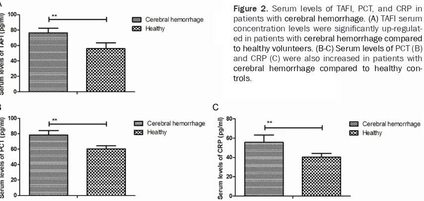
[image:2.612.91.289.97.240.2]Inflammatory cytokines TAFI, PCT, and CRP were analyzed in patients with cerebral hemor-rhage. As shown in Figure 2A, TAFI serum con-centration levels were significantly up-regulat-ed in patients with cerebral hemorrhage com- pared to those in the healthy volunteers. We observed that the serum levels of PCT and CRP were also increased in patients with cerebral hemorrhage compared to those in the healthy control (Figure 2B and 2C). These results indi-cated that serum levels of TAFI, PCT, and CRP were up-regulated in patients with cerebral hemorrhage.
Table 1. Characteristic of patients with cere-bral hemorrhage
Characteristics Patients Health
Number 138 100
Female 65 48
Male 73 52
Age 36.6-66.5 40.5-68.7
Clinical stage
CCH 34 0
ESCH 19 0
SLCH 21 0
ACH 24 0
Correlation analysis between TAFI and inflam-matory cytokines IL-1β, IL-6, IL-17, and TNF-α in patients with cerebral hemorrhage
Associations were analyzed between TAFI and inflammatory cytokines IL-1β, IL-6, IL-17, and TNF-α in patients with cerebral hemorrhage as determined by regression analysis. We show that TAFI concentration is positively correlated with IL-1β in cerebral hemorrhage patients but not in healthy volunteers (Figure 3A). Results present serum levels of IL-6 and IL-17 that were positively correlated with TAFI serum
concen-(Figure 3B and 3C). We also found that TAFI was positively correlated with TNF-α serum levels in patients with cerebral hemorrhage (Figure 3D). These results indicated that serum levels of TAFI are positively correlated with the inflammatory cytokines IL-1β, IL-6, IL-17, and TNF-α in patients with cerebral hemorrhage.
Correlation analysis between TAFI and PCT, CRP in patients with cerebral hemorrhage
[image:3.612.98.520.72.276.2]The associations between TAFI and PCT, CRP were respectively analyzed in patients with Figure 1. Serum levels of inflammatory cytokines IL-1β, IL-6, IL-17,and TNF-α in patients with cerebral hemorrhage. (A-B) Serum levels of IL-1β (A) and IL-6 (B) were increased in patients with cerebral hemorrhage compared to healthy volunteers. (C-D) Serum levels of IL-17 (C) and TNF-α (D) were significantly up-regulated in patients with cerebral hemorrhage.
Figure 2. Serum levels of TAFI, PCT, and CRP in patients with cerebral hemorrhage. (A) TAFI serum concentration levels were significantly up-regulat-ed in patients with cerebral hemorrhage compared to healthy volunteers. (B-C) Serum levels of PCT (B) and CRP (C) were also increased in patients with
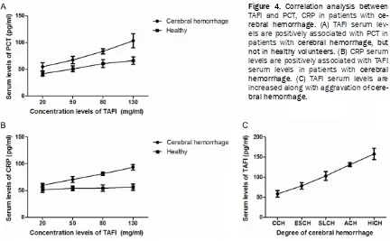
[image:3.612.97.524.350.552.2]trols. Outcomes showed that TAFI serum levels were associated with PCT in patients with cere-bral hemorrhage, but not in healthy volunteers (Figure 4A). Results also revealed that CRP
serum levels were positively associated with TAFI serum levels in patients with cerebral hemorrhage (Figure 4B). Importantly, TAFI serum levels were increased along with
aggra-Figure 3. Correlation analysis between TAFI and inflammatory cytokines IL-1β, IL-6, IL-17, and TNF-α in patients with cerebral hemorrhage (A) TAFI concentration is positively correlated with IL-1β in cerebral hemorrhage patients but not in healthy volunteers. (B-C) Serum levels of IL-6 (B) and IL-17 (C) were positively correlated with TAFI serum concentration levels in cerebral hemorrhage patients. (D) Serum levels of TAFI are positively correlated with TNF-α serum levels in patients with cerebral hemorrhage.
Figure 4. Correlation analysis between
[image:4.612.95.522.73.313.2] [image:4.612.92.524.392.659.2]vation of cerebral hemorrhage (Figure 4C). These results suggest that serum levels of TAFI are positively associated with serum levels of CT, CRP, and illness status of patients with cerebral hemorrhage.
Discussion
Numerous studies have indicated that cerebral hemorrhage causes damage of neuronal cells and further aggravates brain damage, even resulting in contralateral limb dysfunction [15-17]. TAFI plays important roles in the progres-sion and prognosis of cerebral hemorrhage [18-20]. In this study, we analyzed changes of serum levels of TAFI and the relationships between TAFI and cytokines, CT, CRP in patients with cerebral hemorrhage. Although the sys-tematic review and meta-analysis analyzed ge- netic variations in thrombin-activatable fibrino-lysis inhibitor gene and risk of cardiovascular disease [21], the relationships between TAFI and the severity of illness of cerebral hemor-rhage have not been well understood. Findings in this study indicate that serum TAFI is up-reg-ulated in cerebral hemorrhage patients com-pared to that in the healthy volunteers. Results also found inflammatory cytokines and serum TAFI level was positively correlated with inflam-matory cytokines IL-1β, IL-6, IL-17, and TNF-α in patients with cerebral hemorrhage.
IL-1β is increased following hypertensive intra-cerebral hemorrhage (ICH), potentially related to neural damage by cerebral edema and cor-relation between serum IL-1beta levels and cerebral edema extent has been reported in a hypertensive intracerebral hemorrhage rat model [22]. Croci et al. have analyzed serum IL-6 changes in experimental rabbit subarach-noid hemorrhage and the results reveal a sta-tistically significant correlation between IL-6 and ET-1 levels in the CSF [23]. Beeftink et al.
reported a relationship between serum TNF-alpha and TNF-alpha genotype with delayed cerebral ischemia and outcome in subarach-noid hemorrhage [24]. Previous studies have showed that TAFI is increased in patients with cerebral hemorrhage [25-27]. However, the association between TAFI and inflammation cytokines IL-1β, IL-6, IL-17, and TNF-α remains poorly understood. Results in this study show that inflammatory cytokines IL-1β, IL-6, IL-17, and TNF-α serum levels are up-regulated and correlate with TAFI increasing in patients with cerebral hemorrhage.
Previous studies have shown that early system-ic PCT levels are increased in patients with aneurysmal subarachnoid hemorrhage [28]. Festic et al. suggested that the utility of serum PCT of 0.2 ng/mL or greater was demonstrated to be very specific for sepsis among patients with aneurysmal subarachnoid hemorrhage [29]. Additionally, Srinivasan et al. suggested that CRP has a significant independent associ-ation with poor GOS score, indicating the pre-eminence of early cellular response in SAH pathophysiology [30]. Our results indicate that serum levels of PCT and CRP are up-regulated in patients with cerebral hemorrhage. Our find-ings suggest that serum TAFI is positively asso-ciated with serum levels of PCT and CRP in patients with cerebral hemorrhage. Importantly, the results indicated that TAFI serum levels can be responsible for aggravation of cerebral hemorrhage.
In conclusion, although oral anticoagulant or even surgical resection are often used as the mainstay treatment for cerebral hemorrhage in the clinic, effective biomarkers and the prog-nosis of cerebral hemorrhage are insufficient to predict therapeutic effects. Results in this study show that inflammatory cytokines IL-1β, IL-6, IL-17, and TNF-α and PCT and CRP are up-regulated in patients with cerebral hemorrhage. Interestingly, TAFI serum levels are increased and positively correlate with inflammatory cyto-kines, PCT, and CRP in patients with cerebral hemorrhage. These results may provide a cli- nical foundation for patients with cerebral hemorrhage.
Disclosure of conflict of interest
None.
Address correspondence to: Botao Wu, Depart- ments of Neurosurgery, Dongying People’s Hospital, 317 South Road, Dongcheng, Dongying 257091, Shandong, China. Tel: +86-45378653302; E-mail: wubotaodongning@163.com
References
[2] Khatib KI and Baviskar AS. Treatment of cere-bral venous sinus thrombosis with subdural hematoma and subarachnoid hemorrhage. J Emerg Trauma Shock 2016; 9: 155-156. [3] Lin C, Zhao Y, Wan G, Zhu A and Wang H.
Ef-fects of simvastatin and taurine on delayed cerebral vasospasm following subarachnoid hemorrhage in rabbits. Exp Ther Med 2016; 11: 1355-1360.
[4] Lin PY, Hagan K, Fenoglio A, Grant PE and Franceschini MA. Reduced cerebral blood flow and oxygen metabolism in extremely preterm neonates with low-grade germinal matrix- in-traventricular hemorrhage. Sci Rep 2016; 6: 25903.
[5] Isozaki M, Arai Y, Higashino Y, Okazawa H and Kikuta KI. Cerebral hyperperfusion syndrome resulting in subarachnoid hemorrhage after carotid artery stenting. Ann Nucl Med 2016; 30: 669-674.
[6] Jabbarli R, Reinhard M, Roelz R, Kaier K, Wey-erbrock A, Taschner C, Scheiwe C and Shah M. Clinical relevance of anterior cerebral artery asymmetry in aneurysmal subarachnoid hem-orrhage. J Neurosurg 2017; 127: 1070-1076. [7] Mrak G, Duric KS and Nemir J. Middle cereb-
ral artery fusiform aneurysm presented with stroke and delayed subarachnoid hemorrha- ge trapping, thrombectomy, and bypass. Surg Neurol Int 2016; 7: S209-213.
[8] Hasegawa S, Hasegawa Y and Miura M. Cur-rent therapeutic drugs against cerebral va- sospasm after subarachnoid hemorrhage: a comprehensive review of basic and clinical studies. Curr Drug Deliv 2017; 14: 843-852. [9] Marar TT and Boffa MB. Identification of a
thrombomodulin interaction site on thrombin-activatable fibrinolysis inhibitor that mediates accelerated activation by thrombin. J Thromb Haemost 2016; 14: 772-783.
[10] Plug T and Meijers JC. Structure-function rela-tionships in thrombin-activatable fibrinolysis inhibitor. J Thromb Haemost 2016; 14: 633-644.
[11] Yaoita N, Satoh K, Satoh T, Sugimura K, Tatebe S, Yamamoto S, Aoki T, Miura M, Miyata S, Kawamura T, Horiuchi H, Fukumoto Y and Shi-mokawa H. Thrombin-activatable fibrinolysis inhibitor in chronic thromboembolic pulmo-nary hypertension. Arterioscler Thromb Vasc Biol 2016; 36: 1293-1301.
[12] Leung LL, Myles T, Nishimura T, Song JJ and Robinson WH. Regulation of tissue inflamma-tion by thrombin-activatable carboxypeptidase B (or TAFI). Mol Immunol 2008; 45: 4080-4083.
[13] Gad MZ, El-Mesallamy HO and Sanad EF. hsCRP, sICAM-1 and TAFI in hemodialysis patients: linking inflammation and
hypofibri-nolysis to cardiovascular events. Kidney Blood Press Res 2008; 31: 391-397.
[14] Hayes AF and Rockwood NJ. Regression-based statistical mediation and moderation analysis in clinical research: observations, recommen-dations, and implementation. Behav Res Ther 2017; 98: 39-57.
[15] Chamnanvanakij S, Margraf LR, Burns D and Perlman JM. Apoptosis and white matter injury in preterm infants. Pediatr Dev Pathol 2002; 5: 184-189.
[16] Riggs AJ and Riggs JE. Epilepsy’s role in the historical differentiation of religion, magic, and science. Epilepsia 2005; 46: 452-453. [17] Gao F, Guo Y, Zhang H, Wang S, Wang J, Wu JM,
Chen Z and Ding MP. Anterior thalamic nucleus stimulation modulates regional cerebral me-tabolism: an FDG-MicroPET study in rats. Neu-robiol Dis 2009; 34: 477-483.
[18] Philippou H. Thrombin activatable fibrinolysis inhibitor (TAFI): more complex when it meets the clot. Thromb Res 2014; 133: 1-2.
[19] Wang S, Zhao Z, Cong Z and Suo G. Thrombin-activatable fibrinolysis inhibitor is activated in an instant blood-mediated inflammatory reac-tion after intraportal islet transplant. Exp Clin Transplant 2014; 12: 62-66.
[20] Yildirim MN, Selcoki Y, Uysal S, Nacar AB, Demircelik B, Aydin HI and Eryonucu B. Throm-bin activatable fibrinolysis inhibitor: its role in slow coronary flow. Herz 2014; 39: 993-1000. [21] Shi J, Zhi P, Chen J, Wu P and Tan S. Genetic
variations in the thrombin-activatable fibrinoly-sis inhibitor gene and risk of cardiovascular disease: a systematic review and meta-analy-sis. Thromb Res 2014; 134: 610-616. [22] Wei P, You C, Jin H, Chen H and Lin B.
Correla-tion between serum IL-1beta levels and cere-bral edema extent in a hypertensive intracere-bral hemorrhage rat model. Neurol Res 2014; 36: 170-175.
[23] Croci D, Nevzati E, Danura H, Schopf S, Fandi-no J, Marbacher S and Muroi C. The relation-ship between IL-6, ET-1 and cerebral vaso-spasm, in experimental rabbit subarachnoid hemorrhage. J Neurosurg Sci 2016; [Epub ahead of print].
[24] Beeftink MM, Ruigrok YM, Rinkel GJ and van den Bergh WM. Relation of serum TNF-alpha and TNF-alpha genotype with delayed cerebral ischemia and outcome in subarachnoid hem-orrhage. Neurocrit Care 2011; 15: 405-409. [25] Ammollo CT, Semeraro F, Incampo F, Semeraro
N and Colucci M. Dabigatran enhances clot susceptibility to fibrinolysis by mechanisms dependent on and independent of thrombin-activatable fibrinolysis inhibitor. J Thromb Hae-most 2010; 8: 790-798.
the molecular inactivation mechanism of hu-man activated thrombin-activatable fibrino- lysis inhibitor. J Thromb Haemost 2010; 8: 1056-1065.
[27] Qin L, D’Alessandro-Gabazza CN, Aoki S, Gil-Bernabe P, Yano Y, Takagi T, Boveda-Ruiz D, Ramirez Marmol AY, San Martin Montenegro VT, Toda M, Miyake Y, Taguchi O, Takei Y, Mors-er J and Gabazza EC. Pulmonary hypMors-ertension is ameliorated in mice deficient in thrombin-activatable fibrinolysis inhibitor. J Thromb Hae-most 2010; 8: 808-816.
[28] Muroi C, Lemb JB, Hugelshofer M, Seule M, Bellut D and Keller E. Early systemic procalci- tonin levels in patients with aneurysmal sub-arachnoid hemorrhage. Neurocrit Care 2014; 21: 73-77.
[29] Festic E, Siegel J, Stritt M and Freeman WD. The utility of serum procalcitonin in distin-guishing systemic inflammatory response syn-drome from infection after aneurysmal sub-arachnoid hemorrhage. Neurocrit Care 2014; 20: 375-381.