organic papers
o3334
Wardellet al. C13H8INO4 doi:10.1107/S1600536805029272 Acta Cryst.(2005). E61, o3334–o3336
Acta Crystallographica Section E Structure Reports
Online
ISSN 1600-5368
4-Nitrophenyl 2-iodobenzoate: sheets
built from C—H
O hydrogen bonds
and two-centre iodo–nitro interactions
James L. Wardell,aJanet M. S. Skakle,bJohn N. Lowband Christopher Glidewellc*
aInstituto de Quı´mica, Departamento de
Quı´mica Inorgaˆnica, Universidade Federal do Rio de Janeiro, CP 68563, 21945-970 Rio de Janeiro, RJ, Brazil,bDepartment of Chemistry,
University of Aberdeen, Meston Walk, Old Aberdeen AB24 3UE, Scotland, andcSchool of Chemistry, University of St Andrews, Fife KY16 9ST, Scotland
Correspondence e-mail: cg@st-andrews.ac.uk
Key indicators
Single-crystal X-ray study
T= 120 K
Mean(C–C) = 0.004 A˚
Rfactor = 0.026
wRfactor = 0.058
Data-to-parameter ratio = 16.9
For details of how these key indicators were automatically derived from the article, see http://journals.iucr.org/e.
#2005 International Union of Crystallography All rights reserved
Molecules of the title compound, C13H8INO4, are linked into
complex sheets by two C—H O hydrogen bonds and one two-centre iodo–nitro interaction.
Comment
We have recently reported the molecular and supramolecular structures of a wide range of iodoaryl–nitroaryl compounds, including sulfonamides (Kellyet al., 2002), benzylideneanilines (Glidewell, Howie et al., 2002; Wardell et al., 2002), benzyl-anilines (Glidewell, Lowet al., 2002; Glidewell, Low, Skakle, Wardell & Wardell, 2004; Ferguson et al., 2005), phenyl-hydrazones (Glidewell, Low, Skakle & Wardell, 2004; Glide-well et al., 2003), 1,4-diaryl-2,3-diaza-1,3-butadienes (Glidewell, Low, Skakle & Wardell, 2005), N -(iodophenyl)-nitrophthalimides (Glidewell, Low, Skakle, Wardell & Wardell, 2005) and benzoylhydrazones (Glidewell, Low & Wardell, 2005). We have now extended this investigation to include the title ester, 4-nitrophenyl 2-iodobenzoate, (I).
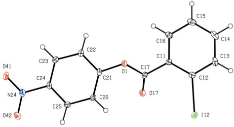
Within the molecule of (I) (Fig. 1), the central ester frag-ment between atoms C11 and C21 is effectively planar, but the iodinated and nitrated aryl rings make dihedral angles with this plane of 39.9 (2) and 42.7 (2), respectively, probably in
[image:1.610.212.449.589.718.2]Received 14 September 2005 Accepted 15 September 2005 Online 21 September 2005
Figure 1
order to minimize the repulsive intramolecular contacts involving the polarized atom O17. The nitro group makes a dihedral angle of 7.4 (2)with the adjacent aryl ring. The bond
distances and inter-bond angles show no unusual values. The molecules are linked into complex sheets, the forma-tion of which is readily analysed in terms of two one-dimen-sional substructures. In the simpler of the two substructures, atom C14 in the iodinated ring of the molecule at (x,y,z) acts as hydrogen-bond donor to carbonyl atom O17 in the mol-ecule at (1
2 + x, 3
2 y, 1
2 + z), thereby forming a C(7)
(Bernstein et al., 1995) chain running parallel to the [101] direction and generated by then-glide plane aty= 0.75 (Fig. 2). The second substructure is built from a combination of a C—H O hydrogen bond and an iodo–nitro interaction. Atom C26 in the nitrated ring of the molecule at (x,y,z) acts as hydrogen-bond donor to nitro atom O42 in the molecule at (1
2x, 1 2+y,
3
2z), so forming aC(6) chain running parallel to
the [010] direction and generated by the 21screw axis along (14,
y,3
4) (Fig. 3). In addition, atom I12 in the molecule at (x,y,z)
forms a short contact with atom O41 in the molecule at (x, 1 +y,z), with I Oi= 3.240 (2) A˚ and C—I Oi= 169.8 (2) [symmetry code: (i)x, 1 +y,z], thus generating by translation aC(11) (Starbucket al., 1999) chain, also running parallel to the [010] direction. The combination of these two interactions then generates a [010] chain of edge-fused R3
3(17) rings
(Fig. 3).
The combination of the [010] and [101] chains generates a (101) sheet in the form of a (4,4)-net. If just the C—H O hydrogen bonds are considered, this sheet is built from two types ofR4
4(38) ring (Fig. 4).
Experimental
A solution containing equimolar quantities (2 mmol of each) of 4-nitrophenol and 2-iodobenzoyl chloride in chloroform (50 ml) was heated under reflux for 1 h; the solvent was removed under reduced pressure and the resulting solid residue was recrystallized from ethanol to yield crystals suitable for single-crystal X-ray diffraction.
Crystal data
C13H8INO4
Mr= 369.10
Monoclinic, P21=n
a= 9.7231 (4) A˚
b= 11.7890 (3) A˚
c= 11.1187 (4) A˚ = 97.363 (2)
V= 1263.98 (8) A˚3
Z= 4
Dx= 1.940 Mg m
3 MoKradiation Cell parameters from 2905
reflections = 3.0–27.5
= 2.54 mm1
T= 120 (2) K Plate, colourless 0.100.080.01 mm
Data collection
Bruker Nonius KappaCCD area-detector diffractometer ’and!scans
Absorption correction: multi-scan (SADABS; Sheldrick, 2003)
Tmin= 0.814,Tmax= 0.975 12630 measured reflections
2905 independent reflections 2570 reflections withI> 2(I)
Rint= 0.036 max= 27.5
h=12!11
k=13!15
l=14!14
organic papers
Acta Cryst.(2005). E61, o3334–o3336 Wardellet al. C
[image:2.610.314.563.75.310.2]13H8INO4
o3335
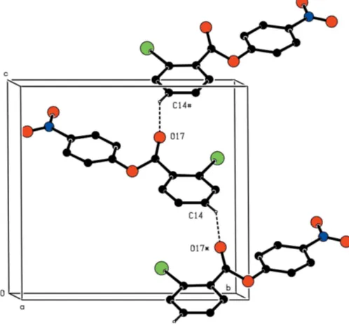
Figure 2
Part of the crystal structure of (I), showing the formation of aC(7) chain along [101]. For the sake of clarity, H atoms not involved in the motif shown have been omitted. Atoms marked with an asterisk (*) or a hash (#) are at the symmetry positions (1
2+x, 3
2y,
1
2+z) and ( 1 2+x,
3 2y, 1
[image:2.610.356.514.421.672.2]2+z), respectively.
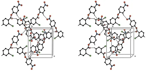
Figure 3
Part of the crystal structure of (I), showing the formation of a chain of edge-fusedR3
3(17) rings along [010]. For the sake of clarity, H atoms not involved in the motif shown have been omitted. Atoms marked with an asterisk (*), a hash (#) or an ampersand (&) are at the symmetry positions (1
2x, 1 2+y,
3
2z), (x, 1 +y,z) and ( 1
2x,
1 2+y,
3
Refinement
Refinement onF2
R[F2> 2(F2)] = 0.026
wR(F2) = 0.058
S= 1.10 2905 reflections 172 parameters
H-atom parameters constrained
w= 1/[2(F
o2) + (0.0125P)2 + 1.8703P]
whereP= (Fo2+ 2Fc2)/3 (/)max= 0.001
max= 0.80 e A˚
3
min=0.91 e A˚
3
Table 1
Hydrogen-bond geometry (A˚ ,).
D—H A D—H H A D A D—H A
C14—H14 O17i
0.95 2.50 3.334 (3) 147
C26—H26 O42ii
0.95 2.54 3.395 (3) 149
Symmetry codes: (i)xþ1 2;yþ
3 2;z
1 2; (ii)xþ
1 2;yþ
1 2;zþ
3 2.
All H atoms were located in difference maps and then treated as riding atoms, with C—H distances of 0.95 A˚ andUiso(H) = 1.2Ueq(C).
Data collection: COLLECT (Hooft, 1999); cell refinement:
DENZO(Otwinowski & Minor, 1997) andCOLLECT; data reduc-tion:DENZOandCOLLECT; program(s) used to solve structure:
OSCAIL (McArdle, 2003) and SHELXS97 (Sheldrick, 1997); program(s) used to refine structure: OSCAIL and SHELXL97
(Sheldrick, 1997); molecular graphics:PLATON(Spek, 2003); soft-ware used to prepare material for publication: SHELXL97 and
PRPKAPPA(Ferguson, 1999).
X-ray data were collected at the EPSRC X-Ray Crystal-lographic Service, University of Southampton, England; the authors thank the staff of the Service for all their help and advice. JLW thanks CNPq and FAPERJ for financial support.
References
Bernstein, J., Davis, R. E., Shimoni, L. & Chang, N.-L. (1995).Angew. Chem. Int. Ed. Engl.34, 1555–1573.
Ferguson, G. (1999).PRPKAPPA. University of Guelph, Canada.
Ferguson, G., Glidewell, C., Low, J. N., Skakle, J. M. S. & Wardell, J. L. (2005).
Acta Cryst.C61, o445–o449.
Glidewell, C., Howie, R. A., Low, J. N., Skakle, J. M. S., Wardell, J. L. & Wardell, S M. S. V. (2002).Acta Cryst.B58, 864–876.
Glidewell, C., Low, J. N., Skakle, J. M. S. & Wardell, J. L. (2003).Acta Cryst.
C59, o98–o101.
Glidewell, C., Low, J. N., Skakle, J. M. S. & Wardell, J. L. (2004).Acta Cryst.
C60, o19–o23.
Glidewell, C., Low, J. N., Skakle, J. M. S. & Wardell, J. L. (2005).Acta Cryst.
C61, o312–o316.
Glidewell, C., Low, J. N., Skakle, J. M. S., Wardell, S. M. S. V. & Wardell, J. L. (2002).Acta Cryst.C58, o487–o490.
Glidewell, C., Low, J. N., Skakle, J. M. S., Wardell, S. M. S. V. & Wardell, J. L. (2004).Acta Cryst.B60, 472–480.
Glidewell, C., Low, J. N., Skakle, J. M. S., Wardell, S. M. S. V. & Wardell, J. L. (2005).Acta Cryst.B61, 227–237.
Glidewell, C., Low, J. N. &. Wardell, J. L. (2005).Acta Cryst.E61, o2438–o2440. Hooft, R. W. W. (1999).COLLECT. Nonius BV, Delft, The Netherlands. Kelly, C. J., Skakle, J. M. S. Wardell, J L., Wardell, S. M. S. V., Low, J. N. &
Glidewell, C. (2002).Acta Cryst.B58, 94–108.
McArdle, P. (2003). OSCAIL for Windows. Version 10. Crystallography Centre, Chemistry Department, NUI Galway, Ireland.
Otwinowski, Z. & Minor, W. (1997). Methods in Enzymology, Vol. 276,
Macromolecular Crystallography, Part A, edited by C. W. Carter Jr & R. M. Sweet, pp. 307–326. New York: Academic Press.
Sheldrick, G. M. (1997).SHELXS97andSHELX97. University of Go¨ttingen, Germany.
Sheldrick, G. M. (2003).SADABS. Version 2.10. University of Go¨ttingen, Germany.
Spek, A. L. (2003).J. Appl. Cryst.36, 7–13.
Starbuck, J., Norman, N. C. & Orpen, A. G. (1999).New J. Chem.23, 969–972. Wardell, J. L., Wardell, S. M. S. V., Skakle, J. M. S., Low, J. N. & Glidewell, C.
(2002).Acta Cryst.C58, o428–o430.
organic papers
o3336
Wardellet al. C [image:3.610.43.298.71.196.2]13H8INO4 Acta Cryst.(2005). E61, o3334–o3336
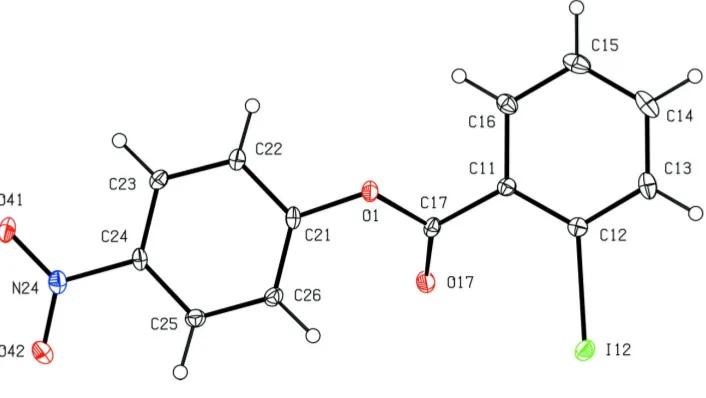
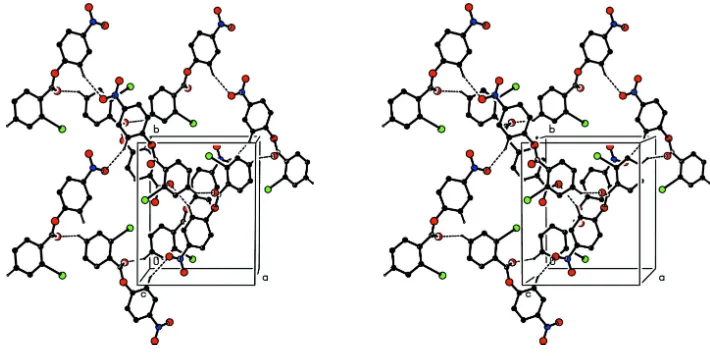
Figure 4
Stereoview of part of the crystal structure of compound (I), showing the formation of a hydrogen-bonded (101) sheet ofR4
supporting information
sup-1
Acta Cryst. (2005). E61, o3334–o3336supporting information
Acta Cryst. (2005). E61, o3334–o3336 [doi:10.1107/S1600536805029272]
4-Nitrophenyl 2-iodobenzoate: sheets built from C
—
H
···
O hydrogen bonds and
two-centre iodo
–
nitro interactions
James L. Wardell, Janet M. S. Skakle, John N. Low and Christopher Glidewell
S1. Comment
We have recently reported the molecular and supramolecular structures of a wide range of iodoaryl–nitroaryl compounds,
including sulfonamides (Kelly et al., 2002), benzylideneanilines (Glidewell, Howie et al., 2002; Wardell et al., 2002), benzylanilines (Glidewell, Low et al., 2002; Glidewell, Low, Skakle, Wardell & Wardell, 2004; Ferguson et al., 2005), phenylhydrazones (Glidewell, Low, Skakle & Wardell, 2004; Glidewell et al., 2003), 1,4-diaryl-2,3-diaza-1,3-butadienes (Glidewell, Low, Skakle & Wardell, 2005), N-(iodophenyl)nitrophthalimides (Glidewell, Low, Skakle, Wardell & Wardell, 2005) and benzoylhydrazones (Glidewell, Low & Wardell, 2005). We have now extended this investigation to
include the title ester 4-nitrophenyl 2-iodobenzoate, (I).
Within the molecule of (I) (Fig. 1), the central ester fragment between atoms C11 and C21 is effectively planar, but the
iodinated and nitrated aryl rings make dihedral angles with this plane of 39.9 (2) and 42.7 (2)°, respectively, probably in
order to minimize the repulsive intramolecular contacts involving the polarized atom O17: the nitro group makes a
dihedral angle of 7.4 (2)° with the adjacent aryl ring. The bond distances and inter-bond angles show no unusual values.
The molecules are linked into complex sheets, whose formation is readily analysed in terms of two one-dimensional
substructures. In the simpler of the two substructures, atom C14 in the iodinated ring of the molecule at (x, y, z) acts as hydrogen-bond donor to carbonyl atom O17 in the molecule at (1/2 + x, 1.5 − y, −1/2 + z), thereby forming a C(7) (Bernstein et al., 1995) chain running parallel to the [101] direction and generated by the n-glide plane at y = 0.75 (Fig. 2).
The second substructure is built from a combination of a C—H···O hydrogen bond and an iodo–nitro interaction. Atom
C26 in the nitrated ring of the molecule at (x, y, z) acts as hydrogen-bond donor to nitro atom O42 in the molecule at (1/2 − x, 1/2 + y, 1.5 − z), so forming a C(6) chain running parallel to the [010] direction and generated by the 21 screw axis
along (1/4, y, 3/4) (Fig. 3). In addition, atom I12 in the molecule at (x, y, z) forms a short contact with atom O41 in the molecule at (x, 1 + y, z), with I···Oi = 3.240 (2) Å and C—I···Oi = 169.8 (2)° [symmetry code: (i) x, 1 + y, z)], thus
generating by translation a C(11) (Starbuck et al., 1999) chain, also running parallel to the [010] direction. The combination of these two interactions then generates a [010] chain of edge-fused R33(17) rings (Fig. 3)
The combination of the [010] and [101] chains generates a (101) sheet in the form of a (4,4)-net: if just the C—H···O
hydrogen bonds are considered, this sheet is built from two types of R44(38) ring (Fig. 4).
S2. Experimental
A solution containing equimolar quantities (2 mmol of each) of 4-nitrophenol and 2-iodobenzoyl chloride in chloroform
(50 ml) was heated under reflux for 1 h; the solvent was removed under reduced pressure and the resulting solid residue
supporting information
sup-2
Acta Cryst. (2005). E61, o3334–o3336S3. Refinement
All H atoms were located from difference maps and then treated as riding atoms, with C—H distances of 0.95 Å and
[image:5.610.130.483.122.321.2]Uiso(H) = 1.2Ueq(C).
Figure 1
The molecule of compound (I), showing the atom-labelling scheme. Displacement ellipsoids are drawn at the 30%
supporting information
[image:6.610.128.486.74.404.2]sup-3
Acta Cryst. (2005). E61, o3334–o3336Figure 2
Part of the crystal structure of (I), showing the formation of a C(7) chain along [101]. For the sake of clarity, H atoms not
involved in the motif shown have been omitted. Atoms marked with an asterisk (*) or a hash (#) are at the symmetry
supporting information
[image:7.610.167.443.71.522.2]sup-4
Acta Cryst. (2005). E61, o3334–o3336Figure 3
Part of the crystal structure of (I), showing the formation of a chain of edge-fused R33(17) rings along [010]. For the sake
of clarity, H atoms not involved in the motif shown have been omitted. Atoms marked with an asterisk (*), a hash (#) or
supporting information
[image:8.610.126.481.71.249.2]sup-5
Acta Cryst. (2005). E61, o3334–o3336Figure 4
Stereoview of part of the crystal structure of compound (I), showing the formation of a hydrogen-bonded (101) sheet of
R44(38) rings. For the sake of clarity, H atoms not involved in the motifs shown have been omitted.
4-Nitrophenyl 2-iodobenzoate
Crystal data
C13H8INO4
Mr = 369.10
Monoclinic, P21/n
Hall symbol: -P 2yn
a = 9.7231 (4) Å
b = 11.7890 (3) Å
c = 11.1187 (4) Å
β = 97.363 (2)°
V = 1263.98 (8) Å3
Z = 4
F(000) = 712
Dx = 1.940 Mg m−3
Mo Kα radiation, λ = 0.71073 Å Cell parameters from 2905 reflections
θ = 3.0–27.5°
µ = 2.54 mm−1
T = 120 K Plate, colourless 0.10 × 0.08 × 0.01 mm
Data collection
Bruker–Nonius 95mm CCD camera on κ
goniostat diffractometer
Radiation source: Bruker-Nonius FR91 rotating anode
Graphite monochromator
Detector resolution: 9.091 pixels mm-1
φ and ω scans
Absorption correction: multi-scan (SADABS; Sheldrick, 2003)
Tmin = 0.814, Tmax = 0.975
12630 measured reflections 2905 independent reflections 2570 reflections with I > 2σ(I)
Rint = 0.036
θmax = 27.5°, θmin = 3.0°
h = −12→11
k = −13→15
l = −14→14
Refinement
Refinement on F2
Least-squares matrix: full
R[F2 > 2σ(F2)] = 0.026
wR(F2) = 0.058
S = 1.10 2905 reflections 172 parameters 0 restraints
Primary atom site location: structure-invariant direct methods
Secondary atom site location: difference Fourier map
Hydrogen site location: inferred from neighbouring sites
supporting information
sup-6
Acta Cryst. (2005). E61, o3334–o3336w = 1/[σ2(F
o2) + (0.0125P)2 + 1.8703P]
where P = (Fo2 + 2Fc2)/3
(Δ/σ)max = 0.001
Δρmax = 0.80 e Å−3
Δρmin = −0.91 e Å−3
Fractional atomic coordinates and isotropic or equivalent isotropic displacement parameters (Å2)
x y z Uiso*/Ueq
I12 0.580705 (19) 0.888948 (14) 0.650830 (16) 0.02111 (7) O1 0.62987 (19) 0.51101 (15) 0.59123 (16) 0.0181 (4)
O17 0.6392 (2) 0.63226 (16) 0.75041 (17) 0.0223 (4)
O41 0.3784 (2) 0.06357 (16) 0.7691 (2) 0.0298 (5)
O42 0.2579 (2) 0.18618 (17) 0.8565 (2) 0.0301 (5)
N24 0.3467 (2) 0.1619 (2) 0.7914 (2) 0.0217 (5)
C11 0.7462 (3) 0.6829 (2) 0.5758 (2) 0.0160 (5)
C12 0.7347 (3) 0.8009 (2) 0.5729 (2) 0.0177 (5)
C13 0.8224 (3) 0.8659 (2) 0.5107 (3) 0.0238 (6)
C14 0.9227 (3) 0.8134 (3) 0.4527 (3) 0.0279 (7)
C15 0.9341 (3) 0.6962 (3) 0.4530 (3) 0.0272 (7)
C16 0.8463 (3) 0.6312 (2) 0.5131 (2) 0.0204 (6)
C17 0.6661 (3) 0.6105 (2) 0.6508 (2) 0.0166 (5)
C21 0.5571 (3) 0.4276 (2) 0.6460 (2) 0.0164 (5)
C22 0.5994 (3) 0.3177 (2) 0.6297 (2) 0.0170 (5)
C23 0.5297 (3) 0.2288 (2) 0.6755 (2) 0.0175 (5)
C24 0.4191 (3) 0.2544 (2) 0.7381 (2) 0.0171 (5)
C25 0.3732 (3) 0.3646 (2) 0.7521 (2) 0.0190 (5)
C26 0.4434 (3) 0.4527 (2) 0.7041 (2) 0.0184 (5)
H13 0.8132 0.9461 0.5082 0.029*
H14 0.9842 0.8579 0.4124 0.033*
H15 1.0022 0.6605 0.4118 0.033*
H16 0.8536 0.5509 0.5121 0.024*
H22 0.6762 0.3032 0.5871 0.020*
H23 0.5567 0.1524 0.6645 0.021*
H25 0.2955 0.3793 0.7936 0.023*
H26 0.4139 0.5290 0.7110 0.022*
Atomic displacement parameters (Å2)
U11 U22 U33 U12 U13 U23
supporting information
sup-7
Acta Cryst. (2005). E61, o3334–o3336C15 0.0207 (15) 0.0418 (18) 0.0200 (14) −0.0022 (13) 0.0058 (11) −0.0070 (13) C16 0.0180 (14) 0.0257 (14) 0.0176 (13) −0.0040 (11) 0.0028 (11) −0.0022 (11) C17 0.0172 (13) 0.0122 (12) 0.0199 (13) 0.0008 (10) −0.0002 (11) 0.0015 (10) C21 0.0182 (13) 0.0133 (12) 0.0164 (12) −0.0048 (10) −0.0021 (10) 0.0017 (10) C22 0.0178 (13) 0.0142 (12) 0.0180 (12) −0.0016 (10) −0.0014 (10) 0.0017 (10) C23 0.0178 (13) 0.0141 (12) 0.0197 (13) 0.0017 (10) −0.0009 (11) −0.0008 (10) C24 0.0165 (13) 0.0147 (12) 0.0193 (13) −0.0047 (10) −0.0008 (10) 0.0033 (10) C25 0.0165 (13) 0.0191 (13) 0.0217 (13) 0.0013 (11) 0.0031 (11) −0.0022 (10) C26 0.0164 (13) 0.0149 (12) 0.0234 (13) 0.0023 (10) 0.0012 (11) 0.0000 (10)
Geometric parameters (Å, º)
C11—C12 1.396 (4) C17—O17 1.198 (3)
C11—C16 1.406 (4) C21—C22 1.378 (4)
C11—C17 1.482 (4) C21—C26 1.382 (4)
C12—C13 1.394 (4) C22—C23 1.381 (4)
C12—I12 2.098 (3) C22—H22 0.95
C13—C14 1.383 (4) C23—C24 1.387 (4)
C13—H13 0.95 C23—H23 0.95
C14—C15 1.386 (4) C24—C25 1.389 (4)
C14—H14 0.95 C24—N24 1.463 (3)
C15—C16 1.381 (4) N24—O42 1.229 (3)
C15—H15 0.95 N24—O41 1.233 (3)
C16—H16 0.95 C25—C26 1.388 (4)
O1—C17 1.371 (3) C25—H25 0.95
O1—C21 1.396 (3) C26—H26 0.95
C12—C11—C16 118.6 (2) C22—C21—C26 122.2 (2)
C12—C11—C17 122.7 (2) C22—C21—O1 115.4 (2)
C16—C11—C17 118.5 (2) C26—C21—O1 122.2 (2)
C13—C12—C11 120.5 (3) C21—C22—C23 119.8 (3)
C13—C12—I12 116.6 (2) C21—C22—H22 120.1
C11—C12—I12 122.87 (19) C23—C22—H22 120.1
C14—C13—C12 119.8 (3) C22—C23—C24 117.9 (2)
C14—C13—H13 120.1 C22—C23—H23 121.0
C12—C13—H13 120.1 C24—C23—H23 121.0
C13—C14—C15 120.4 (3) C23—C24—C25 122.7 (2)
C13—C14—H14 119.8 C23—C24—N24 119.0 (2)
C15—C14—H14 119.8 C25—C24—N24 118.3 (2)
C16—C15—C14 119.9 (3) O42—N24—O41 123.4 (2)
C16—C15—H15 120.0 O42—N24—C24 118.4 (2)
C14—C15—H15 120.0 O41—N24—C24 118.3 (2)
C15—C16—C11 120.6 (3) C26—C25—C24 118.4 (3)
C15—C16—H16 119.7 C26—C25—H25 120.8
C11—C16—H16 119.7 C24—C25—H25 120.8
C17—O1—C21 120.4 (2) C21—C26—C25 118.8 (2)
O17—C17—O1 123.8 (2) C21—C26—H26 120.6
supporting information
sup-8
Acta Cryst. (2005). E61, o3334–o3336O1—C17—C11 110.0 (2)
C16—C11—C12—C13 −1.1 (4) C17—O1—C21—C22 −139.1 (2)
C17—C11—C12—C13 173.4 (2) C17—O1—C21—C26 45.9 (3)
C16—C11—C12—I12 175.25 (19) C26—C21—C22—C23 −2.0 (4)
C17—C11—C12—I12 −10.2 (4) O1—C21—C22—C23 −177.0 (2)
C11—C12—C13—C14 −0.8 (4) C21—C22—C23—C24 −0.7 (4)
I12—C12—C13—C14 −177.3 (2) C22—C23—C24—C25 2.5 (4)
C12—C13—C14—C15 1.9 (4) C22—C23—C24—N24 −177.8 (2)
C13—C14—C15—C16 −1.0 (4) C23—C24—N24—O42 173.0 (2)
C14—C15—C16—C11 −0.9 (4) C25—C24—N24—O42 −7.3 (4)
C12—C11—C16—C15 1.9 (4) C23—C24—N24—O41 −6.6 (4)
C17—C11—C16—C15 −172.8 (3) C25—C24—N24—O41 173.0 (2)
C21—O1—C17—O17 0.1 (4) C23—C24—C25—C26 −1.7 (4)
C21—O1—C17—C11 178.1 (2) N24—C24—C25—C26 178.6 (2)
C12—C11—C17—O17 −37.8 (4) C22—C21—C26—C25 2.8 (4)
C16—C11—C17—O17 136.7 (3) O1—C21—C26—C25 177.5 (2)
C12—C11—C17—O1 144.3 (2) C24—C25—C26—C21 −0.9 (4)
C16—C11—C17—O1 −41.2 (3)
Hydrogen-bond geometry (Å, º)
D—H···A D—H H···A D···A D—H···A
C14—H14···O17i 0.95 2.50 3.334 (3) 147
C26—H26···O42ii 0.95 2.54 3.395 (3) 149