ISSN Online: 2164-3199 ISSN Print: 2164-3180
DOI: 10.4236/ojbd.2018.82004 Jun. 26, 2018 27 Open Journal of Blood Diseases
Role of Immunostaining in Detecting
Extra-Pattern and Subtle Lymphomatous
Infiltration in Bone Marrow Biopsies of NHL
Patients
Salwa S. Khodeir, Tahany A. Elkerdany, Abeer Saad Attia, Yasmin N. El Sakhawy,
Gehan M. Hamed, Noha Bassiouny Hassan
*Department of clinical Pathology, Faculty of Medicine, Ain Shams University Hospital, Cairo, Egypt
Abstract
Introduction: Immunohistochemistry (IHC) enables the examination of a greater number of trephine biopsy levels and is helpful in determining addi-tional scattered malignant cells. The aim of this study is to detect ex-tra-pattern and subtle lymphomatous infiltration in bone marrow biopsies using CD20 and CD3 immunostaining. Patients and Methods: This study was conducted on 100 newly diagnosed Non Hodgkin Lymphoma (NHL) pa-tients. Their bone marrow trephine biopsies were assessed on routine histolo-gy [Hematoxylin and Eosin (H & E)], and were further subjected to IHC using CD20 and CD3. Results: Pattern of involvement by H & E was highlighted by IHC. It showed additional interstitial pattern in 9 cases, parasinusoidal streaks in one case and highlighted a patchy pattern in another case with interstitial involvement on H & E. IHC also detected subtle infiltrations on additional 5.5% cases compared with histology alone. It helped in differentiating reactive (12 cases) and malignant lymphoid infiltration (33 cases). Conclusion: CD20 and CD3 immunostaining performed routinely on bone marrow trephine bi-opsies has the ability to reveal extra-pattern of infiltration and improve detec-tion of subtle lymphoid involvement. A combined procedure identifying sev-eral distinctive features, in particular histotopography and IHC, provides a promising way of discriminating reactive from neoplastic lymphoid infiltrates in bone marrow trephine biopsies.
Keywords
Immunohistochemistry (IHC), Hematoxylin and Eosin (H & E), Non-Hodgkin Lymphoma (NHL)
How to cite this paper: Khodeir, S.S., Elkerdany, T.A., Attia, A.S., El Sakhawy, Y.N., Hamed, G.M. and Hassan, N.B. (2018) Role of Immunostaining in Detect-ing Extra-Pattern and Subtle Lymphomat-ous Infiltration in Bone Marrow Biopsies of NHL Patients. Open Journal of Blood Dis-eases, 8, 27-36.
https://doi.org/10.4236/ojbd.2018.82004
Received: May 11, 2018 Accepted: June 23, 2018 Published: June 26, 2018
Copyright © 2018 by authors and Scientific Research Publishing Inc. This work is licensed under the Creative Commons Attribution International License (CC BY 4.0).
DOI: 10.4236/ojbd.2018.82004 28 Open Journal of Blood Diseases
1. Introduction
The bone marrow (BM) biopsy is a necessary procedure for patients with lym-phomatous neoplasm (NHL) and it’s used for staging and following up purposes and hence has an impact on their prognosis and treatment options [1]. It serves occasionally as a primary diagnostic technique in suspicious clinical settings as those with isolated splenomegaly or inaccessible lymphadenopathy [2]. Five major patterns of BM infiltration can be discerned, namely a nodular (intertra-becular or paratra(intertra-becular), an interstitial, a diffuse and an intrasinusoidal pat-tern. However patterns can change during the course of the disease, and mixed patterns occur frequently [3]. Lymphocyte aggregates have been reported to oc-cur in between 1% and 60% of normal BM specimens. Therefore, neoplastic lymphomatous infiltrates need to be differentiated from benign (reactive) lym-phocytes [4] [5]. Many studies have focused on the use of immunohistochemi-stry (IHC) on BM trephines in ambiguous cases to differentiate between benign and malignant lymphoid aggregates [6] [7]. IHC enables the examination of a greater number of trephine biopsy levels and is helpful in determining additional scattered and clusters of malignant cells. There are exceptional cases that need IHC to detect subtle lymphomatous cells which might be easily missed if only hematoxylin and eosin (H & E) sections are examined as occult interstitial in-volvement which usually occur early in the disease course or after treatment as residual cells, intrasinusoidal pattern and accompanying hemophagocytic syn-drome [8]. Detection of subtle lymphomatous BM involvement hinges on ade-quacy of both routine (H & E) histological diagnosis and IHC analysis [3]. This study aimed to reveal extra-pattern and subtle lymphomatous infiltration in bone marrow biopsies using a limited panel of immunostaining (CD20 and CD3) in comparison to H & E sections, and differentiate benign versus malig-nant lymphoid infiltrates.
2. Patients and Methods
The present study was conducted on one hundred newly diagnosed NHL pa-tients (according to their lymph node biopsies) attended hematology department of Ain-Shams University Hospital (ASUH) from June 2015 to August 2016. This study was approved by the ethical and moral committee of Faculty of Medicine Ain-Shams University and a verbal informed consent was taken from all patients participating in the study. They were further classified according to their lymph node biopsy into 94 with B-NHL while 6 patients with T-NHL, their median age was 49 years, 45 of them were males and 55 were females. For staging purposes, bone marrow biopsies were performed with a mean length of 20 mm and a range of 8 - 30 mm, in addition to the following.
2.1. BM Histology
DOI: 10.4236/ojbd.2018.82004 29 Open Journal of Blood Diseases paraffin, and an attempt was made to stain 5 sections with H & E. Sections with at least 5 well preserved marrow spaces were studied for cellularity, normal he-matological elements, presence of lymphoid infiltrates if any; the extent and his-tological pattern were looked for. All specimens were subjected to appropriate IHC for highlighting the pattern and extent of any lymphomatous infiltration, characterizing their nature, differentiating between benign and malignant lym-phomatous infiltration, in addition to detecting any subtle infiltrates.
2.2. IHC
IHC analysis was performed manually for all cases on formalin fixed, paraffin embedded trephine biopsies. Polymeric HRP (horseradish peroxidase) method was used to immunostain sections using the following monoclonal (mouse ready to use) antibodies: T-cell marker; CD3 (Genemed Biotechnologies, Inc. Code 60-0011-7-USA) and B-cell marker; CD20 (Genemed Biotechnologies; clone L26), in addition to the detection kit (power stain TM 1.0 poly HRP DAB kit for mouse and rabbit). The slides were immersed in heat retrieval solution (Ge-nemed biotechnologies; citrate buffer, pH6) followed by heating in microwave oven adjusted at 800 watt for 10 minutes (2 min for 5 cycles). After cooling of the slides at room temperature, peroxidase block (3% H2O2) was applied to cover
the specimens for 10 - 15 min. Each primary antibody covered the specimens for 60 min at room temperature, followed by poly −HRP conjugate for 25 min and lastly DAB solution for 5 - 10 min in dark place. Between each step, the slides were immersed and washed 3 times by phosphate buffer saline (PBS) with pH (7.4 ± 0.2). Counterstaining by Mayer’s hematoxylin was used for 40 sec if using stock solution or for 3 - 5 min if using working solution. Dehydration step ended by immersion in xylene solution for 3 min followed by mounting step using dis-tyrene, a plasticizer, and xylene (DPX ) after dryness of xylene on slides and then cover slipped. With each run tonsillar tissue was used as positive control, nega-tive control slide was used from the same tissue block, treated as all specimens except for primary antibody which was replaced by PBS. It is particularly im-portant that the negative control slide is subjected to the same retrieval as pri-mary antibody.
DOI: 10.4236/ojbd.2018.82004 30 Open Journal of Blood Diseases that were used to create a visual impression of normal amounts of background T (CD3) and B (CD20) cells [13].
2.3. Statistical Analysis
In addition to descriptive analysis, data were analyzed using SPSS version 22.0.1 (International Business Machines Corporation, NEW YORK, 2009) statistical package.
3. Results
The one hundred NHL patients were further subdivided according to their bone marrow trephine biopsies (H & E) results into: Forty nine cases with obvious lymphoid infiltration, which were further classified by IHC as reactive (10 cases) versus malignant (30 cases) lymphoid infiltration, while two cases showed no in-filtration on deeper sections taken for IHC. Fifty one cases with no obvious infil-tration by H & E, while by IHC; 44 of them showed concordant no infilinfil-tration as H & E while 5 cases with lymphomatous infiltrates by IHC (subtle). Nine cases could not be classified due to detached IHC sections.
[image:4.595.210.531.494.681.2]3.1. Agreement between IHC and H & E Results among All Cases
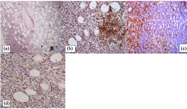
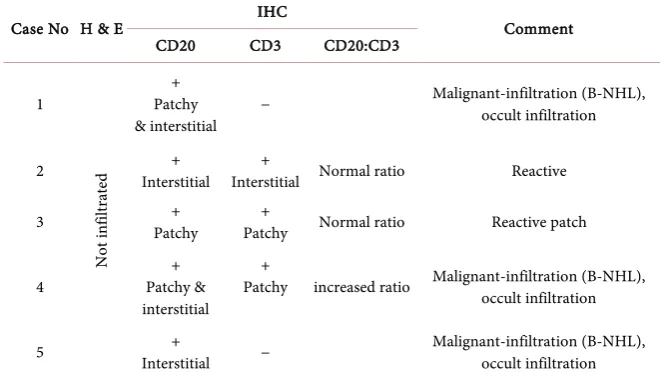
IHC results were available in 91 cases. There was an agreement between H & E and IHC (K = 0.85) concerning final interpretation as 40 (44%) out of 91 cases showed lymphoid infiltration by both H & E and IHC, while 44 (48.3%) out of 91 cases showed no lymphoid infiltration by both of them. IHC helped in iden-tifying 5 (5.5%) additional cases with lymphoid infiltration (2 reactive and 3 ma-lignant lymphomatous infiltrates) that were not seen by routine H & E (Figure 1), while 2 (2.2%) out of 91 cases showed lymphoid infiltration by H & E that were not seen on IHC sections (Table 1 & Table 2).Figure 1. A case of B-NHL showing no obvious lymphoid infiltration in H & E (×10). (a)
DOI: 10.4236/ojbd.2018.82004 31 Open Journal of Blood Diseases
Table 1. Agreement between IHC and H & E for all cases.
Variable
IHC
Agreement
K = p
Lymphomatous
infiltration No lymphomatous infiltration Bone marrow
trephine (H & E)
Not infiltrated 5 44
0.85 <0.001
Infiltrated 40 2
Table 2. Cases with subtle marrow involvement.
Comment IHC
H & E Case No CD20:CD3 CD3 CD20 Malignant-infiltration (B-NHL), occult infiltration − + Patchy & interstitial N ot in filt ra te d 1 Reactive Normal ratio + Interstitial + Interstitial 2 Reactive patch Normal ratio + Patchy + Patchy 3 Malignant-infiltration (B-NHL), occult infiltration increased ratio + Patchy + Patchy & interstitial 4 Malignant-infiltration (B-NHL), occult infiltration − + Interstitial 5
−: no lymphoid infiltration; +: presence of lymphoid infiltration.
3.2. Pattern of Infiltration by H & E versus IHC
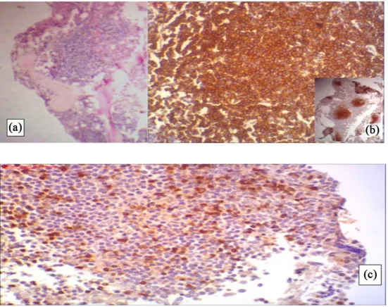
Comparing pattern of lymphoid infiltration between H & E and IHC (Table 3); 10 cases showed diffuse lymphoid infiltration by H & E, however by IHC; only 7/10 cases with the same diffuse pattern, 2/10 cases with multiple patchy pattern while the remaining case showed mixed patchy and interstitial pattern. Twenty cases showed patchy infiltration (Figure 2) by routine H & E while by IHC only 10/20 cases with the same pattern of infiltration, where 9/20 cases showed mixed patchy and interstitial infiltration (here IHC by CD20 detected extra-interstitial pattern), and one case showed interstitial pattern. Six cases showed combined patchy and interstitial infiltration by routine H & E which showed same pattern by IHC markers. Four cases showed interstitial infiltration by H & E, 3/4 cases showed same pattern by IHC (immunostaining by CD20 highlighted pa-ra-sinusoidal streaks of lymphomatous infiltration (Figure 3) in addition to in-terstitial pattern in one of those cases), and the remaining one showed patchy infiltration.
3.3. Significance of Routine Histology (H & E) and IHC Markers
(CD20 & CD3) in Differentiating Reactive and Malignant
Lymphomatous Infiltrates
[image:5.595.208.539.210.399.2]DOI: 10.4236/ojbd.2018.82004 32 Open Journal of Blood Diseases
Figure 2. A case of B-NHL showing patchy lymphoid infiltration in H & E (×10):
[image:6.595.233.514.345.446.2](a) Positive CD20 (×10) (×4); (b) Positive CD3 (×10) (increased CD20: CD3 ratio); (c) Malignant patchy lymphoid infiltration.
Figure 3. Para-sinusoidal streaks detected by CD20 (×10) (extra pattern by IHC).
Table 3. Discrepancy between patterns of infiltration in both H/E versus IHC.
Pattern Diffuse Patchy Combined patchy and interstitial Interstitial No infiltration
H & E 10 20 6 4 49
IHC 7 15 16 6 44
between reactive and malignant lymphomatous infiltrates. Concerning IHC markers used in this study, it was found that CD3 and the ratio between CD20 and CD3 significantly (p = 0.001) differentiate between the two types of lym-phomatous infiltration together with topographic and morphological appear-ance.
4. Discussion
[image:6.595.208.540.500.558.2]DOI: 10.4236/ojbd.2018.82004 33 Open Journal of Blood Diseases [13]. In our study,CD20 revealed additional interstitial pattern in 9/20 cases which showed patchy pattern of lymphomatous infiltration in H & E sections and also has highlighted a patchy pattern in one case with interstitial infiltration on H & E sections and in another case with interstitial infiltration by H & E, immunostaining by CD20 highlighted additionally parasinusoidal streaks of lymphomatous infiltration. This change in pattern of lymphoid infiltration be-tween H & E and IHC was related to the neoplastic cells that are dispersed throughout BM specimen, admixed with haemopoietic cells and may be over-looked by routine H & E and that foci of BM involvement in follicular lympho-ma lympho-may be subtle, hypocellular or fibrotic, delympho-manding careful scrutiny, prompt sectioning and immunohistochemical procedures [14]. In Thiele et al. [15] study, an ill-defined border of a nodular lymphoid aggregate was recognized with spillage of single lymphocytes between adipocytes or an Indian file like in-terstitial extension in NHL that can be shown by applying IHC methods. In this study, the use of IHC routinely on all cases detected 5 additional cases (5.5%) with BM lymphomatous involvement (3 malignant and 2 reactive infiltrations) that weren’t apparent on routine H & E sections. This was lower than the pre-vious study [3] that showed a more modest 11.1% rate of occult involvement by IHC and this was related to the use of extended panel of immunostaining using two B-cell markers (CD20 & CD79a), two T-cell marker (CD3 & CD45RO), and kappa/lambda light chains in addition to examination of additional sections in their immunostaining . In general there is a paucity of data regarding the role of IHC analysis in picking up histological non apparent infiltrate in BM. Fraga et
al.[16] reported that in a case series (n = 42) of anaplastic large cell lymphoma,
routine histology detected marrow involvement in 17% cases. Routine IHC analysis using CD30 detected occult disease in a further 23% cases, the inability of routine histology to detect low level disease was related to the scarcity of ma-lignant cells among normal hematopoietic cells and the difficulty in distinguish-ing them from immature hemopoietic cells .
DOI: 10.4236/ojbd.2018.82004 34 Open Journal of Blood Diseases infiltrations are composed mainly of CD3 positive T lymphocytes with small numbers of CD20 positive B cells as the normal ratio of T to B cells in BM being up to 6:1, these cells are scattered randomly or arranged as small non paratrabe-cular aggregates. In a study conducted by Lee et al.[17] found that considerable number of B lineage cases showed heterogeneous patterns with evidence of BM infiltration. Thus, heterogeneous immunohistochemical staining patterns (par-ticularly in B lineage lymphomas) may signify malignant lymphoma cell infiltra-tion into the BM.
On the other hand, a previous study [5] considered these cases with combined CD20 & CD3 must be interpreted cautiously because a wide variety of reaction patterns can occur with both benign lymphoid aggregates and lymphoma. B cell lymphomas may be accompanied by reactive T-lymphocyte. Therefore, a mix of B and T cells or a predominant of T cells doesn’t rule out a B cell lymphoma so, the use of a more extended panel of antibodies including anti-Ig κ and anti-Ig λ will undoubtedly increase the diagnostic potential but still problematical when using fixed and paraffin wax embedded tissue. Monoclonality of these infiltrates was not established in this study unequivocally by seeking light chain restriction. Monoclonality may present a major argument for NHL infiltrates versus benign lymphatic hyperplasia. This finding should be always interpreted in the context of the other features in making the diagnosis as number of infiltrate in section profile, histotopography and reticulin fibers. IHC uniform pattern with predo-minant of B or T lymphocytes is consistent with malignant lymphoma in con-trast with benign lymphoid aggregates that show polyclonal pattern with mix-ture of B & T lymphocytes [15].
5. Conclusion
In conclusion, our results have demonstrated that using IHC markers (CD20 and CD3), has highlighted extra-pattern and subtle lymphomatous infiltration, in addition to differentiate benign versus malignant infiltrates.
Conflict of Interest
The authors have no conflict of interest.
Acknowledgements
Both professor doctor Salwa Saad and Tahany El-Kerdany had supported the work and helped in results interpretation.
References
[1] Kremer, M., Quintanilla-Martinez, L., Nahrig, J., Von Schilling, C. and Fend, F. (2005) Immunohistochemistry in Bone Marrow Pathology: A Useful Adjunct for Morphologic Diagnosis. Virchows Archiv, 447, 920-937.
https://doi.org/10.1007/s00428-005-0070-8
DOI: 10.4236/ojbd.2018.82004 35 Open Journal of Blood Diseases https://doi.org/10.1016/S0740-2570(03)00026-1
[3] Riley, R.S., Williams, D., Ross, M. and Zhao, S. (2009) A Bone Marrow Aspirate and Biopsy: A Pathologist’s Perspective. Interpretation of the Bone Marrow Aspirate and Biopsies. Journal of Clinical Laboratory Analysis, 23, 259-307.
https://doi.org/10.1002/jcla.20305
[4] Dominis, M., Pešut, A., Boroveèki, A., Marušiæ-Vrsaloviæ, M. and Kušec, R. (2005) Bone Marrow Lymphoid Aggregates in Malignant Lymphomas. Croatian Medical Journal, 46, 410-416.
[5] Maes, B., Achten, R., Demunter, A., Peeters, B., Verhoef, G. and De Wolf-Peeters, C. (2000) Evaluation of B Cell Lymphoid Infiltrates in Bone Marrow Biopsies by Morphology, Immunohistochemistry, and Molecular Analysis. Journal of Clinical Pathology, 53, 835-840. https://doi.org/10.1136/jcp.53.11.835
[6] Palacio, C., Acebedo, G., Navarrete, M., Ruiz-Marcellan, C., Sanchez, C., Blanco, A.,
et al. (2001) Flow Cytometry in the Bone Marrow Evaluation of Follicular and Dif-fuse Large B-Lymphomas. Haematologica, 86, 934-940.
[7] Mazur, G., Halon, A., Wrobel, T., Jelen, M. and Kuliczkowski, K. (2004) Contribu-tion of Flow Cytometric Immunophenotyping and Bone Marrow Trephine Biopsy in the Detection of Lymphoid Bone Marrow Infiltration in Non-Hodgkin’s Lym-phomas. Neoplasma, 51, 159-163.
[8] Treetipsatit, J. (2013) Is Immunohistochemistry Mandatory in Staging Bone Mar-row Trephine Biopsy of Patients with Diffuse Large B-Cell Lymphoma (DLBCL)?
Asian Archives of Pathology, 9, 79-80.
[9] Chu, P.G., Loera, S., Huang, Q. and Weiss, L.M. (2006) Lineage Determination of CD20-B-Cell Neoplasms: An Immunohistochemical Study. American Journal of Clinical Pathology, 126, 534-544. https://doi.org/10.1309/3WG32YRAMQ7RB9D4
[10] West, R.B., Warnke, R.A. and Natkunam, Y. (2002) The Usefulness of Immunohis-tochemistry in the Diagnosis of Follicular Lymphoma in Bone Marrow Biopsy Spe-cimens. American Journal of Clinical Pathology, 117, 636-643.
https://doi.org/10.1309/W3QX-WJ1C-WG2K-225V
[11] Chetty, R., Echezarreta, G., Comley, M. and Gatter, K. (1995) Immunohistochemi-stry in Apparently Normal Bone Marrow Trephine Specimens from Patients with Nodal Follicular Lymphoma. Journal of Clinical Pathology, 48, 1035-1038.
https://doi.org/10.1136/jcp.48.11.1035
[12] Jalal, S.M., Law, M.E., Stamberg, J., Fonseca, R., Seely, J.R., Myers, W.H., et al.
(2001) Detection of Diagnostically Critical, Often Hidden, Anomalies in Complex Karyotypes of Haematological Disorders Using Multicolour Fluorescence in Situ
Hybridization. British Journal of Haematology, 112, 975-980.
https://doi.org/10.1046/j.1365-2141.2001.02630.x
[13] Talaulikar, D., Shadbolt, B., Dahlstrom, J.E. and McDonald, A. (2009) Routine Use of Ancillary Investigations in Staging Diffuse Large B-Cell Lymphoma Improves the International Prognostic Index (IPI). Journal of Hematology & Oncology, 2, 49-58.
https://doi.org/10.1186/1756-8722-2-49
[14] Anagnostou, D. (2005) Pitfalls in the Pattern of Bone Marrow Infiltration in Lym-phoproliferative Disorders. Current Diagnostic Pathology, 11, 170-179.
https://doi.org/10.1016/j.cdip.2005.01.005
DOI: 10.4236/ojbd.2018.82004 36 Open Journal of Blood Diseases
[16] Fraga, M., Brousset, P., Schlaifer, D., Payen, C., Robert, A., Rubie, H., et al. (1995) Bone Marrow Involvement in Anaplastic Large Cell Lymphoma. Immunohisto-chemical Detection of Minimal Disease and Its Prognostic Significance. American Journal of Clinical Pathology, 103, 82-89. https://doi.org/10.1093/ajcp/103.1.82