A Review of Image Registration Methods in Medical
Im-aging
Leeba John
Dept of ICTVeer Narmad South Gujarat University, Surat, Gujarat, India
Lissa John
Dept of ICTVeer Narmad South Gujarat University, Surat, Gujarat, India
ABSTRACT
Image registration is the series of methods which superimpos-es or aligns two or more imagsuperimpos-es of the same picture taken at different instance of time or moment, from different perspec-tive or angle, and/or by different sensors or any external de-vice. Registration makes the pixels in two images precisely coincide to the same points in the scene. After registration of the images, it can be combined or fused in a way that im-proves information extraction. Image registration combines two images, i.e., reference image and sensed image, geometri-cally. There are different approaches of image registration and these approaches are categorized according to their nature that is area based and feature based. Essential steps in Image Reg-istration are feature detection, feature matching, transform model estimation, and image resampling and transformation. This paper focuses on an analysis on Image Registration methods. A literature survey of different research of Medical Image Registration is also presented here. Medical Imaging plays a significant role in medical diagnosis and treatment. It provides a clear view for medical experts in taking the correct decisions on patient’s condition. By combining more than one image obtained from different medical imaging modalities, experts can achieve better image visualization for different human anatomy. The aim of this paper is to provide a source for the researchers involved in Image Registration as well as Medical Image Fusion used in diverse applications.
Keywords
Image Registration; Feature Detection; Feature Matching; Transform Model Estimation; Resampling; Transformation; Medical Imaging; Medical Image Modalities.
1.
INTRODUCTION
Image Registration is an image processing technique in which images can be combined or fused in a way that improves in-formation extraction. It makes the pixels in two images specif-ically relate to the same points in the scene. Therefore it also helps to align multiple scenes into a single integrated homo-geneous image. It is also thought of as a process of superim-posing or aligning of more than two images of the same pic-ture taken at different instance of time or moment, from dif-ferent perspective or angle, and/or by difdif-ferent sensors or any external device. It overcomes issues such as image rotation, scaling, and skewing which are common when superimposing images. The goal of image registration is to align images spa-tially to minimize a desired error with respect to each other. The input for this process is two images: the original image is known as the reference image while the image that will be aligned with the reference image is known as the sensed im-age. It produces more useful and better vivid images based on the input ones as shown in Fig 1. Essential steps in Image Registration are feature detection, feature matching, transform model estimation, and image resampling and transformation.
(a) Image 1 (b) Image 2 (c) Fused Image (Focus on left) (Focus on right) (All focus)
Fig 1: Fusion result for multi-focus images
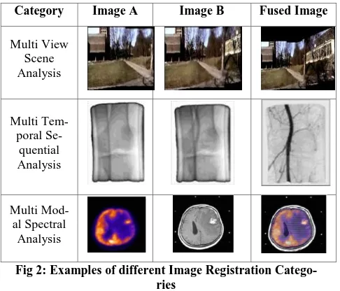
The Application of Image Registration can be arranged into the following categories as discussed below:
i.Various Perspective/Angle (Multi-view Scene Analysis): From multiple perspectives, the images
cap-tured of the same object or views are taken into con-sideration for better representation of the scanned object.
Examples: Image Mosaicing (Panorama view), gesture analysis, face detection etc.
ii. Various Time/Moment (Multi-temporal Sequen-tial
Analysis): At various instance of time / period, images taken of the same object or scene under different circumstances to monitor changes are taken into consideration.
Examples: climate change, biodiversity, agriculture, for-estry, monitoring of tumor evolution.
iii. Various Sensor/Spectral (Multi-modal Spectral Analysis): Acquiring the images captured from various
sensors of the same object or scene is considered to obtain the minutiae of the scanned object.
Category Image A Image B Fused Image
Multi View Scene Analysis
Multi Tem-poral
Se-quential Analysis
[image:2.595.48.291.70.278.2]Multi Mod-al SpectrMod-al Analysis
Fig 2: Examples of different Image Registration Catego-ries
2.
CLASSIFICATION OF IMAGE
REG-ISTRATION METHODOLOGY
Image Registration is a highly vibrant area due to its broad range applicability in remote sensing for weather forecasting, fusion of medical images which are taken from different med-ical imaging modalities like CT-MRI, PET-MRI etc. Image Registration Methodology and steps are discussed here. Ac-cording to Barbara Zitova and Jan Flusser [7] the field of Image Registration can be categorized into two groups: area based methods and feature based methods. Area based meth-ods are used for comparing a pixel in the sensed image to that of the referenced image. It basically uses gray value of the pixels to describe matching entities. On the other side Feature based methods does not uses gray value to describe matching entities, instead it uses image features derived by feature ex-traction algorithm to find the corresponding pairs. Features such as edges, surfaces, corners, point of intersection, con-tours etc, which carries relevant information about images used for matching. Majority of the registration methods con-sist of following four basic steps [8]:
i. Feature Detection: In this step, the significant and appropriate features like Regions, Edges, Corners, etc are identified in both reference image and sensed image automatically or manually by various domain professionals. The above detected features are also known as Control Points Set.
ii. Feature Matching: The mapping of features in the reference and sensed image are considered. Match-ing method is selected on the basis of image content or the Control Points Set.
iii. Transform Model Estimation: It is one of the cru-cial steps where once the selected features are ex-tracted with reference to the position of the sensed image and reference image, the parameters and the type of the transformation is estimated. Some of the transformation functions which can be applied are linear, affine, projective etc.
iv. Image Resampling and Transformation: Once the transformation is estimated, the sensed image is transformed and then is resampled using interpola-tion techniques.
Fig 3: Steps in Image Registration Process
3.
VARIOUS IMAGE REGISTRATION
METHODS
3.1.Extrinsic Methods
In this method external objects or markers are used in the patient body [9-12] to be registered. The goal of this method is to obtain more effectiveness, correctness, consistency, high computational speed by evaluating their matching features as it does not need complex algorithm for implementation. It is used in brain image registration skin markers or stereo-tactic frames are used.
3.2.Moments and Principle Axes Methods
Principal Axes Registration (PAR) is used for global matching of binary volumes from CT, MR or PET images. The inertia is diagonal when computed with respect to principal axes. The centroid and principal axes can describe the orientation of a volume. The principal axes registration can resolve six de-grees of freedom of an object (three rotations and three trans-lations). It can compare the orientations of two binary vol-umes through rotation, translation and scaling [13]. In mo-ment based methods pre – segmo-mentation is done to obtain satisfactory results.
3.3.Mutual Information Based Methods
Mutual Information is applied to measure the statistic depend-ence between the image intensities of corresponding voxels in both images, which is assumed to be maximal if the images are geometrically aligned [14]. It is a measure of how much information one random variable tells about another. For two images, the mutual information is computed from the joint probability distribution of images intensity and gray value. In medical image processing, an application where the alignment is based on images of the same individual is known as intrapatient registration. Matching datasets of different indi-viduals is known as interpatient registration. One of the main advantages of using mutual information is than it can be used to align images of different modalities (e.g. CT to MR-T1, MR-T1 to PET etc).
3.4.Surface Methods
[image:2.595.321.532.71.251.2]3.5.Wavelet Based Methods
The wavelet transform (WT) has gained widespread ac-ceptance in the field of signal processing. The wavelet trans-form is calculated for each segment according to time at dif-ferent frequencies. They are particularly suitable for applica-tions where scalability and tolerable degradation are relevant as it is multi-resolution in nature. In Discrete wavelet trans-form, wavelets are discretely sampled. At each level of de-composition, the signal is split into high frequency and low frequency components. The low frequency can further be decomposed until the desired resolution is achieved. Different types of wavelets like the Haar, Symlet, Daubechies [17] and Coiflets are applied for finding the correspondence with dif-ferent sets of wavelet coefficients.
3.6.Correlation Based Methods
The maximum is searched from the matching or correspond-ing points obtained from window pairs of sensed and refer-ence images. This is eventually used in multi-modal images and for comparison of various images of similar object or scene [18]. There are two techniques namely Cross – correla-tion and Phase – correlacorrela-tion based on Fourier transformacorrela-tions are used for image registration. Fourier-based techniques along with search algorithms have been used to estimate the transformation among the two input images [19].
3.7.Soft Computing Based Methods
It includes Artificial Neural Networks, Fuzzy Sets and several Optimization Heuristics.
3.7.1.
Artificial Neural Networks
Artificial Neural Networks are used to process the information the way biological systems process analog signals like image and sound. There are three layers namely Input Layer, Hidden Layer, Output Layer. In Back propagation algorithm, infor-mation about errors is filtered back through the system and is used to adjust the connections between the layers, thus im-proving performance. The algorithm is used to update weights and bias of the neural networks. Weight and bias decides the functionality of the network. Value of the weight and bias elements are calculated during training phase. Neural Net-works are used in the medical field for solving mono-modal and multi-modal image registration problems [20].
3.7.2.
Optimization Heuristics
Optimization Problems may be unconstrained or constrained having both continuous as well as discrete variables. With many limitations being present at the points of global optima the task of finding the optimal solutions is hard. Meta-heuristics include Genetic Algorithm (GA) [21], Gravitational Search Algorithm (GSA), Ant Colony Optimization (ACO), Particle Swarm Optimization (PSO) [22], Stimulated Anneal-ing (SA), and Plant Propagation Algorithm (PPA) and so on. These heuristic methods are being used by image registration problems for finding out the best possible parameters neces-sary for designing a transformation problem [23].
3.7.3.
Fuzzy Sets
Fuzzy Sets are the extension of conventional (crisp) set theo-ry. It handles the concept of partial truth using a membership function or degrees. It extends conventional Boolean logic to find out partial truths and uncertainties. It is a dominant means to represent and process human understanding in form of fuzzy if-then rules. It is a group of all approaches that un-derstand and process the images, their sections and features are fuzzy sets. This permits Fuzzy sets to deal with vagueness and incorrectness. Fuzzy Sets have been applied in the field of
Medical Image registration also [24].
[image:3.595.308.547.125.767.2]Summary of Various Image Registration Methods are listed below in Table 1.
Table 1: Presents a summarized view of various Image Registration Methods.
Methods Key Points Application
Extrinsic Methods
External objects or markers are used in the patient body to be
registered.
Brain image regis-tration skin markers
or stereo-tactic frames. Moments and Princi-ple Axes Methods
It requires the compu-tation of the centroid and the three principal
axes for each of the two scans.
This method is ap-plied in radiation treatment planning based on combined CT/MR images, in study of orientation of the scaphoid bone
in the wrist and in analyzing PET im-ages of
schizophren-ics.
Mutual In-formation Based Methods
Measures the statistic dependence between the image intensities of corresponding voxels in both imag-es, which is assumed
to be maximal if the images are
geometri-cally aligned.
It is highly reliable and effective meth-od of the multimmeth-odal
images registration. Surface Methods Surface-based image registration methods involve determining corresponding surfac-es in various imagsurfac-es (and/or physical space) and computing
the Transformation that best aligns these
sur-faces.
Used in Multimodal-ity Brain image.
Wavelet Based Methods
It usually extracts a large number of con-trol points. The can-didate control points
are extracted using the local maxima of
the wavelet coeffi-cients.
It has been used for Electrocardiography, functional neuro-imaging including positron emission tomography and functional MRI. Correlation Based Methods
Eventually used in multi-modal images and for comparison of
various images of similar object or sce-ne. Extracted features
from the images are used to obtain the cross – correlation coefficients for image
registration.
It has been used to register tomographic brain and abdominal
images.
Artificial Neural
Net-It is formulated based on biological neural
works networks. and multi-modal medical image
regis-tration problems.
Optimization Heuristics
Genetic Algorithm (GA) is used to de-termine which such features are the most
predictive.
It showed good re-sults in diagnosing patient of liver, thy-roid disorder, and
cancer.
Fuzzy Sets
It is a dominant means to represent and process human understanding in form of fuzzy if-then rules. This permits Fuzzy
sets to deal with vagueness and
incor-rectness.
It showed good re-sults on retinal im-ages. Bio-medical images made use of
Neuro fuzzy ap-proach.
4.
MEDICAL IMAGING
APPLICA-TIONS
[image:4.595.48.289.71.267.2]For acquiring high resolution and more edifying analysis of human anatomies and its functions becomes possible due to the rapid advances in medical imaging technology. Such de-velopment motivates the researchers in the medical image analysis field. Medical Image registration methods can be grouped based on some criterion [25] listed below in Table 2.
Table 2: Classification of Medical Registration Methods
Criteria
Method-ologies Examples
Dimension-ality
2D - 2D, 2D - 3D, 3D - 3D
2D – 2D: registration of temporal mammograms. 3D – 2D: 3D computed
to-mography images and 2D portal images. 3D – 3D: registration of two tomographic datasets,
moni-toring of tumor growth.
Domain of
Transfor-mation
Global
Image of the entire head based on computation done in the area of facial surface
only.
Local Individual vertebrae in an image of a spinal column.
Type of Transfor-mation
Rigid
Head of the same patient, the skull, falx, tentorium, cardiac images and postoperative
images etc. Affine
Projective Non Linear
Parameters of
Registra-tion
Parameters computed
directly
- Parameters
searched for
Subject of Registration
Intra
sub-ject 3D / 3D MR or CT brain image application. Inter
sub-ject Atlas
Object of Registration
Head Brain or Skull, Eye, Dental. Abdomen General, Kidney, Liver.
Thorax Entire, Cardiac, Breast. Limbs General, Femur, Hemurus,
Hand.
Nature of Registration
Extrinsic
Stereotactic frame, Fiducials (screw markers), Dental
adapter etc. Intrinsic Image Formation Non Image
Based
Position of surgical tools mounted on robot arm to
images.
Interaction
Interactive
- Semi
au-tomatic Automatic
Modalities involved
Mono Modal
Two myocardial SPECT images are acquired of the patient under rest and stress
conditions. Multi
Modal
PET images are registered to an MR image.
Patient to Modality
In radiotherapy treatment, patient can be positioned with the aid of registration of in – position X-ray simulator images to a pre-treatment
anatomical image.
Image Registration Techniques are discussed in the fields of Diabetic Retinopathy and Lesion Detection in Cervix. Other diseases along with its diagnostic modalities used and regis-tration / fusion classes are listed out in Table 3.
5.
DIABETIC RETINOPATHY
[image:4.595.318.543.654.741.2]Diabetic Retinopathy is a microvasculature complication of diabetes in retina, causing damage to the blood vessels of the light sensitive tissue at retina, which may also lead to blind-ness [26]. Eventually due to the increased permeability of the capillary walls, mircroaneurysms along with haemorrhages are formed in retina [27]. As there is an increase of sugar in the blood, it can lead to blockage of the blood vessels which causes microinfarcts called soft exudates in the retina. Clinical signs observed by color fundus photographs include mircroaneurysms, haemorrhages, exudates and intra-retinal micro-vascular abnormalities [26].
Longitudinal Registration method is used to detect and moni-tor retinal change in Diabetic Retinopathy development and progression. It makes use of Feature based registration meth-ods to extract a set of feature points from each of the reference and floating images, which are then matched to find the trans-formation. The features are normally vessel structures, branching, bifurcation points, cross-over points etc. which are extracted manually or automatically.
Other Retinal Image Registration methods that are considered are cross-modality registration, spatial registration and tem-poral registration. When the ophthalmologists wants to use two various imaging modalities to get complementary infor-mation about retinal vessel disorder cross-modality registra-tion is considered and when the ophthalmologist’s wishes to merge narrow-field images together to produce a wider field view of the retina, spatial registration is considered. The need
for temporal registration occurs when it is required to track the changes of retina eventually.
6.
LESIONS DETECTION IN CERVIX
[image:5.595.49.545.257.769.2]The abnormality found in cervical cells may develop into Cervical Cancer if left untreated. Temporal image registration method is used to detect precancerous lesions of the cervix. The images are obtained from colposcopy examinations. The proposed approach yielded the time series for all pixels in the initial image of the sequence because such values form the basis to establish the similarity. The Temporal method pro-vided the best tradeoff between low error values and time required to carry out the registration process. In addition to it, the temporal method is the most robust among other methods.
Table 3: The current diseases based fusion works
Disease / Disorder/ Surgery Fusion Classes Diagnostic Modality
Head Arterial steno-occlusive disease of the head to
study head motion scanning errors [28]
Rigid
PET Monomodal
Pituitary adenoma [29] Rigid MR
Monomodal Brain
Huntington’s disease [30] Nonrigid MR
Monomodal
Parkinson’s Disease [31] Nonrigid + Rigid MR
Multimodal SPECT
Eye
Eye fundus (ocular pathologic conditions) [32] Rigid Ophthalmological images Multimodal
Retinal diseases [33] Rigid Ophthalmological images
Multimodal Oral
Tongue disorders [34] Nonrigid hMR
Multimodal Cine MR
Lung Assist in detecting and diagnosing lung cancer
[35]
Nonrigid
Chest Radiographs Monomodal
Cardiac
Coronary artery diseases [36]
Rigid X-ray
Multimodal CT
Nonrigid
MR Monomodal
Ischemic heart disease [37] Rigid + Nonrigid CT
Monomodal Liver
Hepatocellular carcinoma [38] Nonrigid CT
Some of the contributions done in this field along with its best registration / fusion technique are listed out in Table 4. Table 4: Presents some recent work in medical image fusion for diverse applications
Work Fusion
Tech-nique
Level of
Fusion Modality Organ Contribution
“Extraction of brain regions affected by Alzheimer disease via fusion of brain
multispec-tral MR images” [39].
Dual tree wave-let transform
(DTWT)
Pixel
based MRI Brain
Proposed a new technique for extrac-tion of affected regions by Alzheimer
disease from multispectral medical images by means of fusion and
seg-mentation methods. “Multimodal medical image
fusion using modified fusion rules and guided filter” [40].
Gaussian de-composition, guided filters, modified
salien-cy and weight maps
Window
based MRI Brain
Proposed a modified fusion algorithm to reduce the contrast reduction and
halo artifacts.
“Multimodal medical image sensor fusion framework using
cascade of wavelet and contourlet transform domains”
[41]
PCA Pixel
based MRI, CT Brain
Proposed a multimodal fusion algo-rithm that consists of two stages: stationary wavelet transform (SWT)
and non sub-sampled contourlet transform (NCST) to enhance the
shift variance, directionality and phase information. “Medical image fusion by
combining SVD and shearlet
transform” [42]. Maximum fu-sion rule
Pixel
based MRI, PET Brain
Proposed a two stage fusion tech-nique using shearlet transform and then applying SVD on low pass sub-bands before fusion to provide higher
image quality. “Free-breathing diffusion
ten-sor imaging and tractography of the human heart in healthy volunteers using wavelet based
image fusion” [43].
Wavelet based image fusion
algorithm
Window based
Diffusion Tensor
Imag-ing(DTI)
Heart
Proposed a technique to provide 3D fiber architecture properties of the
human heart.
“Spine medical image fusion using wiener filters in shearlet
domain” [44]. Maximum fu-sion rule
Pixel
based MRI, CT Spine
Proposed an algorithm that provides both functional and anatomical struc-ture for spine by applying three steps:
Shearlet transform, Wiener filter, Fusion of low and high pass sub
bands. “Multi focus and multi modal
image fusion using wavelet transform” [45].
Dual tree dis-crete wavelet
transform(DT-DWT)
Pixel
based MRI, CT Brain
Proposed a multifocus and multimod-al image fusion techniques using DT-DWT then applying fuzzy logic clus-tering for segmentation that helps in
tumor identification.
7.
CONCLUSION
To extract information from different images which comes from different sources, Image Registration plays a crucial role. This paper presented a survey on various Image Regis-tration methods as well as Medical Image RegisRegis-tration and/or fusion. Fundamentals of Image Registration methods and techniques were discussed followed by the application areas where it is used. Objective of Image Registration is to com-bine two or more images and extract information from them. Then a discussion for Medical Imaging Modalities and the Medical Image Registration was been done. In today’s con-text, Medical Image Registration is considered relevant for the medical experts to make decisions regarding the patient’s condition. This paper also covers some recent contribution of Medical Image fusion for diverse applications.
8.
REFERENCES
[1] Manjusha Deshmukh, Udhav Bhosle "A survey of image registration", "International Journal of Image Processing (IJIP), Volume (5): Issue (3), 2011".
[2] D.L.G. Hill, P.G. Batchelor, M. Holden, D.J. Hawkes, Medical image registration, Physics in Medicine and Bi-ology 46 (2001) R1–R45.
[3] H. Lester, S.R. Arridge, A survey of hierarchical non-linear medical image registration, Pattern Recognition 32 (1999) 129–149.
image registration, Medical Image Analysis 2 (1998) 1– 36.
[6] Damas, Sergio, Oscar Cordón, and Jose Santamaría. "Medical image registration using evolutionary computa-tion: An experimental survey." IEEE Computational In-telligence Magazine 6.4 (2011): 26-42.
[7] Barbara Zitova, Jan Flusser "A survey on image registra-tion methods", "Image and vision computing 21(2003) 977-1000".
[8] Alexander Wong, David A. Clausi "ARRSI: Automatic Registration of Remote-Sensing Images", " IEEE Trans-actions on Geoscience and Remote Sensing, volume(45), No. 5, May 2007".
[9] K. P. Gall and L. J. Verhey, “Computer-assisted posi-tioning of radiotherapy patients using implanted radioopaque fiducials”, Medical physics, 1993, 1152– 1159.
[10]C. R. Maurer, G. B. Aboutanos, B. M. Dawant, R. A. Margolin, R. J. Maciunas and J. M. Fitzpatrick., “Regis-tration of CT and MR brain images using a combination of points and surfaces”, Medical imaging: image pro-cessing, volume 2434, Bellingham, WA, 1995. SPIE Press, 109-123.
[11]A. C. Evans, S. Marrett, J. Torrescorzo, S. Ku, and L.Collins, “MRI-PET correlation in three dimensions us-ing a volume of interest (VOI) atlas”, Journal of cerebral blood flow and metabolism , 11, A69–A78, 1991. [12]W. D. Leslie, A. Borys, D. McDonald, J. O. Dupont and
A. E. Peterdy, “External reference markers for the cor-rection of head rotation in brain single-photon emission tomography”, European journal of nuclear medicine, 22(4):351–355, 1995.
[13]J. Flusser, T. Suk, A moment-based approach to registra-tion of images with affine geometric distorregistra-tion, IEEE Transactions on Geoscience and Remote Sensing 32 (1994) 382–387.
[14]P.Viola, W.M. Wells, “Alignment by maximization of mutual information”, International Journal of Computer Vision 24, (1997), 137–154.
[15]S.M. Yamany, A.A. Farag, “Free-form surface registra-tion using surface signatures” Proceedings of the Seventh IEEE International Conference on Computer Vision, Vol. 2, 1999, 1098–1104.
[16]Chi Kin Chow, Hung Tat Tsui, Tong Lee, “Surface regis-tration using a dynamic genetic algorithm”, Pattern Recognition 37, (2004), 105-117.
[17]J. le Moigne, “Parallel registration of multi-sensor re-motely sensed imagery using wavelet coefficients”, Pro-ceedings of the SPIE: Wavelet Applications, Orlando, Florida, 2242, (1994), 432–443.
[18]B.K. Ghaffary, A.A. Sawchuk, “A survey of new tech-niques for image registration and mapping”, Proceedings of the SPIE: Applications of Digital Image Processing 432 (1983) 222–239.
[19]Samritjiarapon O. Chitsobhuk O. , “An FFT-Based Technique and Best-first Search for Image Registration”, International Symposium on Communications and In-formation Technologies, ISCIT 2008.
[20] Lifeng Shang, Jian Cheng Lv, Zhang Yi, “Rigid medical image registration using PCA neural network”, Neurocomputing 69 (2006), 1717–1722.
[21] S.-J. Wu and P.-T. Chow, Genetic algorithms for nonlin-ear mixed discrete-integer optimization problems via me-ta-genetic parameter optimization, Engineering Optimi-zation, vol. 24, no. 2, pp. 137–159, 1995.
[22] R. Eberhart and J. Kennedy, A new optimizer using par-ticle swarm theory, in Proceedings of the 6th Internation-al Symposium on Micro Machine and Human Science (MHS ’95), pp. 39– 43, IEEE, Nagoya, Japan, October 1995.
[23] J.M. Rouet, J.J. Jacq, and C. Roux, "Genetic algorithms for a robust 3-D MR-CT registration," IEEE Trans. In-form. Technol. Biomed., vol. 4, (Jun. 2000), 126-136. [24] Ramirez L. Durdle N.G. Raso V.J. “A Parameters
Selec-tion Scheme for Medical Image RegistraSelec-tion”, Fuzzy In-formation Processing Society, 2006. NAFIPS 2006. An-nual meeting of the North American (June2006), 505-510.
[25] J. B. Antoine Maintz and Max A. Viergever "An over-view of medical image registration methods", "August 1998 UUCS-1998-22 ISSN: 0924-3275".
[26] H. Narasimha-Iyer, C. Ali, R. Badrinath, V.S. Charles, et al.Robust detection and classification of longitudinal changes in color retinal fundus images for monitoring di-abetic retinopathy IEEE Trans. Biomed. Eng., 53 (6) (2006), pp. 1084-1098.
[27] T. Kauppi, V. Kalesnykiene, J.K. Kamarainen, et al.Diaretdb1 diabetic retinopathy database and evaluation protocol Medical Image Understanding and Analysis (MIUA’ 07)(2007), pp. 61-65.
[28] Matsubara K, Ibaraki M, Nakamura K, Yamaguchi H, Umetsu A, Kinoshita F, et al. Impact of subject head mo-tion on quantitative brain 15O PET and its correcmo-tion by image-based registration algorithm. Ann Nucl Med 2013; 27(4):335–45.
[29] Ringstad G, Emblem K, Holland D, Dale A, Bjornerud A, Hald J. Assessment of pituitary adenoma volumetric change using longitudinal MR image registration. Neuro-radiology 2011; 54():435–43.
[30] Modat M, Taylor Z, Ridgway G, Barnes J, Wild E, Hawkes D, et al. Nonlinear elastic spline registration: evaluation with longitudinal Huntington’s disease data. In: Biomedical image registration; 2010. p. 128–39. [31] Lee J, Huang C, Chen C, Weng Y, Lin K, Chen C. A
brain MRI/SPECT registration system using an adaptive similarity metric: application on the evaluation of Par-kinson’s disease. In: Computer vision/computer graphics collaboration techniques; 2007. p. 235–46.
[32] Bernardes R, Guimaraes P, Rodrigues P, Serranho P. Fully-automatic multimodal co-registration of retinal fundus images.In: IFMBE proceedings; 2014. p. 248–51. [33] Ghassabi Z, Shanbehzadeh J, Sedaghat A, Fatemizadeh
E. An efficient approach for robust multimodal retinal image registration based on UR-SIFT features and PIIFD descriptors.EURASIP J Image Video Process 2013; 2013(1):25.
high-resolution and Cine MR tongue images. Lect Notes Comput Sci 2011:556–63.
[35]Li M, Castillo E, Luo H, Zheng X, Castillo R, Meshkov D, et al.Deformable image registration for temporal sub-traction of chest radiographs. Int J CARS 2013; 9(4):513–22.
[36]Moosavi Tayebi R, Wirza R, Sulaiman P, Dimon M, Khalid F,Al-Surmi A, et al. 3D multimodal cardiac data reconstruction using angiography and computerized tomographic angiography registration. J Cardiothorac Surg 2015; 10(1):1–25.
[37]Zuluaga M, Hernandez Hoyos M, Davila J, Uriza L, Orkisz M. A fast lesion registration to assist coronary heart disease diagnosis in CTA images. In: Computer vi-sion and graphics; 2012. p. 710–7.
[38]Xu H, Gong G, Wei H, Chen L, Chen J, Lu J, et al. Fea-sibility and potential benefits of defining the internal gross tumor volume of hepatocellular carcinoma using contrast-enhanced 4D CT images obtained by deforma-ble registration. Radiat Oncol2014; 9(1):221.
[39]Tannaz Akbarpour, Mousa Shamsi, “Extraction of Brain Regions Affected by Alzheimer Disease Via Fusion of Brain Multispectral MR Images”, International Confer-ence on Information and Knowledge Technology, pp. 1-6,2015.
[40] Pritika, Sumit Budhiraja, “Multimodal Medical Image Fusion Using Modified Fusion Rules and Guided Filter”, International Conference on Computing, Communication and Automation (ICCCA), pp. 1067–1072, ISBN 978-1-4799-8889-1, 2015.
[41] Vikrant Bhateja, Aimé Lay-Ekuakille, “Multimodal Medical Image Sensor Fusion Framework Using Cascade of Wavelet and Contourlet Transform Domains”, IEEE Sensors Journal, vol. 15, no. 12, December 2015. [42] Biswajit Biswas, Somoballi Ghoshal, “Medical Image
Fusion by Combining SVD and Shearlet Transform”, 2nd International Conference on Signal Processing and Integrated Networks (SPIN), 2015.
[43] Hongjiang Wei, Magalie Viallon, “Free-Breathing Diffu-sion Tensor Imaging and Tractography of the Human Heart in Healthy Volunteers Using Wavelet-Based Image Fusion”, IEEE Transactions on Medical Imaging, Vol. 34, No. 1, January 2015.
[44] Biswajit Biswas, Amlan Chakrabarti, “Spine Medical Image Fusion Using Wiener Filter in Shearlet Domain”, IEEE 2nd International Conference on Recent Trends in Information Systems, 2015.