Characterisation of circulating tumour cells in
men with advanced prostate cancer and
correlation with numbers of circulating and
tissue-based inflammatory cells
A thesis submitted to the University of Dublin
for the degree of Doctor in Medicine (M.D.)
2019
D
R
B
RIAN
H
AYES
BM
ED
S
C
MB
BC
H
BAO
FRCP
ATH
FFP
ATH
Page | i
DECLARATION
I declare that this thesis has not been submitted as an exercise for a degree at this or any other university and it is entirely my own work.
I agree to deposit this thesis in the University’s open access institutional repository or allow the Library to do so on my behalf, subject to Irish Copyright Legislation and Trinity College Library conditions of use and acknowledgement.
Page | ii
SUMMARY
Despite the widespread availability of PSA screening for prostate cancer many men present with advanced stage disease, and in these men treatment is directed towards improved survival and quality of life rather than cure. Exercise therapies are recognised to have benefits in quality-of-life, all-cause and cancer-specific mortality in cancer patients, and prostate cancer is no exception. Obesity and metabolic syndrome are encountered with increasing frequency in prostate cancer patients, either as pre-existing comorbidities or as a side-effect of androgen deprivation therapy. The systemic inflammatory milieu which underpins the obese state has profoundly negative effects on cancer survivorship, but data show that control of obesity, including through managed exercise interventions, can improve outcomes.
Blood-borne circulating tumour cells are considered an intermediate step in the metastatic cascade and therefore a potentially useful target for therapy. Interventions (including exercise) targeted at improving the deleterious pro-oncogenic systemic consequences of obesity may in part exert their effects by interfering with interactions between platelets, circulating tumour cells and cells of the immune system. Adhesion of platelets to circulating tumour cells may impair the ability of NK-cells to destroy them, and it has been hypothesised that enhanced platelet cloaking of circulating tumour cells in obese men with prostate cancer, due to increased systemic inflammation, is a mechanism underlying the worse prognosis of cancer in these patients.
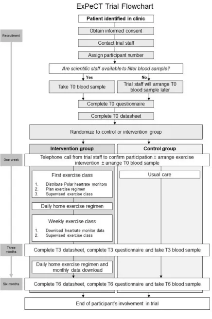
In order to test this hypothesis the interational multidisciplinary ExPeCT clinical trial (“Examining Exercise, Prostate Cancer and Circulating Tumour Cells”) was established. This recruited men with advanced prostate cancer from centres in Dublin and London and randomised them either to participation in a formal six-month supervised walking intervention (exercise group), or to usual care (control group). Blood samples were taken, quality-of-life questionnaires were completed and clinical data (including blood pressure, body mass index, waist circumference etc) were gathered at the time of recruitment (T0) and after three (T3) and six (T6) months. Participants were divided into “exposed” and “non-exposed” groups based on baseline body mass index ≥ 25 kg/m2 or < 25kg/m2 respectively. Blood samples were passed through microporous ScreenCell®
Page | iii cytometry for numbers and subsets of circulating lymphocytes was undertaken in a subset of Dublin-based participants.
Participant accrual to the ExPeCT trial was slower than expected, with 67 men recruited over the course of the trial, and 61 having at least a T0 blood sample drawn. 30 participants were randomized to the exercise group, 31 to the control group. There were 11 participants in the non-exposed group and 50 in the non-exposed group. Circulating tumour cells were identified on the vast majority of ScreenCell® filters, and there was a significant reduction in their numbers between T0 and T3, with the change driven by differences in the control group and the exposed group. Platelet cloaking was significantly more frequently seen in blood draws from the control group than the exercise group participants, which might suggest that the exercise intervention played a role in altering platelet adhesion to those men who participated in the exercise programme. However morphological assessment of platelet cloaking was found to be difficult, as the majority of circulating tumour cells present on the filters lacked cytoplasm, perhaps consequent upon the shear forces exerted on the cells during the filtration process.
A higher circulating fraction of CD3-positive T-lymphocytes and a lower circulating fraction of B-lymphocytes and NK-cells were found in the exercise group when compared with the control group. Linear correlations were identified between circulating tumour cell numbers on the one hand and platelet count, total lymphocyte count, CD4-positive T-lymphocyte count and NK-cell count on the other hand, relationships which were found to be independent of one another by multiple regression analysis. The demonstration of a relationship with platelet count is the first such report in men with prostate cancer, providing clinical evidence to support the abundant published scientific data that circulating tumour cells interact with platelets in various ways to enhance their metastatic potential.
Atrophic prostatic lesions were identified in 43% of biopsy specimens, and 65% had an infiltrate of chronic inflammatory cells in the benign background prostatic tissue. 91% of biopsies had at least mild chronic inflammation within the tumour, with T-lymphocytes being the predominant cell type. No correlation was seen between circulating tumour cell numbers and the density of inflammatory cell subsets in core biopsy tissue.
Page | iv
CONTENTS
DECLARATION ... (i)
SUMMARY ... (ii)
DEDICATION ... (ix)
FIGURES ... (x)
TABLES ... (xii)
ABBREVIATIONS ... (xiii)
PUBLICATIONS ... (xv)
ACKNOWLEDGEMENTS ... (xvi)
CHAPTER 1 – INTRODUCTION... 1
1.1 GENERAL INTRODUCTION ... 1
1.2 THE PROSTATE GLAND... 3
1.2.1 ANATOMY AND HISTOLOGY ... 3
1.2.2 PROSTATIC HYPERPLASIA ... 4
1.2.3 PROSTATITIS AND PROSTATIC ATROPHY ... 4
1.2.4 PROSTATE CARCINOMA ... 6
1.2.4.1 Epidemiology ... 6
1.2.4.2 PSA ... 6
1.2.4.3 Pathological features ... 7
1.2.4.4 Prognosis ... 8
1.2.4.5 Management strategies ... 8
1.3 CIRCULATING TUMOUR CELLS ... 10
1.3.1 DEFINITION ... 10
1.3.2 STEMNESS AND CTCS ... 10
1.3.3 MEANS OF IDENTIFICATION AND ENUMERATION ... 10
1.3.4 PROGNOSTIC SIGNIFICANCE ... 12
1.4 COAGULATION ... 14
1.4.1 OVERVIEW OF COAGULATION ... 14
1.4.2 PLATELETS – STRUCTURE AND PHYSIOLOGY ... 14
1.4.3 PLATELETS AND CANCER ... 16
1.5 THE IMMUNE SYSTEM ... 17
Page | v
1.5.2 INNATE IMMUNITY ...17
1.5.3 LYMPHOCYTES ...17
1.5.3.1 T-lymphocytes ...18
1.5.3.2 B-lymphocytes ...18
1.5.3.3 Natural killer cells ...19
1.6 OBESITY ... 21
1.6.1 DEFINITION ...21
1.6.2 EPIDEMIOLOGY ...22
1.6.3 AETIOLOGY ...23
1.6.4 COMPLICATIONS ...23
1.6.5 ASSOCIATIONS WITH SYSTEMIC INFLAMMATION...24
1.6.6 ADIPOKINES ...25
1.6.7 THE ROLE OF SKELETAL MUSCLE IN OBESITY ...26
1.6.8 OBESITY AND PROSTATE CANCER ...26
1.7 EXERCISE THERAPY ... 28
1.7.1 FOR NON-NEOPLASTIC DISORDERS ...28
1.7.2 IN CANCERS ...28
1.7.2.1 Primary prevention ...28
1.7.2.2 Secondary and tertiary prevention ...28
1.7.2.3 Quality-of-life...29
1.7.3 PROSTATE CANCER AND BENEFITS OF EXERCISE ...29
1.7.4 EXERCISE AND SYSTEMIC INFLAMMATION ...30
1.8 RESEARCH TECHNIQUES USED IN THIS PROJECT ... 32
1.8.1 LIGHT MICROSCOPY ...32
1.8.2 HISTOLOGY ...34
1.8.2.1 History of histology ...34
1.8.2.2 Current techniques in histology ...34
1.8.2.3 Immunohistochemistry ...35
1.8.3 CYTOPATHOLOGY ...36
1.8.3.1 History of cytopathology ...36
1.8.3.2 Specimen acquisition and preparation ...36
1.8.3.3 Staining ...37
1.8.4 FLOW CYTOMETRY ...39
1.9 SUMMARY OF PROJECT BACKGROUND... 41
Page | vi
2.1 GENERAL AIMS AND OBJECTIVES ... 43
2.2 SPECIFC AIMS AND OBJECTIVES ... 44
CHAPTER 3 – METHODS ... 45
3.1 STUDY DESIGN AND ACQUISITION OF FUNDING ... 45
3.2 ETHICAL APPROVAL ... 51
3.3 CIRCULATING TUMOUR CELLS ... 52
3.3.1 ACQUISITION OF SPECIMENS ... 52
3.3.2 FILTRATION OF BLOOD SAMPLES ... 52
3.3.3 STAINING, STORAGE AND TRANSPORTATION OF FILTERS ... 53
3.3.4 MICROSCOPIC ASSESSMENT OF FILTERS ... 54
3.3.5 MORPHOLOGICAL DEFINITION OF CTCS, CLUSTERS AND PLATELET CLOAKING ... 55
3.3.6 DETAILED MORPHOLOGICAL ASSESSMENT OF SELECTED CTC-RICH FILTERS ... 57
3.4 PERIPHERAL BLOOD FLOW CYTOMETRY ... 59
3.4.1 BLOOD SAMPLE ACQUISITION ... 59
3.4.2 FLOW CYTOMETRY METHOD ... 59
3.5 NEEDLE CORE BIOPSIES... 61
3.5.1 ACQUISITION OF MATERIAL FROM DIFFERENT SITES ... 61
3.5.2 CUTTING AND STAINING ... 61
3.5.3 MICROSCOPIC EXAMINATION ... 61
3.6 STATISTICAL METHODS... 68
CHAPTER 4 – RESULTS ... 69
4.1 ACCRUAL AND RANDOMIZATION OF PARTICIPANTS ... 69
4.2 BASELINE CLINICOPATHOLOGICAL FEATURES OF TRIAL PARTICIPANTS ... 70
4.3 CIRCULATING TUMOUR CELLS ... 72
4.3.1 GENERAL FEATURES OF FILTERS AND CTCS ... 72
4.3.2 CTC ENUMERATION PER FILTER ... 73
4.3.2.1 Overall and subgroup analysis ... 73
4.3.2.2 Timepoint analysis ... 73
4.3.3 CTC ENUMERATION PER BLOOD DRAW... 75
Page | vii
subgroup analysis ...75
4.3.3.3 Timepoint analysis ...76
4.3.4 PLATELET CLOAKING ...76
4.3.5 CTC CLUSTERS ...77
4.3.6 DETAILED CTC MORPHOLOGY IN SELECTED LONDON FILTERS ...77
4.4 PERIPHERAL BLOOD FLOW CYTOMETRY ... 81
4.4.1 OVERALL FINDINGS ...81
4.4.2 TIMEPOINT ANALYSIS ...81
4.4.3 SUBGROUP ANALYSIS ...82
4.5 PROSTATE BIOPSY INFLAMMATION AND ATROPHY ... 84
4.5.1 SPECIMENS FOR REVIEW ...84
4.5.2 PROSTATE ATROPHIC LESIONS ...84
4.5.3 MORPHOLOGICAL ASSESSMENT OF INFLAMMATION IN BENIGN PROSTATE ...84
4.5.4 ASSESSMENT OF INFLAMMATION IN PROSTATE CANCER TISSUE ...85
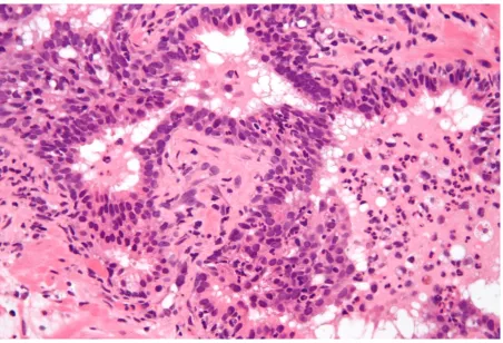
4.5.4.1 H&E tumour inflammation ...85
4.5.4.2 T-lymphocytes ...85
4.5.4.3 Natural killer cells ...86
4.5.4.4 Macrophages ...86
4.6 CORRELATIONS ... 87
4.6.1 CORRELATION BETWEEN CTC NUMBERS AND CIRCULATING LYMPHOCYTES ...87
4.6.2 CORRELATION BETWEEN CTC NUMBERS AND PLATELET COUNTS ....91
4.6.3 CORRELATION BETWEEN CTC NUMBERS, LYMPHOCYTE SUBGROUPS AND PLATELET COUNTS ...95
4.6.4 CORRELATION BETWEEN CTC NUMBERS AT T0 AND NCB INFLAMMATORY CELLS ...96
CHAPTER 5 – DISCUSSION ... 98
5.1 MAIN FINDINGS ... 98
5.2 PARTICIPANT ACCRUAL ... 99
5.3 BASELINE CLINICOPATHOLOGICAL FEATURES OF PARTICIPANTS ... 100
5.4 CIRCULATING TUMOUR CELLS ... 101
5.4.1 SCREENCELL® CYTO FILTERS ...101
Page | viii 5.4.3 COMPARISON OF CTC NUMBERS BETWEEN GROUPS AND ACROSS
TIMEPOINTS ... 106
5.4.4 PLATELET CLOAKING ... 107
5.4.5 DETAILED CTC MORPHOLOGY ... 108
5.5 PERIPHERAL BLOOD FLOW CYTOMETRY ... 110
5.6 NEEDLE CORE BIOPSIES... 112
5.6.1 ACUTE INFLAMMATION ... 112
5.6.2 PROSTATE ATROPHIC LESIONS ... 112
5.6.3 INFLAMMATION IN BENIGN PROSTATE TISSUE ... 112
5.6.4 INFLAMMATION IN PROSTATE CANCER TISSUE ... 112
5.7 CORRELATIONS ... 115
5.7.1 CTC / PLATELETS ... 115
5.7.2 CTC / LYMPHOCYTE SUBSETS ... 117
5.7.3 CTC / INFLAMMATION IN NCBS ... 118
CHAPTER 6 – CONCLUSIONS ... 120
Page | ix This work is dedicated to my wife Aoife and to my daughters Anna and Eleanor
Page | x
FIGURES
Figure 1.1: The prostate gland ... 3
Figure 1.2: Acute prostatitis ... 4
Figure 1.3: Simple atrophy and simple atrophy with cyst formation ... 5
Figure 1.4: Morphological features of prostatic adenocarcinoma. ... 7
Figure 1.5: Platelets and erythrocytes ... 15
Figure 1.6: Light microscope ... 33
Figure 1.7: Paraffin-embedded tissue blocks ... 35
Figure 1.8: Mesothelial cells, Pap stain ... 37
Figure 1.9: Keratising squamous cell carcinoma, Pap stain ... 38
Figure 1.10: Thyroid follicular epithelial cells, MGG stain ... 39
Figure 1.11: Flow cytometer schematic diagram ... 40
Figure 3.1: ExPeCT trial flow chart ... 46
Figure 3.2: ExPeCT trial project 1 ... 47
Figure 3.3: ExPeCT trial project 2 ... 48
Figure 3.4: ExPeCT trial project 3 ... 48
Figure 3.5: ExPeCT trial project 4 ... 49
Figure 3.6: ScreenCell® Cyto filtration unit ... 53
Figure 3.7: ScreenCell® Cyto filters and storage box ... 54
Figure 3.8: Circulating tumour cell bare nuclei on ScreenCell® filter (MGG, 40x) ... 55
Figure 3.9: Circulating tumour cell with cytoplasm on ScreenCell® filter (MGG, 40x) ... 56
Figure 3.10: Circulating tumour cell cluster on ScreenCell® filter (MGG, 20x) ... 56
Figure 3.11: Circulating tumour cell with adherent platelets on ScreenCell® filter (MGG, 40x) ... 57
Figure 3.12: FACSCanto II flow cytometer readout example ... 60
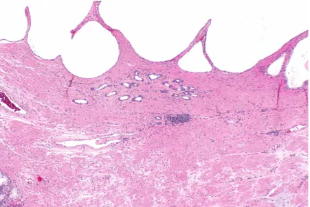
Figure 3.13: Grade 3 stromal inflammation in benign prostate tissue (H&E, 20x). ... 62
Figure 3.14: Grade 0 tumour chronic inflammation(H&E, 20x) ... 64
Figure 3.15: Grade 1 tumour chronic inflammation (H&E, 10x) ... 64
Figure 3.16: Grade 2 tumour chronic inflammation (H&E, 10x) ... 65
Figure 3.17: Area with Grade 0 CD3+ T-lymphocytes (CD3, 10x)... 66
Figure 3.18: Area with Grade 1 CD3+ T-lymphocytes (CD3, 10x)... 66
Figure 3.19: Area with Grade 2 CD3+ T-lymphocytes (CD3, 10x)... 67
Figure 4.1: BMI categories of ExPeCT trial participants ... 70
Page | xi
Figure 4.3: CTC with attached cytoplasm (MGG, 40x)...73
Figure 4.4: Nuclear diameter of CTCs in selected CTC-rich filters ...78
Figure 4.5: CTC with nuclear indentation, radiating from the overlain filter pore (MGG, 40x) ...79
Figure 4.6: Correlation between total lymphocyte count and mean CTC count at a given timepoint...88
Figure 4.7: Correlation between absolute NK-cell count and mean CTC count at a given timepoint...89
Figure 4.8: No correlation between CD4:CD8 ratio and mean CTC count at a given timepoint ...90
Figure 4.9: Correlation between platelet count and mean CTC count at a given timepoint in all groups ...91
Figure 4.10: Correlation between platelet count and mean CTC count at a given timepoint in control group ...92
Figure 4.11: No correlation between platelet count and mean CTC count at a given timepoint in non-exposed group ...93
Figure 4.12: Correlation between platelet count and mean CTC count at a given timepoint in blood draws which lacked platelet-cloaking ...94
Figure 4.13: No correlation between platelet count and mean CTC count at a given timepoint in blood draws with platelet-cloaking ...95
Figure 4.14: No correlation between intratumoural NK-cell count and mean CTC count at T0 ...97
Figure 5.1: Fungal organisms on ScreenCell® filter ...103
Figure 5.2: Anucleate squamous cell on ScreenCell® filter ...104
Figure 5.3: Contaminant fibres on ScreenCell® filter ...104
Figure 5.4: Non-cellular debris on ScreenCell® filter ...105
Figure 5.5: Abundant white blood cells on ScreenCell® filter ...105
Figure 5.6: CD56 expression in benign atrophic prostate epithelial cells ...113
Page | xii
TABLES
Table 1.1: Definition of obesity and overweight ... 21
Table 1.2: Criteria for diagnosis of metabolic syndrome ... 22
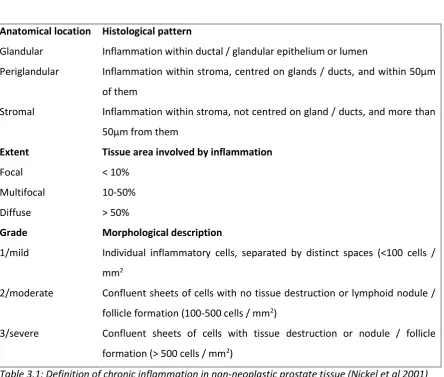
Table 3.1: Definition of chronic inflammation in non-neoplastic prostate tissue ... 62
Table 3.2: Inflammatory / Atrophic Lesions Definition ... 63
Table 4.1: CTC numbers per filter, timepoint analysis ... 74
Table 4.2: CTC numbers per blood draw, timepoint analysis ... 76
Table 4.3: Morphological features of CTCs in selected CTC-rich filters ... 80
Table 4.4: ANOVA of flow-cytometry derived lymphocyte counts per timepoint ... 82
Table 4.5: Comparison of lymphocyte counts between exercise and control groups ... 83
Table 4.6: Intensity and distribution of chronic inflammation in benign regions of prostate NCBs ... 85
Table 4.7: CTC number / absolute lymphocyte count correlations ... 87
Table 4.8: CTC number / lymphocyte fraction correlations ... 90
Table 4.9: Multiple regression analysis of mean CTC count per blood draw against absolute lymphocyte and platelet counts ... 96
Page | xiii
ABBREVIATIONS
ADP Adenosine diphosphate ADT Androgen deprivation therapy AMP Adenosine monophosphate ANOVA Analysis of variance
ATP Adenosine triphosphate BMI Body mass index
BPH Benign prostatic hyperplasia CEC Circulating epithelial cell CRP C-reactive protein CSC Cancer stem cell CTC Circulating tumour cell DRE Digital rectal examination
EDTA Ethylene diamine tetra-acetic acid EGF Epidermal growth factor
EMT Epithelial-mesenchymal transition
ExPeCT Exercise, Prostate Cancer and Circulating Tumour Cells FNA Fine needle aspiration
H&E Haematoxylin and Eosin HDL High density lipoprotein
HGPIN High grade prostatic intraepithelial neoplasia HIF Hypoxia-inducible factor
HPF High power (40x) field
ICH-GCP International Conference on Harmonisation – Good Clinical Practice IFNγ Interferon gamma
IGF Insulin-like growth factor
IGFBP Insulin-like growth factor binding protein IHC Immunohistochemistry
IL Interleukin
lcSFA Long chain saturated fatty acid LPS Lipopolysaccharide
MGG May Grunwald Giemsa
Page | xiv NCB Needle core biopsy
NFκb Nuclear factor kappa-light-chain-enhancer of activated B cells NK Natural killer
NO Nitric oxide PA Partial atrophy
PAH Postatrophic hyperplasia PD Pore diameter
PDGF Platelet derived growth factor PI3K Phosphoinositide-3-kinase
PIA Proliferative inflammatory atrophy PIN Participant Identifier Number PLR Platelet lymphocyte ratio PrCa Prostate cancer
PSA Prostate specific antigen QoL Quality of Life
SA Simple atrophy
SACF Simple atrophy with cyst formation SCID Severe combine immune deficiency SII Systemic immune-inflammation index T0 Timepoint 0 months
T3 Timepoint 3 months T6 Timepoint 6 months
TCIPA Tumour cell induced platelet aggregation TCR T-cell receptor
TF Tissue factor
TGFβ Transforming growth factor beta TLR4 Toll-like receptor 4
TNFα Tumour necrosis factor alpha
TURP Transurethral resection of the prostate VEGF Vascular endothelial growth factor vWF Von Willebrand Factor
Page | xv
PUBLICATIONS
Exercise and prostate cancer: evidence and proposed mechanisms for disease modification. Hayes BD, Brady L, Pollak MN, Finn S.
Cancer Epidemiol Biomark Prev 2016;25:1281-1288 [Published OnlineFirst July 7, 2016; doi: 10.1158/1055-9965.EPI-16-0223].
The ExPeCT (Examining Exercise, Prostate Cancer and Circulating Tumour Cells) trial: study protocol for a randomized controlled trial.
Sheill G, Brady L, Guinan E, Hayes B, Casey O, Greene J, Vlajnic T, Cahill F, Van Hemelrijck M, Peat N, Rudman S, Hussey J, Cunningham M, Grogan L, Lynch T, Manecksha RP, McCaffrey J, Mucci L, Sheils O, O’Leary J, O’Donnell DM, McDermott R, Finn S.
Trials 2017;18:456
Examining the link between obesity, inflammation, and exercise in patients with metastatic prostate cancer—An interim analysis from the ExPeCT trial.
Lauren Brady, Grainne Sheill, Anne-Marie Baird, Emma H. Allott, Tatjana Vlajnic, John Greene, Orla Casey, Brian Hayes, Emer Guinan, Juliette Hussey, Fidelma Cahill, Mieke Van Hemelrijck, Nicola Peat, Sarah Rudman, Moya Cunningham, Liam Grogan, Thomas Lynch, Rustom P. Manecksha, John McCaffrey, Orla Sheils, Dearbhaile M. O’Donnell, John O’Leary, Ray McDermott and Stephen P. Finn.
Cancer Research 2018; 78: 16 Suppl: pp. A057. DOI: 10.1158/1538-7445.PRCA2017-A057
(Published abstract)
Lifestyle and health-related quality of life in men with metastatic prostate cancer.
Gráinne Sheill, Lauren Brady, Emer Guinan, Juliette Hussey, David Hevey, Tatjana Vlajnic, Orla Casey, Anne-Marie Baird, Fidelma Cahill, Mieke Van Hemelrijck, Nicola Peat, Sarah Rudman, Thomas Lynch, Rustom P. Manecksha, Brian Hayes, Moya Cunningham, Liam Grogan, John McCaffrey, Dearbhaile M. O’Donnell, Ray McDermott, John O Leary and Stephen P. Finn.
Page | xvi
ACKNOWLEDGEMENTS
The work presented in this thesis is but one angle of the large, international, multidisciplinary ExPeCT trial, and many people were involved in its delivery. I would like to thank in particular Stephen Finn who conceived and led this project. His encouragement to me at the early stages was really very valuable, in particular when we failed in our initial attempt to acquire funding and he refused to be disheartened. His mentoring and guidance at a formative stage of my career was tremendously important. In addition Orla Sheils and John O’Leary were very important in the initial planning and funding acquisition of the ExPeCT trial. Orla was a constant friendly and supportive presence during my years as a lecturer in Trinity. John took me under his wing as a first-year histopathology SpR and encouraged me to see myself as a researcher, and opened my eyes to all of the possibilities that an enquiring mind can see. The six months I spent with him in the Coombe were some of the most enjoyable of my training.
Thanks also to Lauren Brady, who did most of the filtering and staining of the Dublin ScreenCell® filters on-site in St James’s, and was endlessly patient when her own work was delayed by my tardiness in producing results! She and Tatiana Vlajnic did much early work in cell-line spiking experiments to validate the use of the ScreenCell® filters. Anne-Marie Baird was also closely involved in specimen filtration and staining. Lauren and Anne-Marie also acquired and organised staining of the paraffin tissue blocks from the participants’ needle core biopsies. Emer Guinan and Grainne Sheill ran the exercise component of the ExPeCT trial in Dublin and acquired blood samples. Jean Dunne and Dean Holden performed the flow cytometry analysis at the Department of Immunology at St James’s Hospital, and the staff of the histopathology labs at St James’s Hospital Dublin undertook H&E and immunohistochemistry staining work. I am very grateful for all of their help. In addition I would like to thank all other members of the ExPeCT team in Ireland (particularly Juliette Hussey, Moya Cunningham, Liam Grogan, Thomas Lynch, Rustom Manecksha, John McCaffrey, Dearbhaile O’Donnell, Ray McDermott), the UK (particularly Fidelma Cahill, Mieke Van Hemelrijck, Sarah Rudman, Nicola Peat) and the USA (particularly Lorelei Mucci and Bryan Stanfill, who provided invaluable statistical support). Thanks also to Ronan Leen for help with proofreading.
Finally my thanks to all of the men who participated in the ExPeCT trial, giving selflessly of themselves in order that we might make one small step further along the road to beating cancer.
Page | 1
1. INTRODUCTION
1.1 GENERAL INTRODUCTION
Prostate cancer (PrCa), the most frequently diagnosed male cancer in the developed world (Jemal et al 2011), is a leading cause of male cancer death. Although prostate specific antigen (PSA) screening identifies many early cancers, numerous men still present with locally advanced or metastatic disease for whom radical surgery with curative intent is inappropriate. In this setting increased disease-free and overall survival and improved quality-of-life (QoL) are the primary management objectives, and new therapies and lifestyle alterations which can assist are increasingly needed.
The incorporation of exercise programmes into cancer care is increasingly recognised to have beneficial effects. A systematic review of exercise interventions in cancer survivors found improvements in strength, fatigue, fitness, functional QoL, self esteem and anxiety (Speck et al 2010). Much of this research relates to colon and breast cancer but increasing evidence demonstrates improvements in symptom control, all-cause mortality and cancer-specific mortality in PrCa also. Improved cancer-specific mortality suggests the potential for exciting developments in our understanding of the biology and aggressiveness of PrCa, together with novel therapeutic opportunities.
In those PrCa patients with potentially curable disease, obesity and its complications may make surgery impractical. Androgen deprivation therapy (ADT), the mainstay of systemic treatment for hormone responsive advanced PrCa, itself causes obesity and metabolic syndrome (MS). As medical therapy for obesity-related cardiovascular risk factors improves, aggressiveness of PrCa becomes more important than cardiovascular complications in determining the cause of mortality in these men. We know that obese men have a worse outlook regarding cancer-related mortality than non-obese men. The combination of an aging population with an increased PrCa incidence, increasing obesity prevalence and improved management of cardiovascular risk factors means that in the future more men are going to die as a result of the deleterious effect of overweight in advanced PrCa.
Page | 3
1.2 THE PROSTATE GLAND
1.2.1 ANATOMY AND HISTOLOGY
The prostate gland is the largest accessory gland in the male reproductive system, and normally weighs approximately 20g. It is situated in the pelvis and shaped like an inverted pyramid, with its base applied to the trigone of the bladder and its apex pointing inferiorly. The prostatic urethra runs through the prostate gland. The glandular tissue can be divided into four regions on the basis of anatomy and function: the central zone, transitional zone, peripheral zone and an anterior region of fibromuscular stroma.
Figure 1.1: The prostate gland. Henry Gray. Anatomy of the Human Body. 1918 (source: Wikimedia commons)
[image:21.595.116.539.261.611.2]Page | 4 highlighted with immunohistochemical stains (e.g. p63), and are a useful diagnostic adjunct in the assessment of individual glands as malignant or benign, as invasive glands lack a basal cell layer.
1.2.2 PROSTATIC HYPERPLASIA
Nodular hyperplasia is a very common disorder, generally affecting men over the age of 50, with the potential for compressive obstruction of the urethra and associated lower urinary tract symptoms. Microscopic evidence of hyperplasia rises in incidence from 20% at age 40 to 90% at age 70, but direct correlation with symptomatology is not a feature (Kumar et al 2005). Nodular hyperplasia (benign prostatic hyperplasia – BPH) tends to arise in the transitional zone of the gland. Surgical treatment of BPH by transurethral resection (TURP) can give rise to the incidental diagnosis of prostate cancer in the resected tissue.
[image:22.595.60.513.316.624.2]1.2.3 PROSTATITIS AND PROSTATIC ATROPHY
Figure 1.2: Acute prostatitis (source: Wikimedia commons)
Page | 5 resistant to systemic antimicrobial treatment, or can frequently be abacterial. Granulomatous prostatitis can be caused by tuberculosis, Bacille Calmette-Guerin treatment of bladder cancer, or can be nonspecific and idiopathic.
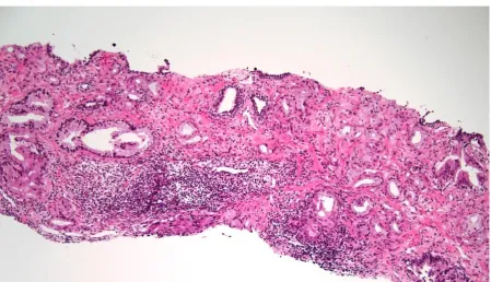
Figure 1.3: Simple atrophy (centre) and simple atrophy with cyst formation (top) (source: Wikimedia commons)
[image:23.595.104.556.137.439.2]Page | 6 Prostatic atrophy is often linked with chronic inflammation, to such an extent that SA and PAH are termed “proliferative inflammatory atrophy” (PIA). They are thought by some to be associated with high grade prostatic intraepithelial neoplasia (HGPIN), a probable precursor lesion for carcinoma (De Marzo et al 1999, Putzi and De Marzo 2000). The occurrence of most PIA lesions in the peripheral zone and the morphological identification of “merging” of PIA lesions with HGPIN are presented as supportive evidence (Wang et al 2009). In addition various somatic and epigenetic alterations seen in HGPIN are also seen in PIA lesions (Nakayama et al 2003). Many studies however show no association between atrophic lesions and carcinoma (Servian et al 2015), PrCa lethality (Davidsson et al 2011) or even show that baseline atrophy is protective against the subsequent development of malignancy (Moreira et al 2015).
1.2.4 PROSTATE CARCINOMA
1.2.4.1 Epidemiology
Prostatic acinar adenocarcinoma, by far the most common malignant neoplasm of the prostate gland, is a very common disease, but often clinically undetected. Autopsy studies have shown foci of carcinoma in completely-embedded prostate glands of up to 40% of men in western countries (Breslow et al 1977). An American man aged more than 50 years has a 9.5% lifetime risk of clinical detection of PrCa, and although many cancers never present clinically, PrCa is the second most common male malignancy worldwide, and the fifth most common cause of male cancer death (Humphrey et al 2016, Bray et al 2018). In Ireland, most Western European countries and most countries in North and South America, PrCa is the most frequently diagnosed cancer in men in 2018 (Bray et al 2018). There is a dramatic variation worldwide however, and while the age-standardised incidence rate is 85.7 per 100,000 in Western Europe, it is only 5 per 100,000 in south central Asia (Bray et al 2018). Currently identified risk factors for the development of PrCa are predominantly dietary and include ingestion of red meat and animal fats, and milk and dairy products. Tomatoes, fish, cauliflower and broccoli may be protective (Humphrey et al 2016).
1.2.4.2 PSA
Page | 7
1.2.4.3 Pathological features
Tissue can be acquired for pathological diagnosis of PrCa through fine needle aspiration (FNA - no longer recommended for routine use), needle core biopsy (NCB - 10-12 systematic 18-gauge ultrasound-guided cores, with additional targeting of any clinically or ultrasonographically suspicious areas), transurethral resection (typically performed for management of BPH, with incidental detection of carcinoma in a minority of cases) or simple prostatectomy (for treatment of BPH). Radical prostatectomy as definitive treatment for PrCa is not undertaken without pre-operative histological diagnosis. Sometimes the primary diagnosis is made at the time of sampling of metastatic sites, such as targeted biopsy of bony lesions, or detection of carcinoma in bone reamings following pathological fracture.
Figure 1.4: Morphological features of prostatic adenocarcinoma. Small malignant glands, closely packed, nucleolated, with blue luminal mucin (source: Wikimedia commons)
Page | 8 adenocarcinoma exist, some of which have a worse prognosis (signet-ring carcinoma, pleomorphic giant cell carcinoma, sarcomatoid carcinoma – Humphrey et al 2012) and some of which can histologically mimic benign prostate epithelium (atrophic and foamy gland variants). In small foci, the absence of p63-positive, 34βE12-positive basal cells, combined with cytoplasmic positivity for P504S (AMACR / racemase), can support morphological assessment in making the diagnosis. Sometimes broader immunohistochemical panels are required to distinguish poorly-differentiated prostatic carcinoma from urothelial or colorectal carcinoma (Epstein et al 2014).
1.2.4.4 Prognosis
Various prognostic features, clinical, histological and molecular, have been identified. Gleason grading, first developed in the late 1960s (Gleason and Mellinger, 1974) remains the most useful histological factor. Gleason grading is based entirely on architectural rather than cytological features, and the Gleason score is assigned based on the two worst grades present. On NCB, the number of cores involved by tumour and the percentage of tissue involved also have prognostic significance (Srigley et al 2009). Pathological stage, primarily determined in radical prostatectomy specimens, is the second most useful pathological feature. The AJCC TNM 8th edition staging
system is based on the presence and volume of tumour on one or both sides, extraprostatic extension (including seminal vesicle involvement) and invasion of adjacent structures (Amin et al 2017).
1.2.4.5 Management strategies
Management of PrCa is stratified depending on risk categories, usually defined as follows (D’Amico et al 1998, Heidenreich et al 2014, Heidenreich et al 2014):
Low risk: cT1-T2a and Gleason score ≤ 6 and PSA < 10μg/L
Intermediate risk: cT2b-T2c or Gleason score = 7 or PSA 10-20μg/L High risk: cT3a, Gleason score 8-10 or PSA > 20μg/L
Very high risk: cT3b-T4 or cN1
Page | 10
1.3 CIRCULATING TUMOUR CELLS
1.3.1 DEFINITION
Circulating tumour cells (CTCs) are an intermediate stage in the process of metastasis, whereby a cancer can spread from a primary site to set up secondary malignant growths at anatomically distant sites. In order to establish a metastatic deposit cancer cells need to invade into the circulation at the primary site, embolise within the blood and survive the various immunological threats posed to them in doing so, lodge within the circulation at a capillary bed within a distant site, invade into the surrounding tissues and grow within these tissues, whose microenvironment may be less conducive to neoplastic growth than the primary site. As metastases become more established or following resection of a primary tumour CTCs may increasingly originate within metastatic deposits rather than within the primary tumour itself (Micalizzi et al 2017). The CTC stage is a potentially useful target for therapy as intravascular cells are vulnerable to the cellular and humoral processes of the innate and adaptive immune systems. Haematogenous dissemination of CTCs should be considered separately from the process of metastasis to local / regional lymph nodes, by which malignant cells spread through lymphatic vessels.
1.3.2 STEMNESS AND CTCS
The cancer stem cell (CSC) model posits that carcinomas develop from mutated adult stem cells with high tumorigenicity, capacity for self-renewal and ability to recapitulate the heterogeneity of the original tumour when transplanted. CSCs represent a minority of cells within a tumour but are responsible for its aggressive behaviour (Lang et al 2009). ALDH1A1, part of the differentiation-related retinoic acid pathway, is found in CSCs in haematopoietic and lung cancers, and it has also been isolated from PrCa cell lines (Li et al 2010). ALDH1A1+ cells show many convincing features of PCa CSCs, including the capacity to recapitulate a heterogeneous tumour population when serially passaged in vivo. High ALDH1A1 expression was associated with poor cancer-specific survival.
1.3.3 MEANS OF IDENTIFICATION AND ENUMERATION
Page | 11 The most popular and FDA-approved system for CTC enrichment involves CTC separation from leucocytes by virtue of their affinity for certain specific antibodies, for example EpCAM. Epithelial cells (with an EpCAM+, CD45- immunophenotype) rarely exceed one cell per 7.5ml of blood in patients without malignancy (Allard et al 2004). The CellSearch® Circulating Tumor Cell Kit contains a ferrofluid-based capture reagent, with a magnetic core and anti-EpCAM antibodies. Following immunomagnetic capture fluorescent reagents are added for identification of CTCs, including anti-CK-Phycoerythrin (specific for the intracellular protein cytokeratin, characteristic of epithelial cells), DAPI (which stains the cell nucleus), and anti-CD45-Allophycocyanin (specific for leukocytes). The reagent/sample mixture is dispensed by a proprietary CellTracks® AutoPrep® System into a cartridge that is inserted into a MagNest® cell presentation device, within which a strong magnetic field attracts the magnetically labeled epithelial cells to the cartridge surface. An analyser then scans the entire surface of the cartridge, acquires images and displays any event to the user with co-location of CK-PE and DAPI fluorescence. The CellSearch® system classifies an event as a CTC when its morphology is consistent with that of a tumor cell and it exhibits an EpCAM+, CK+, DAPI+ and CD45- phenotype.
Epithelial-mesenchymal transition (EMT) involves cellular loss of epithelial differentiation and switching towards a mesenchymal phenotype. It occurs in embryogenesis but also in the development of invasion in early stage carcinomas. The loss of epithelial surface markers by EMT-switched tumour cells invading the circulation may impede CTC identification by the CellSearch® system. Cancer cells which have acquired EMT share CSC characteristics and express CSC markers (Kong et al 2010). A drawback of CellSearch® is that cells which have successfully invaded the circulation will have undergone EMT, often losing epithelial surface antigens such as EpCAM in the process. It is thought that unfortunately many of the circulating CSC-like cells, which would be the fraction most likely to be biologically significant, undergo EMT as part of the metastatic cascade and thus slip beneath the radar (Chen et al 2012, Yu et al 2013). Other markers such as cell-surface vimentin have been reported to be expressed in CTCs which have undergone EMT, increasing the sensitivity of immunoaffinity-based assays (Satelli et al 2015).
Page | 12 EMT gene expression. ScreenCell® filters use a microporous membrane filter and a vacuum tube to allow leucocytes to pass through the 7.5μm filter pores, while trapping the larger and less deformable CTCs on the filter (Desitter et al 2011). Several devices are available from ScreenCell® for isolating CTCs for different purposes – the “Cyto” filter isolates fixed cells for morphological assessment, including immunocytochemistry or FISH studies, the “CC” filter allows isolation of live cells for culture and the MB filter allows filtration of cells suitable for extraction of nucleic acids and further molecular genetic analysis. An additional advantage of size-based enrichment techniques such as this is the ability to detect CTC clusters, which may have greater capacity to establish metastases (Aceto et al 2014).
Flow cytometry, with its ability to characterise in detail the immunophenotype of small numbers of cells in fluid phase specimens, is an attractive method for detection of CTCs, and significant progress has been made in this area (Ulrich and Tarnok 2014). Both in cell line spiking experiments (Takao and Takeda 2011) and in clinical specimens from patients with head and neck squamous cell carcinoma (Hristozova et al 2012) flow cytometry techniques with antibodies against EpCAM and CK7 were found to be sensitive. A novel platform allows flow cytometric isolation of both single and clustered CTCs, followed by their molecular characterisation (Bhagwat et al 2018), in a mouse model of pancreatic cancer. Flow cytometry is likely in the future to be a powerful tool in translational and clinical research into CTCs in cancer patients.
1.3.4 PROGNOSTIC SIGNIFICANCE
Page | 14
1.4 COAGULATION
1.4.1 OVERVIEW OF COAGULATION
The normal functioning of the circulatory system requires that blood should be a free-flowing liquid within the blood vessels, but as these blood vessels are vulnerable to trauma a mechanism for plugging leaks in the system is required. Following vascular injury there is a localised but transient arteriolar vasoconstrictive response (Mitchell 2005). Exposure to the circulation of subendothelial extracellular matrix allows platelet adherence and activation, and further aggregation, to rapidly form a haemostatic plug. At the same time tissue factor (TF) release from damaged tissue and from platelets initiates a series of enzymatic conversions of proenzymes into active enzymes (the coagulation cascade), which ultimately leads to the activation of thrombin, which in turn converts the soluble plasma protein fibrinogen into insoluble fibrin. A permanent plug is formed by polymerised fibrin and platelets, and at this point regulatory systems are activated to ensure that coagulation is limited to the site of injury and does not become widespread. A balance between pro- and anticoagulant forces is required for normal circulatory function and in order to prevent spontaneous coagulation there are a variety of protective systems at play. Production of nitric oxide (NO) and prostaglandin I2 by intact endothelial cells
prevents inappropriate activation of platelets. Anticoagulant molecules (antithrombin III, protein C, thrombomodulin) which interfere with the coagulation cascade are expressed by endothelial cells. Endothelial cells also produce tissue plasminogen activator, which promotes enzymatic breakdown of polymerised fibrin from endovascular surfaces.
1.4.2 PLATELETS – STRUCTURE AND PHYSIOLOGY
Platelets (thrombocytes) are cytoplasmic fragments derived from bone marrow megakaryocytes. When unactivated they are lenticular discs, 2-3μm in diameter, and they lack nuclei. On a May-Grunwald-Giemsa (MGG) stained smear of peripheral blood they appear as dark purple spots (see image). Although first identified in the first half of the 19th century James Homer Wright coined
Page | 15
Figure 1.5: Platelets and erythrocytes, MGG, 60x. Individual platelets are visible at centre and upper right. Clumps of platelets are also present at bottom left (this specimen is derived from a thyroid FNA procedure and some platelet aggregation has occurred in the short time between the needle being inserted into the thyroid and its contents being expelled onto a glass slide)
Page | 16 by thrombin at the end of the coagulation cascade, which forms an insoluble net of fibres which tie the platelet mass together.
1.4.3 PLATELETS AND CANCER
Page | 17
1.5 THE IMMUNE SYSTEM
1.5.1 ADAPTIVE IMMUNITY.
The immune system has evolved over millions of years in an ongoing effort to protect the body from a variety of threats, both external (viral, bacterial and fungal infections) and internal (malignancies). It comprises a complex series of subsystems which can be conceptually divided into innate and adaptive immunity. Adaptive immunity requires antibody-mediated recognition of non-self antigens, and their destruction by a number of lymphocyte-mediated mechanisms, both cellular and humoral. It is extremely powerful in combating infection and by means of plasma cells and memory cells can provide ongoing immunity to an infectious agent following an episode of acute infection (Abbas 2005).
1.5.2 INNATE IMMUNITY
The innate immune system is not organism-specific in its anti-infective mechanisms and can be considered analogous to a first responder to an emergency rather than the well-equipped hospital emergency department to which a casualty is subsequently transported. The innate, pre-existing defences against infection include the mechanical barriers which prevent ingress or proliferation of micro-organisms into the body (e.g. an intact layer of skin or gastrointestinal mucosa, functional cilia-driven clearance of entrapped micro-organisms from respiratory mucus, complete emptying of the bladder following micturition), the malfunctioning of which can predispose to infection. In addition phagocytic cells such as neutrophils (the effector cells of the acute inflammatory reponse) and macrophages can ingest and destroy micro-organisms. Circulating complement proteins can provide a humoral but non-antibody-mediated weapon with which to lyse micro-organisms – these proteins can also be activated as part of the adaptive immune response. The component of the innate immune response with which the current study is primarily concerned is the natural killer (NK) cell.
1.5.3 LYMPHOCYTES
Page | 18 cytoplasm, easily recognised on Giemsa staining. Plasma cells have more abundant amphophilic cytoplasm with a paranuclear pale zone or “hoff”, corresponding to an expanded smooth endoplasmic reticulum. NK-cells are a little larger than circulating B- and T-lymphocytes, have more cytoplasm and contain azurophilic granules, hence the haematological term “large granular lymphocyte”. During acute infections reactive lymphocytes with enlarged nuclei and increased cytoplasm can be seen on a blood smear. Large B-lymphocytes (such as marginal zone lymphocytes with more abundant cytoplasm, or germinal centre B-lymphocytes which can have convoluted or large nucleolated nuclei) can be found within the lymphoid tissues, but tend to be scanty in the peripheral blood.
1.5.3.1 T-lymphocytes
Each T-lymphocyte bears a T-cell receptor (TCR), composed of either α and β or γ and δ polypeptide chains. The TCR allows recognition by the T-lymphocyte of a specific antigen, which involves major histocompatibility complex (MHC)-mediated binding to a peptide in the case of αβ lymphocytes and non-MHC-mediated binding to peptides and lipids in the case of γδ lymphocytes. Rearrangements of the genes coding for the TCR occur during the process of T-lymphocyte maturation, resulting in the formation of TCRs which are specific for a huge variety of potential antigens, and the presence of a TCR gene rearrangement in a lymphocyte indicates that it is a T-lymphocyte. Part of signal transduction from the TCR into the T-lymphocyte nucleus following antigen binding is mediated by the CD3 molecule, whose presence as determined by immunophenotyping (immunohistochemistry or flow cytometry) is a useful T-lineage specific marker for identification of T-lymphocytes. Other accessory functional molecules such as CD7 and CD2 are also helpful in this regard, and CD4 and CD8 expression divides αβ T-lymphocytes into CD3+CD4+T-helper cells and CD3+CD8+ cytotoxic T-lymphocytes. T-helper cells, whose CD4 molecule binds to MHC class II molecules during T-lymphocyte activation, are primarily responsible for secreting cytokines which modulate the activation and function of other cellular and humoral components of the immune system. T-helper-1 cells secrete interleukin-2 and interferon gamma, and T-helper-2 cells secrete interleukins 4, 5 and 13. Cytotoxic T-lymphocytes, whose CD8 molecule binds only to MHC class I molecules, can secrete T-helper-1 cytokines but mainly function as cytotoxic killers, analogous to the NK-cells of the innate immune system.
1.5.3.2 B-lymphocytes
Page | 19 bound. A rearranged immunoglobulin gene in a lymphocyte indicates that it is a B-lymphocyte, and detection by polymerase chain reaction of monoclonal immunoglobulin gene rearrangements in a B-lymphocyte population is a useful indicator in clinical practice of neoplastic transformation. Following antigen binding and activation B-lymphocytes can transform into plasma cells and secrete immunoglobulins which, when circulating, form the essential component of adaptive humoral immunity. Useful markers of mature B-lymphocytes include the cell-surface proteins CD19 and CD20. Immature and mature B-lymphocytes express CD79a and plasma cells express CD38 and CD138. Nuclear transcription markers also make attractive targets for immunohistochemistry, with PAX5 and OCT2 expressed in mature B-lymphocytes and MUM1 expressed in plasma cells.
1.5.3.3 Natural killer cells
NK-cells, which constitute 10-15% of peripheral blood lymphocytes, are larger than most circulating T- and B-lymphocytes. They do not have rearranged TCR or immunoglobulin genes and are negative for surface CD3 (although they can express CD3 subchains in their cytoplasm) and for B-lineage specific markers. They can bind and lyse cells which have been opsonised with IgG molecules, derived from the adaptive humoral immune system, but they do not themselves require activation by an intrinsic antigen-antibody reaction in order to undertake this “antibody-dependent cell mediated cytotoxicity”. NK-cell killing is mediated by perforins, molecules which when released close to a targeted cell form channels in the target cell membrane, allowing penetration by proteases such as granzyme, leading to apoptosis or lysis. NK-cells avoid killing cells which express MHC class I molecules (i.e. all normal “self” cells), but viral infection or neoplastic change can reduce MHC class I expression and facilitate NK-cell mediated killing, a process which is assisted by activating receptors such as NCR. NK-cells also secrete cytokines (interferon gamma [IFNγ], tumour necrosis factor alpha [TNFα]) and are activated by cytokines (IL-2, IL-12, IL-15) (Abbas 2015). CD56 (also known as neural cell adhesion molecule) is widely used for immunohistochemical identification of NK-cells, but it is not specific, being also expressed on γδ T-lymphocyte, CD8-positive cytotoxic T-lymphocytes, dendritic cells and a variety of non-lymphoid cells such as neurones and neuroendocrine epithelial cells.
Page | 20 (Lautenback et al 2011). The NK-cell fraction of blood is sensitive to exercise (reviewed Timmons et al 2008), and five-fold increases in NK-cell concentrations following exercise have been noted. Brief exercise upregulates molecular pathways in circulating NK-cells associated with cancer and cell communication (Radom-Azik et al 2013). In healthy young men, hypoxic exercise training leads to enhanced in vitro NK-cell cytotoxicity (Wang et al 2011).
Page | 21
1.6 OBESITY
1.6.1 DEFINITION
Obesity in adults is defined by the World Health Organisation (WHO - World Health Organisation 2018) on the basis of body mass index (BMI), measured as height (in meters) divided by the square of weight (in kilograms – see table 1.1). In children, obesity is defined by WHO according to standard deviations from the mean for a given age and sex or by the Centres for Disease Control according to percentiles. While useful as a population-based metric, BMI is less suitable for indiviudalised risk assessment, as it takes no account of body constituents – for example, a professional rugby player 1.78m in height weighing 98kg would have an obese-range BMI. Other measure such as waist-to-hip ratio, waist circumference and waist-to-height ratio may provide more individualised risk assessment for the complications of obesity (Lee et al 2008)
CLASSIFICATION BMI (KG/M2)
Underweight <18.5
Normal weight 18.5-24.9
Overweight 25.0-29.9
Obese Class I 30.0-34.9
Obese Class II 35.0-39.9
Obese Class III ≥40
Table 1.1: Definition of obesity and overweight
Page | 22
MEASURE DEFINITION
Elevated waist circumference Population and country-specific definitions
Elevated triglycerides
(or on drug treatment for same)
≥150mg/dL (≥1.7mmol/L)
Reduced HDL cholesterol
(or on drug treatment for same)
<40mg/dl (<1.0 mmol/L) in males <50mg/dL (<1.3 mmol/L) in females
Elevated blood pressure
(or on drug treatment for same)
Systolic ≥ 130 mmHg and/or diastolic ≥ 85 mmHg
Elevated fasting glucose
(or on drug treatment for same)
≥ 100 mg/dL
Table 1.2: Criteria for diagnosis of metabolic syndrome
Despite the close association of obesity with MS, the WHO definition of obesity does not figure in the diagnostic criteria for MS. Rather it is central adiposity which appears to confer a great degree of the cardiovascular risk in obesity, and hence the inclusion of waist circumference as a surrogate marker for this. In European populations a waist circumference of ≥102cm in men or ≥88cm in women is recommended by the European Cardiovascular Societies as the diagnostic threshold for abdominal obesity (Graham et al 2007)
1.6.2 EPIDEMIOLOGY
Page | 23 1.6.3 AETIOLOGY
Obesity and overweight are fundamentally a consequence of an imbalance in caloric intake on the one hand and energy expenditure on the other. Individual choices and activities clearly play a role in the development of obesity. However individuals can only act and make choices within the environment they inhabit and public policy can therefore play an important role in tackling obesity at a population level. For example the wide availability of energy-dense foods rich in refined carbohydrates and saturated fats, and the relative lack of availability of fresh fruit and vegetables in many areas (Larsen et al 2009) contribute to obesity. The increasingly sedentary nature of work and leisure time and decreases in routine exercise are long-term societal shifts which make obesity a problem which requires increasingly active measures to combat. At the individual level there are many recognised risk factors for obesity. Modifiable risk factors include lack of physical activity, unhealthy eating behaviours, sleep deprivation and psychological stress. Exposure to obesogenic chemicals (including cigarette smoke, air pollution and bisphenol A in childhood) may be avoidable. Non-modifiable risk factors include family history, certain genetic conditions (e.g. Praeder-Willi syndrome), race (obesity in America is most prevalent among black and Hispanic people) and age.
1.6.4 COMPLICATIONS
Page | 24 1.6.5 ASSOCIATIONS WITH SYSTEMIC INFLAMMATION
Obesity is well-recognised to be associated with an inflammatory state in metabolic tissues (reviewed by Gregor and Hotamisligil 2011). The distinction of “insulin-dependent” from “non-insulin-dependent” patients with diabetes mellitus led to a recognition that while some patients lacked insulin entirely, others were resistant to its effects. This latter group were often overweight or obese and were hyperinsulinaemic (Yalow et al 1965). Patients with infections or other states causing hypercortisolaemia sometimes developed insulin resistance, which was presumed to be related to hypothetical circulating insulin antagonist molecules. In a seminal paper, TNFα was shown to be constitutively expressed in adipose tissue and hyperexpressed in obesity (Hotamisligil et al 1993), as are a number of other inflammatory mediators (e.g. 6, IL-1β, CCL2). In addition to increased cytokine expression in adipose tissue obese individuals also have increased numbers of inflammatory cells (particularly pro-inflammatory M1 macrophages, NK-cells and mast cells) in these tissues, both in mouse models (Liu et al 2009, Ohmura et al 2010) and humans (Harman-Boehm et al 2007). The T-lymphocyte populations also alter, with a decreased CD4:CD8 ratio in adipose tissue. These cellular and molecular inflammatory changes in adipose tissue are chronic in duration rather than acute, and do not seem to lead to the resolution and tissue remodelling and fibrosis which is the typical sequela of chronic inflammation.
The systemic inflammation induced by obesity has effects on many different tissues. For example in the adipose tissue TNFα induces insulin resistance in adipocytes (Hotamisligil et al 1994). Signalling abnormalities are also seen in the liver, with steatosis and (if the inflammatory process escalates) steatohepatitis. In muscle tissue inflammatory cytokines can induce insulin resistance (Plomgaard et al 2005), reducing glucose uptake by this metabolically active tissue and tending towards hyperglycaemia and the diabetic paradigm of “starvation in the midst of plenty”. The pancreas also shows increased pro-inflammatory cytokine and macrophage numbers during the development of obesity-induced insulin resistance (Ehses et al 2007). Alterations in gut microbiota with associated inflammatory changes are also seen (Gregor and Hotamisligil 2011).
low-Page | 25 level peaks of inflammation resolve when the nutrients have been metabolised (reviewed by Boutagy et al 2016). Some nutrients also appear to have an intrinsic ability to activate components of the immune system, suggesting that when consumed in excess (as in obesity) they may directly drive the inflammatory response to obesity. For example toll-like-receptor 4 (TLR4), the receptor for lipopolysaccharide (LPS) which is important in initiating the innate immune reponse of macrophage phagocytosis, appears to be activated by long-chain saturated fatty acids (lcSFAs). However TLR4 may not act directly as a receptor for fatty acids, rather “TLR4-dependent priming alters cellular metabolism, gene expression, lipid metabolic pathways, and membrane lipid composition, changes that are necessary for lcSFA-induced inflammation” (Lancaster et al 2018).
Apart from its metabolic effects, insulin mediates many other processes, including platelet inhibition by increasing NO expression, anti-inflammatory activity by decreasing nuclear factor kappa-light-chain-enhancer of activated B cells (NFκb) and C-reactive protein (CRP), and anti-thrombotic activity by reducing TF activity. In a rat model, rises in serum IL-6 and TNFα were suppressed by insulin (Dandona et al 2005). Insulin resistance is characteristic of MS. Therefore, obese men tend to be in a pro-inflammatory and pro-thrombotic state.
TF functions as the principal initiator of the coagulation cascade, and also as a pro-inflammatory mediator, triggering signaling through G-protein–coupled receptors. TF appears to have a critical role at the crossroads of obesity, inflammation and thrombosis (Badeanlou et al 2011). In mice, inhibition of TF-mediated signalling in haematopoietic cells reduced high-fat-diet-related insulin resistance (through downregulation of TNFα and 6 and upregulation of anti-inflammatory IL-10), without impact on body weight. In adipose tissue, inhibition of TF-mediated signaling led to reduced weight gain.
1.6.6 ADIPOKINES
anti-Page | 26 apoptotic protein transcription. Nuclear expression of NFκb is associated with nodal metastasis in PrCa (Ismail et al 2004).
1.6.7 THE ROLE OF SKELETAL MUSCLE IN OBESITY
Skeletal muscle plays an important role in counteracting the pro-inflammatory effects of obesity. Contracting skeletal muscles release myokines which act as antagonists to the generally pro-inflammatory adipokines (Reviewed Pedersen et al 2011). For example, although muscles produce IL-6, a pro-inflammatory mediator which contributes to the deleterious effects of MS when chronically elevated, they do so in a TNFα-independent manner. Physically active people have low basal levels of IL-6. High basal levels are associated with MS. This suggests a role for muscle-derived IL-6 in metabolism rather than inflammation, and IL-6 stimulates insulin-mediated glucose uptake in muscle cells in-vitro. Other myokines include IL-15, which decreases lipid deposition in preadipocytes, and myostatin, whose expression is reduced by aerobic and strength exercise. The overall effects of the skeletal muscle secretome involve muscle hypertrophy, adipose tissue oxidation, increasing insulin sensitivity, increasing osteogenesis, reducing inflammation, increasing antitumour activity and increasing pancreatic function. Obese men are characterised by sarcopenic obesity, and their reduced muscle mass contributes substantially to insulin resistance and MS. Skeletal muscle-derived factors interact substantially with those derived from adipose tissue, and increasing skeletal muscle mass as well as reducing adiposity is likely to be of benefit in reducing platelet cloaking of CTCs. The sarcopenia caused by ADT may be responsive to antagonists of myostatin (Padhi et al 2014). Myostatin negatively regulates skeletal muscle growth and is a member of the TGFβ superfamily. Chronic myostatin exposure can cause apoptosis in cancer cells, through a shift from oxidative phosphorylation to glycolysis (Liu et al 2013).
1.6.8 OBESITY AND PROSTATE CANCER
Page | 27 testosterone [Tande et al 2006]. A separate study of 1880 men found a relative risk for PrCa development of 2 in overweight men and 3 in obese men [Laukkanen et al 2004]. However, the evidence that overweight and obesity confer a worse prognosis in PrCa is more definitive. One study of a cohort of 1554 men from the RTOG 92-02 trial found that 210 deaths were due to PrCa, and that overweight conferred a hazard ratio for PrCa-related death of 1.77 [Smith 2008].
IL-6 and TNFα are both raised in the serum of patients with metastatic carcinoma, compared to patients without metastases. Both are elevated in metastatic PrCa in direct proportion to disease stage, and increases occur at the time of biochemical (PSA) disease progression (Michalaki et al 2004). TNFα enhances the invasion of PrCa cell lines through synthesis of selectin ligands (Radhakrishnan et al 2011).
Page | 28
1.7 EXERCISE THERAPY
1.7.1 FOR NON-NEOPLASTIC DISORDERS
Exercise has well-documented benefits in improving cardiovascular risk and bone mineral density and in treating obesity and diabetes (reviewed, Warburton et al 2006), the details of which are beyond the scope of this discussion. It also has proven benefits in treatment of a variety of chronic musculoskeletal, nervous, respiratory and cardiovascular diseases (Smidt et al 2005).
1.7.2 IN CANCERS
The diagnosis of cancer may provide a “teachable moment” at which time men are more open to undertaking lifestyle changes to improve their health. Exercise following the diagnosis of cancer can reduce cardiovascular risk factors (Ligibel et al 2008) and improve general QoL even without significant reduction in body weight. Observational studies in breast (McTiernan et al 2010) and colon cancer (Meyerhardt et al 2009) suggest that more physical activity is associated with lower cancer-specific mortality. A Cochrane review (Mishra et al 2012) found that exercise may have beneficial effects at varying follow-up periods on health-related QoL on patients with cancer during active treatment.
1.7.2.1 Primary prevention
Regular exercise is important for primary prevention of many cancers. It is associated with decreased risk of endometrial (Voskuil et al 2007), colon (Boyle et al 2012), bladder (Keimling et al 2014), renal (Behrens et al 2013), gastrooesophageal (Behrens et al 2014) and breast cancers, with a dose-effect relationship in the latter in some studies (Wu et al 2013). Evidence for exercise in reduction of haematological cancer risk is not convincing (Jochem et al 2014).
1.7.2.2 Secondary and tertiary prevention
Page | 29
1.7.2.3 Quality-of-life
Exercise helps ameliorate non-specific cancer-related symptoms, reduces cardiovascular risk factors (Ligibel et al 2008) and improves general QoL, even without reduction in body weight. A meta-analysis (Tomlinson et al 2014) found moderately reduced cancer-related fatigue and improved symptoms of depression and sleep disturbance. A Cochrane review (Cramp et al 2012) found that “aerobic exercise can be regarded as beneficial for individuals with cancer-related fatigue during and post-cancer therapy”. A separate Cochrane review (Mishra et al 2012) found improvements in body image/self-esteem, sexuality, social functioning, anxiety and pain. These QoL indicators are particularly important in patients with advanced cancers for whom treatment with the aim of cure is inappropriate.
1.7.3 PROSTATE CANCER AND BENEFITS OF EXERCISE
PrCa is a heterogenous disease, and risk factor associations for total non-aggressive disease are different from aggressive / lethal disease (Giovannucci et al 2007). A retrospective questionnaire-based study of 988 cancer patients (T2 or greater) and 1063 controls found that vigorous physical activity and physical activity over the first 18 years of life decreased cancer risk (Friedenreich et al 2004). A large prospective study found no association between occupational or leisure time activity and PrCa incidence (Johnsen et al 2009), although occupational activity was associated with lower risks of advanced stage PrCa. In the Health Professionals Follow-up Study (Giovannucci et al 2005) there was a lower risk of advanced, high Gleason grade or fatal PrCa for men over 65 years of age undertaking the highest category of vigorous activity. There was no association with PrCa incidence for total, vigorous and nonvigorous activity overall. Most other population-based studies show similar findings, with little effect of exercise on overall incidence but some association with reduced aggressive cancers (Patel et al 2006, Littman et al 2006, Nilsen et al 2006). Some studies find increased risk of PrCa in selected groups of men undertaking exercise (Wiklund et al 2008, Zeegers et al 2005), which underlines the complexity of the issues involved and the difficulty in controlling for quantity and type of exercise in large-scale observational studies.
Page | 30 body mass, and fatigue (Gardner et al 2014). Among 66 men undergoing radiotherapy there was significantly reduced rectal toxicity following 30 minutes of aerobic walking exercise, three times per week, for four weeks (Kapur et al 2010). Among 121 men receiving radiotherapy and / or androgen blockade, both resistance and aerobic exercise improved fatigue, and resistance exercise also improved QoL, strength, triglycerides and body fat (Segal et al 2009). Another study found significant changes in waist circumference following a 16-week intervention, although with high drop-out rates (Culos-Reed et al 2010).
Accumulating data suggest that exercise can modify the biology of PrCa, in addition to improving QoL-related parameters. In a follow-up study of 2705 men with non-metastatic PrCa who survived at least four years following diagnosis, vigorous exercise (cycling, swimming, jogging) for more than three hours weekly led to lower all-cause and cancer-specific mortality (Kenfield et al 2011). Brisk walking trended towards lower cancer-specific mortality without achieving statistical significance. Another study found 57% reduced progression rates among men with clinically localised PrCa who walked briskly for more than three hours weekly. Faster walking pace was associated with decreased risk of progression independent of duration (Richman et al 2011). Several more recent large follow-up studies provide further evidence for physical activity in reducing PrCa-specific mortality (Bonn et al 2015, Friedenreich et al 2016).
1.7.4 EXERCISE AND SYSTEMIC INFLAMMATION
Page | 32
1.8 RESEARCH TECHNIQUES USED IN THIS PROJECT
1.8.1 LIGHT MICROSCOPY
People have noted since prehistory the refraction of light as it passes through a curved surface into a substance of different density. The earliest lenses were probably made from polished quartz or other crystal and have been dated to the 8th century BCE in the case of the
Nimrud-Layard lens (Nimrud-Layard 1853). Writings on optics by Ptolemy and Euclid dating from Greece in the 2nd
and 3rd century CE established the basis of understanding of the properties of refracted light, and
the theoretic basis of optics was greatly expanded by Islamic scholars between the 9th and 13th
centuries CE. Simple magnifying glasses and eyeglasses were being used in northern Italy in the 13th century, but the much higher quality of lenses necessary for microscopic visualisation of
organic tissue was such that the first detailed written account was not published until the 1640s, when Giambattista Odierna (whose primary interest was astronomy) described the microanatomy of the eye of a fly in his work “L’occhio della mosca”. Natural scientists in Italy and northern Europe further described the microscopic structure of organic tissue over the following decades, including Marcello Malpighi’s work on the lungs and skin (1685), Robert’s Hooke’s beautifully illustrated Micrographia in which the term “cell” was first coined (1665) and Antonie van Leeuwenhoek’s descriptions of red blood cells, spermatozoa and micro-organisms.
Page | 33
Figure 1.6: Light microscope
Page | 34 1.8.2 HISTOLOGY
Histology is the study of the microscopic anatomy of normal cells, tissues and organs, whereas histopathology is the stu