Comparative Analysis of Pathogenicity Assays and PCR for
Listeria
monocytogenes
Isolated from Animal, Raw Food
and Environmental Sources in Korea
Sanghun Park1*, Jihun Jung1, Younghee Oh1, Jibho Lee1, Seunghee Ryu1, Hyowon Jung1,
Sunhee Park1, Miok Song1, Gunyong Park1, Sungmin Choi1, Sangmi Lee1, Junghun Kim1,
YoungZoo Chae1, Byungyeol Jung2, Myunghun Lee3, Hyunsoo Kim4
1Seoul Metropolitan Government Research Institute of Public Health and Environment, Yangjae-Dong, Seocho-Gu, Seoul, Korea 2Animal Disease Diagonsis Division, National Veterinary Research and Quarantine Service,
Anyang 6-Dong, Anyang City, Korea
3Veterinary Pharmacology Division, National Veterinary Research and Quarantine Service,
Anyang 6-Dong, Anyang City, Korea
4College of Veterinary Medcine, Chungnam National University, Daejeon, Korea
Email: *sanghun93@seoul.go.kr
Received February 13, 2013; revised March 11, 2013; accepted April 11, 2013
Copyright © 2013 Sanghun Park etal. This is an open access article distributed under the Creative Commons Attribution License,
which permits unrestricted use, distribution, and reproduction in any medium, provided the original work is properly cited.
ABSTRACT
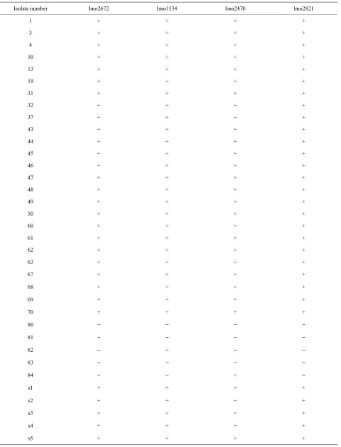
Results of PCR with oligonucleotide primers were designed from the assembled panel of four potential virulence genes (two of internalin gene and two of transcriptional regulator gene). Most of the isolates including reference strains were reactive by PCR, whereas the other strains (No.80, 81, and 83) isolated from pork, were non-reactive by PCR. In par-ticular, all pork isolates were PCR-negative for two primers (lmo2672 and 2821) sets tested. However, No.82 was posi-tive for lmo1134 primer, and No.84 was posiposi-tive for lmo2470 of pork isolates. It was observed that all Listeria mono-cytogenes (L. monocytogenes) penetrate Vero cells, although the invasion efficiency of each strain varied (between 0.5 and 18.9%). When compared in cell assay with PFGE, the results were shown that the mean invasion efficiency for lineage II isolate (2.6%) was significantly lower (ANOVA-test, p < 0.05) than that for lineage I (12.9%) and III isolates (10.3%).
Keywords: PFGE; Vero Cell; Listeriamonocytogenes; inVitro Assay
1. Introduction
Listeria monocytogenes (L. monocytogenes) are charac-terized as gram-positive and rod-shaped bacteria. Liste-riosis caused by L. monocytogenes is a life-threatening disease in fetuses, newborns, immunocompromised peo-ple and the elderly [1,2]. L. monocytogenes continues to be a major food safety concern, a recent report from the Centers for Disease Control and Prevention (CDC) esti-mates 1600 cases and 255 deaths per year in the United States [3]. The most common route of infection by L.
monocytogenes is via the gastrointestinal tract [4]. As
evidenced by several outbreaks of listeriosis caused by the ingestion of contaminated food materials [5]. Nu-merous reports have been published dealing with the prevalence of L. monocytogenes in food products [6].
Despite frequent exposure of the population to this bac-terium, the probability of contracting listeriosis is low, since the incidence of the disease ranges from 1.6 to 6 cases per million populations [7,8]. It has long been as-sumed that all L. monocytogenes strains are virulent. However, many strains in the environment may lack cer-tain virulence properties [9]. Within the L. monocyto-genes species there is great variability between the sero-types found in the environment or food. For example, 64% of the clinical isolates occurring in the UK (United Kingdom) are serotype 4b whereas 4% - 6% of this sero-type is found in the environment [10]. The virulence of L. monocytogenes strains also varies within the same sero-type [11].
Methods that have been developed for L.
monocyto-genes virulence assessment in vivo models frequently
used to determine L. monocytogenes virulence include
determination of 50% lethal dose in mice and recovery of the livers and spleens of immunocompetent or immuno-compromised mice following intravenous or oral inocu-lation [9,12]. However, mice and rats are not natural hosts for L. monocytogenes [13]. Futhermore, mouse and rat E-cadherin differ from human E-cadherin at an im-portant amino acid residue that renders cells resistant to InlA-mediated invasion [14]. Therefore, the use of labo-ratory animals is impractical for a particular study, or the best available animal models have significant limitations. Since evaluation of the role of strain differences in hu-man must be important meaning for the protection of listeriosis and presumption of public health measures, it is advisable to develop and easy, fast laboratory test that can reliable verify the virulence of a given strain. In Vitro virulence test using cell culture models that have been used to characterize L. monocytogenes virulence include the plaque-forming assay, cytotoxicity-based assays, and invasion and multiplication assay [15]. Also, in vitro assays that characterize virulence are often relatively fast and simple to perform.
No reliable molecular method for prediction of L.
monocytogenes virulence has been developed. But, PCR
detection of known L. monocytogenes genes, such as in-ternalin and transcriptional regulator have the potential to provide an alternative method for distinguishing virulent from avirulent isolates [16].
PFGE is a method with high discriminatory power and it has shown to be very accurate and reproducible for fine structure comparison and molecular typing of L. mono-cytogenes [17]. PFGE type and invasion into cell culture assay have considerable correlation [18].
In this study, we tested the virulence of L.
monocyto-genes strains found in Korea by using PCR primers that
were derived from four L. monocytogenes and to com-pare invasion and multiplication in the Vero cell to PFGE types on 35 strains from food, animal, and environmental isolates.
2. Materials and Methods
2.1. Bacterial StrainsA total of 35 L. monocytogenes isolates were used for experiment. 16 L. monocytogenes isolates were obtained from Dr. Byeong Yeal Jung (National Veterinary Re-search & Quarantine Service, Anyang). In particular, L.
monocytogenes obtained from different animal species
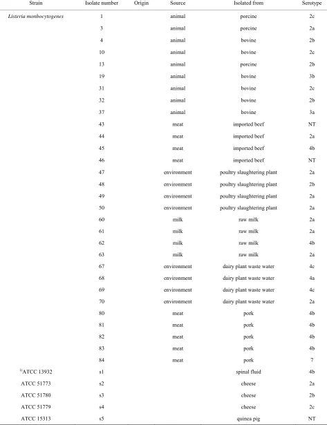
(porcine and bovine), environment (poultry slaughtering plant and dairy plant waste water) and meat (imported beef and pork) and from different foods (milk and cheese) from 1997 to 2007, were used. And five additional strains were purchased from the ATCC. The Listeria strains and their virulence, serotype and origin are in presented in
Table 1. Bacteria for the experiments were pre-cultivated
in brain heart infusion (BHI; Difco) agar or BHI broth at 37˚C for 18 h. Cell lines: Vero cells (African green mon- key kidney) were cultured in Eagle’s Minimum Essential Medium (MEM, Gibco BRL) supplemented with 5% fetal bovine serum (Intergen) at 37˚C in a 5% CO2
at-mosphere.
2.2. Cell Invasion Assay
To assess the abilities of the invasion and muliplication in cell, we used an intracellular growth assay described [15,19,20]. Briefly, cell monolayers of each cell line grown in 24-well tissue culture plates for 72 h were washed twice with Phospahte Buffered Salines (PBS; JBI, Korea) before inoculation of the bacteria. Simultaneously, after trypsin treatment, cells collected from a separate part of the culture plate were used for determination of the cell number using a haemocytometer. For invasion and intracellular growth assays, 18 h pre-cultivated bac-teria were inoculated into brain heart infusion (BHI) broth (Merk, USA) incubated at 37˚C for 13 h, washed twice with PBS, diluted with Minimum Essential Media Eagle (MEM), and then the appropriate bacterial count (5 × 10⁵ - 5 × 10⁷/ml) was inoculated onto the cell monolayer.
The inoculated bacterial number was obtained from the colony count (colony forming units, CFU) after incuba-tion on trypticase soy agar (TSA; Oxoid, Korea), con-taining 0.6% (w/v) yeast extract, for 48 h at 37˚C. Bacte-rial suspensions (0.3 ml per well) were added to the pre-pared cell monolayer, incubated at 37˚C for 2 h, washed twice with PBS and then incubated in 1 ml of MEM sup-plemented with 40 μg/ml of gentamicin at 37˚C for 2 h to kill extracellular bacteria. Monolayers were washed twice with PBS and cells lysed in 1 ml of distilled water con-taining 0.5% Triton X-100 on ice for 10 min. To measure invasion, the number of bacteria released from the cells was determined by plating serial dilutions onto TSA plates. To measure intracellular growth (Multiplication), MEM supplemented with 40 μg/ml of gentamicin was used as the growth medium after infection and infected cells were incubated fro 18 h after removal of extracellu-lar bacteria. The number of intracelluextracellu-lar bacteria was then enumerated at 18 h post-infection as described above and intracellular doubling times between 3.5 and 21.5 h post-infection were calculated. The experiment was re-peated two times.
2.3. PFGE
Table 1. List of the strains used in this study.
Strain Isolate number Origin Source Isolated from Serotype
Listeria monbocytogenes 1 animal porcine 2c
3 animal porcine 2a
4 animal bovine 2b
10 animal bovine 2c
13 animal porcine 2b
19 animal bovine 3b
31 animal bovine 2c
32 animal bovine 2b
37 animal bovine 3a
43 meat imported beef NT
44 meat imported beef 2a
45 meat imported beef 4b
46 meat imported beef NT
47 environment poultry slaughtering plant 2a
48 environment poultry slaughtering plant 2b
49 environment poultry slaughtering plant 2a
50 environment poultry slaughtering plant 2a
60 milk raw milk 2a
61 milk raw milk 2a
62 milk raw milk 4b
63 milk raw milk 2a
67 environment dairy plant waste water 4c
68 environment dairy plant waste water 4a
69 environment dairy plant waste water 4c
70 environment dairy plant waste water 2a
80 meat pork 4b
81 meat pork 4b
82 meat pork 4b
83 meat pork 4b
84 meat pork 7
1)ATCC 13932 s1 spinal fluid 4b
ATCC 51773 s2 cheese 2a
ATCC 51780 s3 cheese 2b
ATCC 51779 s4 cheese 2c
ATCC 15313 s5 quinea pig NT
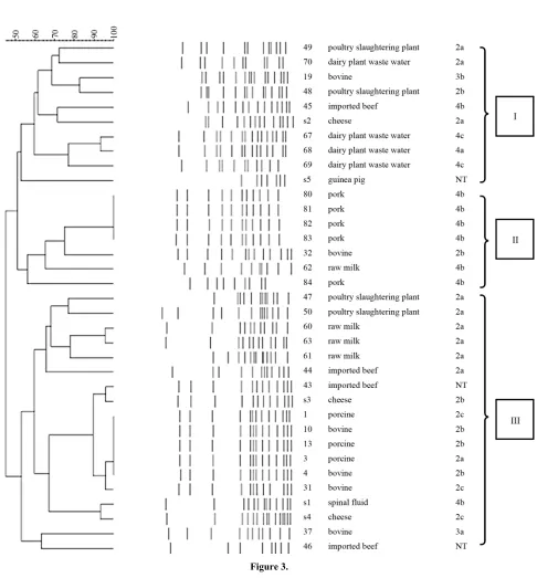
Germany) (SSP), lysed, washed, and digested with the restriction enzymes ApaI for 5 h at 3.0˚C. The DNA re-striction fragments in plugs were separated by electro-phoresis through 1% SeaKem Gold agarose gel in 0.5 × solution of Tris-borate-EDTA (prepared by diluting 10 × TBE) buffer (Bioneer, Korea) at 14˚C in a CHEF-Map- per PFGE apparatus (Bio-Rad, USA). The electropho-retic parameters used were as follows; initial switch time, 4.0 s; final switch time, 40.0 s. PFGE profiles were sepa-rated in to three main lineages showing about 50% ge-netic similarity.
2.4. PCR
Extraction, purification and quantification of DNA from each strain were performed as previously described. All of the primers used for specific PCR amplifications of virulent genes are reported in Table 2 [16]. PCR was performed in a PCR 9600 thermal cycler (Perkin-Elmer Corporation). A 50-ul aliquot contained buffer (10 mM Tris-HCl, 50 mM KCl, 2.5 mM MgCl2 [pH 8.3]), the
dNTP mixture (TaKaRa, Japan) 2.5 mM each, 10 pM primer, 25 ng of DNA, and 0.8 U of Taq DNA poly-merase (TaKaRa, Japan). The cycling conditions were the template DNA was denatured at 94˚C for 2 min fol-lowed by 25 cycles of amplification (each cycle con-sisted of denaturation at 94˚C for 20 s, annealing at 60˚C for 20 s and elongation at 72˚C for 45 s) and finally 72˚C for 2 min. 5 microlitre of the amplified products was separated by electrophoresis in 1.5% agarose gel con-taining ethidium bromide, and visualized under UV.
2.5. Statistical Analysis
Differences among results from PFGE lineage and cell culture assay were evaluated with one-way ANOVA test in SPSS ver10.0. Differences were considered significant at p < 0.05.
3. Results
Results of PCR with oligonucleotide primers were de-
signed from the assembled panel of four potential viru-lence genes (Table 3). Most of the isolates including reference strains were reactive by PCR, whereas the other strains (No.80, 81, and 83) isolated from pork, were non-reactive by PCR. In particular, all pork isolates were PCR-negative for two primers (lmo2672 and 2821) sets tested. However, No.82 was positive for lmo1134 primer, and No.84 was positive for lmo2740 of pork isolates.
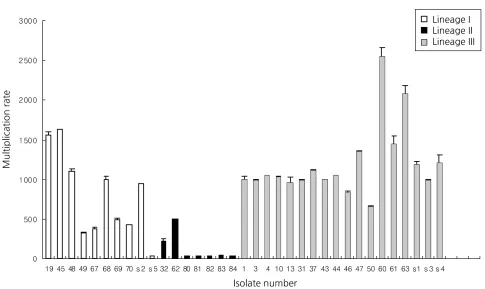
The ability of L. monocytogenes isolates to invade and multiply was evaluated by using and in vitro model with Vero cells (Figures 1 and 2). We previously studied that three types of PFGE pattern showed at 50% relative ge-netic similarity in L. monocytogenes isolates (Figure 3). As the number of deposited bacteria varied with the strain, a percentage of entry and an index of multiplica-tion were determined. Under these condimultiplica-tions, it was observed that all L. monocytogenes penetrate Vero cells, although the invasion efficiency of each strain varied (between 0.5% and 18.9%). Besides, these strains multi-plied broadly the number of bacteria was enhanced 22 to 2548 times 3.5 and 21 h of infection. Entry and intracel-lular multiplication proficiency varied greatly among L. monocytogenes isolates.
The mean invasion efficiency for lineage II isolate (2.6%) was significantly lower (ANOVA-test, p < 0.05) than that for lineage I (12.9%) and III isolates (10.3%). And the mean multiplication rate for lineage II isolate (124) was significantly lower (ANOVA-test, p < 0.05) than that for lineage I (801) and III isolates (1188).
4. Discussion
It has long been presumed that all L. monocytogenes strains are pathogens. However, much evidence to sug-gest that, under natural conditions, some L. monocyto-genes strains may lack virulence properties [22]. In this study, the virulence of various isolates of the L.
monocy-togenes was compared to that of the well-known
[image:4.595.56.539.598.736.2]refer-ence strains to know their capacity to invade and multi-plicate in Vero cell and using PCR of putative virulence gene. We use Vero cell to determine for virulence of L.
Table 2. List of L. monocytogenes and primers used in the PCR assay.
Gene Putative function Primer sequences (5’→ 3’) Size of PCR product (bp)
CGGCACACTTGGATTCTCAT lmo2672 Transcriptional regulator
AGGGCTAGTGACGGATGCTA 481
ACCCGATAGCAAGGAGGAAC lmo1134 Transcriptional regulator
AACTTCTCTCGATACCCATCCA 367
TGATTCCATGCAATTACTAGAACG lmo2470 Internalin
AGGATTCTAAACTAGGTAAGTTGGTG 545
TGTAACCCCGCTTACACAGTT lmo2821 Internalin
Table 3. PCR results using primers derived from four putative L. monocytogenes transcriptional regular and internalin genes.
Isolate number lmo2672 lmo1134 lmo2470 lmo2821
1 + + + +
3 + + + +
4 + + + +
10 + + + +
13 + + + +
19 + + + +
31 + + + +
32 + + + +
37 + + + +
43 + + + +
44 + + + +
45 + + + +
46 + + + +
47 + + + +
48 + + + +
49 + + + +
50 + + + +
60 + + + +
61 + + + +
62 + + + +
63 + + + +
67 + + + +
68 + + + +
69 + + + +
70 + + + +
80 − − − −
81 − − − −
82 − + − −
83 − − − −
84 − − + −
s1 + + + +
s2 + + + +
s3 + + + +
s4 + + + +
Figure 1. Distribution of invasion efficiency of selected lineage I, II, and III isolates in Vero cells.
Figure 2. Distribution of multiplication rate of selected lineage I, II and III isolates in Vero cells.
monocytogenes isolated from various source. Our results
indicated that most of isolates had a significantly higher
[image:6.595.56.541.394.683.2]Figure 3.
80, 81, 82, 83 and 84) in cell culture assay.
A number of studies conducted on food and clinical isolates using in vitro tests produced a pattern of results similar to those of the in vivo studies [23]. Another study demonstrated that two human feces carriage isolates of L.
monocytogenes were compared to reference strains with
regard to their invasiveness towards Caco-2 cells and strains virulent entered Caco-2 cells with great penetra-tion efficiency, whereas strains with attenuated virulence
pro-teins such as cytokines are disrupted [26]. However, be-cause of the high cost associated with knockout mice, these models were difficult to conduct.
More recently, test methods based on detection of spe-cific virulence genes may offer alternative approaches for identifying of L. monocytogenes virulence [15]. We se-lected two genes (lmo2672 and lmo1134) encoding tran-scriptional regulator gene which had the lowest nucleo-tide identity with any sequences from the L. innocua ge-nome (non-virulent strain) [16]. Results of PCR using lmo2672 were correlated with invasion assay in Vero cell but not using lmo1134. Especially, lmo2672 allow pork isolates to be identified negative results of PCR. PCR results also correlated with ATCC reference, which iso-lated from an outbreak of listeriosis. However, our result showed that ATCC 15313, which was avirulent isolate, was positive for lmo2672 and lmo1134. That was con-sistent with D. Liu etal. [16]. D. Liu etal., characterize to virulent and avirulent L. monocytogenes strains by PCR amplification of four putative transcriptional regu-lator genes. And it demonstrated that ATCC 15313 has not maintained its ability to cause cytopathogenic effects in Caco-2 monolayer but positive for all tested primers. ATCC 15313 was originally isolated from an infected rabbit during an outbreak of listeriosis, but it later be-came avirulent after successive laboratory subculturing. It has been demonstrated that ATCC 15313 does not ex-press listeriolysin, a well-known virulence factor this oc-curred spontaneously after its original isolation [27]. So, it was assumed that ATCC 15313, which is derived from a virulent L. monocytogenes isolates, would retain many of its virulence properties [16].
We also selected two genes (lmo2470 and lmo2821) encoding putative internalin genes. Results of PCR using lmo2821 were positive for most isolates but not for pork samples. Lmo2470 gene was detected in most L. mono-cytogenes but not pork isolates (No.80, 81, 82, and 83). These results were correlated with invasion assay in Vero cell but not using lmo2470. PCR results also correlated with ATCC reference, which isolated from an outbreak of listeriosis. However, our result showed that ATCC 15313, which was avirulent isolate, was positive for lmo2470 and lmo2821. The presence of virulence genes
in L. monocytogenes isolates appears to be so common
that such tests are relatively poor discriminators of rela- tive disease-causing potential [15].
We also compare levels of virulence of three different PFGE lineages of L. monocytogenes isolates using cell culture assay. PFGE lineage II expressed a significantly higher level of invasion rate in cell culture assays than the others (p < 0.05). The same results showed in multi-plication assay. These noted variations in invasive and multiplication assay might account for why certain PFGE types were more virulent than others. In other previous
studies [18], PFGE type and invasion into cell culture assay have considerable correlation. Thus, combining as-say using PFGE and invasion asas-say in L. monocytogenes would seem to be of crucial value for the interpretation of the virulence.
The four primer sets and Invasion assay using Vero cell were evaluated by PCR and cell culture system against Listeria isolated various source. Most of the iso- lates were positive for at least one PCR primer. However, some pork isolates (No.80, 81, and 83) were not positive any of the primer. Therefore, most of isolates are consid-ered to be more invasive than some pork isolates and could be potential food-borne pathogens. It was insisted that a single molecular, cell culture or animal assay can-not detect all virulence-attenuated isolates. Rather, it is a combination of cell culture and molecular screening as-says that would offer the best chance of most accurately and reliably identifying naturally virulence-attenuated L. monocytogenes isolates [28].
Therefore, combining studies of differences in the ex- pression of virulence genes with the present study could give more information on the complexity of virulence of
L. monocytogenes. Invasion assay using Vero cells and
PCR-based assay for putative virulence genes could be used as an alternative to test for determent the patho- genicity of Listeria and reduce expensive and time-con- suming animal testing.
REFERENCES
[1] A. Schuchat, B. Swaminathan and C. V. Broome, “Epi- demiology of Human Listeriosis,” ClinicalMicrobiology Reviews, Vol. 99, No. 4, 1991, pp. 169-183.
[2] H. Hof, “History and Epidemiology of Listeriosis,” FEMS Immunology andMedicalMicrobiology, Vol. 35, No. 3,
2003, pp. 199-202. doi:10.1016/S0928-8244(02)00471-6 [3] A. R. Datta, P. Laksanalamai and M. Solomotis, “Recent
Developments in Molecular Sub-Typing of Listeria mo- nocytogenes,” FoodAdditivesandContaminantsPartA,
2012, pp. 1-9.
[4] W. F. Schlech, “New Perspectives on the Gastrointestinal Mode of Transmission in Invasive Listeria monocyto- genes Infection,” Clinical and Investigative Medicine,
Vol. 7, No. 4, 1984, pp. 321-324.
[5] W. F. Schlech, P. M. Lavigne, R. A. Bortolussi, A. C. Allen, E. V. Haldane, A. J. Wort, A. W. Hightower, S. E. Johnson, S. H. King, E. S. Nicholls and C. V. Broome, “Epidemic Listeriosis-Evidence for Transmission by Food,”
NewEnglandJournalofMedicine, Vol. 308, No. 4, 1983,
pp. 203-206. doi:10.1056/NEJM198301273080407 [6] B. Swaminathan and P. Gerner-Smidt, “The Epidemio-
logy of Human Listeriosis,” MicrobesandInfection, Vol.
9, No. 10, 2007, pp. 1236-1243. doi:10.1016/j.micinf.2007.05.011
nocytogenes,” JournalofFoodProtection, Vol. 61, No. 2,
1998, pp. 244-248.
[8] J. Rocourt, C. Jacquet and A. Reilly, “Epidemiology of Human Listeriosis and Seafoods,” International Journal ofFoodMicrobiology, Vol. 62, No. 3, 2000, pp. 197-209.
doi:10.1016/S0168-1605(00)00336-6
[9] H. Hof, “Virulence of Different Strains of Listeria mono-cytogenes Serovar 1/2a,” MedicalMicrobiologyand Im-munology, Vol. 173, No. 4, 1984, pp. 207-218.
doi:10.1007/BF02122112
[10] K. G. Kerr, P. Kite, J. Heritage and P. M. Hawkey, “Typ-ing of Epidemiologically Associated Environmental and Clinical Strains of Listeria monocytogenes by Random
Amplification of Polymorphic DNA,” Journal of food protection, Vol. 58, No. 6, 1995, pp. 609-613.
[11] H. Barbour, A. Rampling and C. E. Hormaeche, “Com-parison of the Infectivity of Isolates of Listeria monocy-togenes Following Intragastric and Intravenous Inocula-tion in Mice,” Microbial pathogenesis, Vol. 20, No. 4,
1996, pp. 247-253. doi:10.1006/mpat.1996.0023
[12] H. Barbour, A. Rampling and C. E. Hormaeche, “Varia-tion in the Infectivity of Listeria monocytogenes Isolates
Following Intragastric Inoculation of Mice,” Infection andImmunity, Vol. 69, No. 7, 2001, pp. 4657-4660.
doi:10.1128/IAI.69.7.4657-4660.2001
[13] M. Pentecost, G. Otto, J. A. Theriot and M. R. Amieva, “Listeria monocytogenes Invades the Epithelial Junctions
at Sites of Cell Extrusion,” PLOSPathogens, Vol. 2, No. 1, 2006, p. e3. doi:10.1371/journal.ppat.0020003
[14] M. Lecuit, S. Dramsi, C. Gottardi, M. Fedor-Chaiken, B. Gumbiner and P. Cossart, “A Single Amino Acid in E- Cadherin Responsible for Host Specificity towards the Human Pathogen Listeria monocytogenes,” The EMBO Journal, Vol. 18, No. 4, 1999, pp. 3956-3963.
doi:10.1093/emboj/18.14.3956
[15] R. B. Raybourne, “Virulence Testing of Listeria monocy-togenes,” JournalofAOACInternational, Vol. 85, No. 2,
2002, pp. 516-523.
[16] D. Liu, A. J. Ainsworth, F. W. Austin and M. L. Law-rence, “Characterization of Virulent and Avirulent Lis-teria monocytogenes Strains by PCR Amplification of
Putative Transcriptional Regulator and Internalin Genes,”
JournalofMedicalMicrobiology, Vol. 52, No. 12, 2003,
pp. 1065-1070. doi:10.1099/jmm.0.05358-0
[17] J. Bille and J. Rocourt, “WHO International Multicenter
Listeria monocytogenes Subtyping Study-Rationale and
Set-Up of the Study,” InternationalJournalofFood Mi-crobiology, Vol. 32, No. 3, 1996, pp. 251-262.
doi:10.1016/S0168-1605(96)01140-3
[18] C. N. Larsen, B. Norrung, H. M. Sommer and M. Ja-kobsen, “In Vitro and in Vivo Invasiveness of Different
Pulsed-Field Gel Electrophoresis Types of Listeria mono- cytogenes,” Applied and Environmental Microbiology,
Vol. 68, No. 11, 2002, pp. 5698-5703. doi:10.1128/AEM.68.11.5698-5703.2002
[19] Z. Xiaohui, J. Xinan and M. Wiedmann, “Listeria mono-cytogenes in the Chinese Food System: Strain Charac-terization through Partial actA Sequencing and Tissue- Culture Pathogenicity Assays,” Journal ofMedical Mi-crobiology, Vol. 54, No. 3, 2005, pp. 217-224.
doi:10.1099/jmm.0.45882-0
[20] F. Yamada, F. Ueda, Y. Ochiai, M. Mochizuki, H. Shoji, K. Ogawa-Goto, T. Sata, K. Ogasawara, A. Fujima and R. Hondo, “Invasion Assay of Listeria monocytogenes Using Vero and Caco-2 Cells,” Journal of Microbiological Methods, Vol. 66, No. 1, 2006, pp. 96-103.
doi:10.1016/j.mimet.2005.10.017
[21] L. M. Graves and B. Swaminathan, “PulseNet Standard-ized Protocol for Subtyping Listeria monocytogenes by Macrorestriction and Pulsed-Field Gel Electrophoresis,”
InternationalJournalofFoodMicrobiology, Vol. 65, No. 1-2, 2001, pp. 55-62.
doi:10.1016/S0168-1605(00)00501-8
[22] L. N. Van, E. Bottreau, S. Bailly, M. Tabouret, J. Marly, P. Pardon and P. Velge, “Tissue Culture Assays Using Caco-2 Cell Line Differentiate Virulent from Non- Viru-lent Listeria monocytogenes Strains,” JournalofApplied Microbiology, Vol. 85, No. 2, 1998, pp. 337-346. doi:10.1046/j.1365-2672.1998.00515.x
[23] A. Gattuso, M. Gianfranceschi, R. Sessa, F. Taggi, M. Pourshaban and P. Aureli, “In Vivo and in Vitro
Assess-ment of the Virulence of Listeria monocytogenes Strains,” NewMicrobiologica, Vol. 23, No. 3, 2000, pp. 289-295.
[24] M. Olier, F. Pierre, J. P. Lemaitre, C. Divies, A, Rousset and J. Guzzo, “Assessment of the Pathogenic Potential of Two Listeria monocytogenes Human Faecal Carriage Iso- lates,” Microbiology, Vol. 148, No. 6, 2002, pp. 1855-
1862.
[25] A. Schuchat, K. A. Deaver, J. D. Wenger, B. D. Plikaytis, L. Mascola, R. W. Pinner, A. L. Reingold and C. V. Bro- ome, “Role of Foods in Sporadic Listeriosis. I. Case- Control Study of Dietary Risk Factors. The Listeria Study Group,” ThejournaloftheAmericanMedicalAssociation,
Vol. 267, No. 15, 1992, pp. 2041-2045. doi:10.1001/jama.1992.03480150047035
[26] R. U. Emil, “Why Listeriosis? A Perspective on Cellular Immunity to Infection,” Immunological Reviews, Vol.
158, No. 1, 1997, pp. 5-9.
doi:10.1111/j.1600-065X.1997.tb00987.x
[27] S. Kathariou and L. Pine, “The Type Strain(s) of Listeria monocytogenes: A Source of Continuing Difficulties,” In- ternational Journal ofSystematicand Evolutionary Mi-crobiology, Vol. 41, No. 2, 1991, pp. 328-330.
[28] A, Roberts, Y. Chan and M. Wiedmann, “Definition of Genetically Distinct Attenuation Mechanisms in Naturally Virulence-Attenuated Listeria monocytogenes by
Com-parative Cell Culture and Molecular Characterization,”
AppliedandEnvironmentalMicrobiology, Vol. 71, No. 7,
2005, pp. 3900-3910.