Bis(triethylammonium) chloranilate
Kazuma Gotoh, Shinpei Maruyama and Hiroyuki Ishida*
Department of Chemistry, Faculty of Science, Okayama University, Okayama 700-8530, Japan
Correspondence e-mail: ishidah@cc.okayama-u.ac.jp
Received 19 July 2013; accepted 1 August 2013
Key indicators: single-crystal X-ray study;T= 180 K; mean(C–C) = 0.002 A˚; Rfactor = 0.044;wRfactor = 0.121; data-to-parameter ratio = 24.1.
In the crystal structure of the title compound [systematic name: bis(triethylammonium) 2,5-dichloro-3,6-dioxocyclo-hexa-1,4-diene-1,4-diolate], 2C6H16N
+
C6Cl2O4 2
, the chlora-nilate anion lies on an inversion center. The triethylammonium cations are linked on both sides of the anion via bifurcated N—H (O,O) and weak C—H O hydrogen bonds to give a centrosymmetric 2:1 aggregate. The 2:1 aggregates are further linked by C—H O hydrogen bonds into a zigzag chain running along [011].
Related literature
For related structures, see: Dayanandaet al.(2012); Gotohet al.(2009, 2010); Yang (2007).
Experimental
Crystal data
2C6H16N+C6Cl2O42
Mr= 411.37 Triclinic,P1 a= 7.7347 (5) A˚ b= 8.5151 (8) A˚ c= 9.3913 (7) A˚
= 64.388 (4) = 68.435 (3)
= 79.060 (5)
V= 518.36 (7) A˚3 Z= 1
MoKradiation
= 0.34 mm1 T= 180 K
0.650.310.21 mm
Data collection
Rigaku R-AXIS RAPID II diffractometer
Absorption correction: numerical (NUMABS; Higashi, 1999) Tmin= 0.858,Tmax= 0.932
10264 measured reflections 3012 independent reflections 2561 reflections withI> 2(I) Rint= 0.078
Refinement
R[F2> 2(F2)] = 0.044 wR(F2) = 0.121 S= 1.06 3012 reflections 125 parameters
H atoms treated by a mixture of independent and constrained refinement
max= 0.48 e A˚
3
min=0.26 e A˚
3
Table 1
Hydrogen-bond geometry (A˚ ,).
D—H A D—H H A D A D—H A
N1—H1 O1 0.867 (18) 1.942 (18) 2.7601 (15) 156.9 (15) N1—H1 O2i 0.867 (18) 2.339 (17) 2.9377 (14) 126.4 (14)
C5—H5C O1 0.98 2.57 3.2911 (19) 131
C9—H9B O1ii
0.98 2.55 3.5315 (17) 177
Symmetry codes: (i)xþ1;yþ2;z; (ii)xþ1;yþ1;zþ1.
Data collection: PROCESS-AUTO (Rigaku/MSC, 2004); cell refinement: PROCESS-AUTO; data reduction: CrystalStructure
(Rigaku/MSC, 2004); program(s) used to solve structure:SHELXS97
(Sheldrick, 2008); program(s) used to refine structure:SHELXL97
(Sheldrick, 2008); molecular graphics: ORTEP-3 for Windows
(Farrugia, 2012); software used to prepare material for publication:
CrystalStructure(Rigaku/MSC, 2004) andPLATON(Spek, 2009).
This work was supported by a Grant-in-Aid for Scientific Research (C) (No. 22550013) from the Japan Society for the Promotion of Science.
Supplementary data and figures for this paper are available from the IUCr electronic archives (Reference: LH5635).
References
Dayananda, A. S., Butcher, R. J., Akkurt, M., Yathirajan, H. S. & Narayana, B. (2012).Acta Cryst.E68, o1037–o1038.
Farrugia, L. J. (2012).J. Appl. Cryst.45, 849–854.
Gotoh, K., Maruyama, S. & Ishida, H. (2010).Acta Cryst.E66, o3255. Gotoh, K., Nagoshi, H. & Ishida, H. (2009).Acta Cryst.C65, o273–o277. Higashi, T. (1999).NUMABS. Rigaku Corporation, Tokyo, Japan.
Rigaku/MSC (2004).PROCESS-AUTOandCrystalStructure. Rigaku/MSC Inc., The Woodlands, Texas, USA.
Sheldrick, G. M. (2008).Acta Cryst.A64, 112–122. Spek, A. L. (2009).Acta Cryst.D65, 148–155. Yang, D.-J. (2007).Acta Cryst.E63, o2600.
Acta Crystallographica Section E
Structure Reports Online
supporting information
Acta Cryst. (2013). E69, o1400 [doi:10.1107/S1600536813021405]
Bis(triethylammonium) chloranilate
Kazuma Gotoh, Shinpei Maruyama and Hiroyuki Ishida
S1. Comment
The title compound was prepared in order to extend our study on D—H···A hydrogen bonding (D = N, O, or C; A = N, O
or Cl) in amine–chloranilic acid systems (Gotoh et al., 2009, 2010). For the tertiary amine–chloranilic acid systems,
crystal structures of bis(hexamethylenetetraminium) chloranilate tetrahydrate (Yang, 2007), triethylammonium hydrogen
chloranilate (Gotoh et al., 2010) and triprolidinium dichloranilate–chloranilic acid–methanol–water (2/1/2/2) (Dayananda
et al., 2012) have been reported.
In the crystal structure of the title compound, an acid-base interaction involving proton transfer is observed between
chloranilic acid and triethylamine, and one chloranilate anion and two triethylammnoium cations are linked by bifurcated
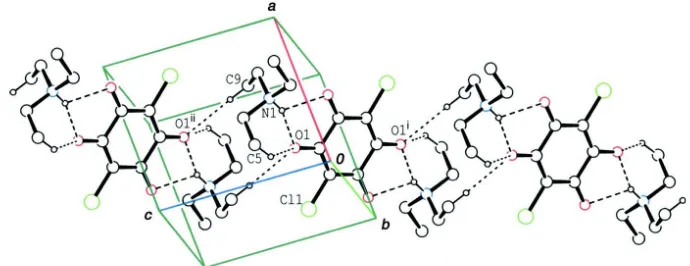
N—H···O and weak C—H···O hydrogen bonds (Table 1) to afford a centrosymmetric 2:1 aggregate (Fig. 1). The 2:1
aggregates are further linked by intermolecular C—H···O hydrogen bonds, forming a zigzag chain running along the
[011] direction (Fig. 2).
S2. Experimental
Single crystals were obtained by slow evaporation from an acetonitrile solution (100 ml) of chloranilic acid (100 mg) and
triethylamine (97 mg) at room temperature.
S3. Refinement
C-bound H atoms were positioned geometrically (C—H = 0.98 or 0.99 Å) and refined as riding, allowing for free rotation
of the methyl group. Uiso(H) values were set at 1.2Ueq(C) or 1.5Ueq(methyl C). The N-bound H atom was found in a
Figure 1
The molecular structure of the title compound, with the atom-labeling. Displacement ellipsoids of non-H atoms are drawn
at the 30% probability level. The dashed lines indicate N—H···O and C—H···O hydrogen bonds. [Symmetry code: (i) -x +
1, -y + 2, -z.]
Figure 2
A partial packing diagram of the title compound. The dashed lines indicate N—H···O and C—H···O hydrogen bonds. H
atoms of the ethyl groups not involved in the C—H···O hydrogen bonds have been omitted. [Symmetry codes: (i) -x + 1,
-y + 2, -z; (ii) -x + 1, -y + 1, -z.]
Bis(triethylammonium) 2,5-dichloro-3,6-dioxocyclohexa-1,4-diene-1,4-diolate
Crystal data
2C6H16N+·C6Cl2O42−
Mr = 411.37 Triclinic, P1 Hall symbol: -P 1 a = 7.7347 (5) Å b = 8.5151 (8) Å c = 9.3913 (7) Å α = 64.388 (4)° β = 68.435 (3)° γ = 79.060 (5)° V = 518.36 (7) Å3
Z = 1
F(000) = 220.00 Dx = 1.318 Mg m−3
Mo Kα radiation, λ = 0.71075 Å Cell parameters from 8559 reflections θ = 3.1–30.1°
µ = 0.34 mm−1
T = 180 K Block, brown
[image:3.610.131.476.301.434.2]Data collection
Rigaku R-AXIS RAPID II diffractometer
Detector resolution: 10.00 pixels mm-1
ω scans
Absorption correction: numerical (NUMABS; Higashi, 1999) Tmin = 0.858, Tmax = 0.932 10264 measured reflections
3012 independent reflections 2561 reflections with I > 2σ(I) Rint = 0.078
θmax = 30.0°, θmin = 3.1°
h = −10→10 k = −11→11 l = −13→13
Refinement
Refinement on F2 Least-squares matrix: full R[F2 > 2σ(F2)] = 0.044
wR(F2) = 0.121
S = 1.06 3012 reflections 125 parameters 0 restraints
Primary atom site location: structure-invariant direct methods
Secondary atom site location: difference Fourier map
Hydrogen site location: inferred from neighbouring sites
H atoms treated by a mixture of independent and constrained refinement
w = 1/[σ2(F
o2) + (0.0654P)2] where P = (Fo2 + 2Fc2)/3 (Δ/σ)max = 0.001
Δρmax = 0.48 e Å−3 Δρmin = −0.26 e Å−3
Special details
Geometry. All e.s.d.'s (except the e.s.d. in the dihedral angle between two l.s. planes) are estimated using the full covariance matrix. The cell e.s.d.'s are taken into account individually in the estimation of e.s.d.'s in distances, angles and torsion angles; correlations between e.s.d.'s in cell parameters are only used when they are defined by crystal symmetry. An approximate (isotropic) treatment of cell e.s.d.'s is used for estimating e.s.d.'s involving l.s. planes.
Refinement. Refinement of F2 against ALL reflections. The weighted R-factor wR and goodness of fit S are based on F2, conventional R-factors R are based on F, with F set to zero for negative F2. The threshold expression of F2 > σ(F2) is used only for calculating R-factors(gt) etc. and is not relevant to the choice of reflections for refinement. R-factors based on F2 are statistically about twice as large as those based on F, and R- factors based on ALL data will be even larger.
Fractional atomic coordinates and isotropic or equivalent isotropic displacement parameters (Å2)
x y z Uiso*/Ueq
Cl1 0.14870 (4) 0.93266 (4) 0.32667 (3) 0.03021 (13)
O1 0.44257 (12) 0.67475 (10) 0.24001 (11) 0.0322 (2)
O2 0.26077 (12) 1.26690 (10) 0.03171 (10) 0.0326 (2)
N1 0.67048 (13) 0.38363 (13) 0.23709 (12) 0.0242 (2)
C1 0.46352 (15) 0.82853 (14) 0.13459 (13) 0.0237 (2)
C2 0.34397 (15) 0.96966 (14) 0.14976 (13) 0.0235 (2)
C3 0.36699 (15) 1.13964 (14) 0.02453 (14) 0.0235 (2)
C4 0.54491 (18) 0.26759 (16) 0.40054 (15) 0.0313 (3)
H4A 0.5321 0.3103 0.4868 0.038*
H4B 0.6024 0.1485 0.4333 0.038*
C5 0.35317 (19) 0.25958 (19) 0.39536 (18) 0.0396 (3)
H5A 0.2712 0.1966 0.5086 0.059*
H5B 0.3625 0.1988 0.3247 0.059*
H5C 0.3018 0.3782 0.3494 0.059*
C6 0.69491 (17) 0.33514 (16) 0.09491 (15) 0.0297 (3)
H6B 0.5708 0.3319 0.0884 0.036*
C7 0.7960 (2) 0.1614 (2) 0.1098 (2) 0.0435 (4)
H7A 0.8200 0.1442 0.0075 0.065*
H7B 0.7191 0.0679 0.2051 0.065*
H7C 0.9143 0.1595 0.1265 0.065*
C8 0.85542 (16) 0.39642 (17) 0.24970 (15) 0.0295 (3)
H8A 0.9018 0.2780 0.3091 0.035*
H8B 0.9459 0.4451 0.1360 0.035*
C9 0.84408 (19) 0.50912 (17) 0.34006 (17) 0.0342 (3)
H9A 0.9693 0.5226 0.3355 0.051*
H9B 0.7662 0.4542 0.4567 0.051*
H9C 0.7894 0.6239 0.2868 0.051*
H1 0.618 (2) 0.487 (2) 0.209 (2) 0.034 (4)*
Atomic displacement parameters (Å2)
U11 U22 U33 U12 U13 U23
Cl1 0.02658 (19) 0.03015 (19) 0.02264 (18) 0.00392 (12) −0.00412 (13) −0.00561 (13)
O1 0.0340 (5) 0.0216 (4) 0.0265 (4) 0.0031 (3) −0.0046 (4) −0.0025 (3)
O2 0.0326 (5) 0.0247 (4) 0.0290 (5) 0.0080 (3) −0.0055 (4) −0.0078 (4)
N1 0.0252 (5) 0.0216 (5) 0.0220 (4) 0.0037 (3) −0.0086 (4) −0.0062 (4)
C1 0.0254 (5) 0.0227 (5) 0.0216 (5) 0.0019 (4) −0.0092 (4) −0.0072 (4)
C2 0.0235 (5) 0.0233 (5) 0.0196 (5) 0.0023 (4) −0.0067 (4) −0.0063 (4)
C3 0.0246 (5) 0.0219 (5) 0.0230 (5) 0.0030 (4) −0.0094 (4) −0.0082 (4)
C4 0.0327 (6) 0.0267 (6) 0.0254 (6) 0.0000 (4) −0.0062 (5) −0.0054 (5)
C5 0.0352 (7) 0.0392 (7) 0.0414 (7) −0.0081 (5) −0.0058 (6) −0.0159 (6)
C6 0.0326 (6) 0.0317 (6) 0.0267 (6) 0.0028 (5) −0.0139 (5) −0.0114 (5)
C7 0.0479 (8) 0.0454 (8) 0.0561 (9) 0.0166 (6) −0.0296 (7) −0.0339 (7)
C8 0.0262 (6) 0.0355 (6) 0.0272 (6) 0.0026 (4) −0.0106 (5) −0.0128 (5)
C9 0.0355 (7) 0.0370 (7) 0.0330 (6) −0.0009 (5) −0.0120 (5) −0.0159 (6)
Geometric parameters (Å, º)
Cl1—C2 1.7390 (11) C5—H5B 0.9800
O1—C1 1.2502 (13) C5—H5C 0.9800
O2—C3 1.2403 (13) C6—C7 1.5103 (18)
N1—C4 1.4943 (15) C6—H6A 0.9900
N1—C6 1.5002 (15) C6—H6B 0.9900
N1—C8 1.5056 (15) C7—H7A 0.9800
N1—H1 0.867 (18) C7—H7B 0.9800
C1—C2 1.3960 (15) C7—H7C 0.9800
C1—C3i 1.5394 (16) C8—C9 1.5054 (17)
C2—C3 1.4079 (15) C8—H8A 0.9900
C3—C1i 1.5394 (16) C8—H8B 0.9900
C4—C5 1.517 (2) C9—H9A 0.9800
C4—H4A 0.9900 C9—H9B 0.9800
C4—H4B 0.9900 C9—H9C 0.9800
C4—N1—C6 113.81 (10) H5B—C5—H5C 109.5
C4—N1—C8 111.29 (9) N1—C6—C7 113.74 (10)
C6—N1—C8 111.25 (9) N1—C6—H6A 108.8
C4—N1—H1 108.1 (10) C7—C6—H6A 108.8
C6—N1—H1 103.7 (10) N1—C6—H6B 108.8
C8—N1—H1 108.2 (10) C7—C6—H6B 108.8
O1—C1—C2 125.01 (10) H6A—C6—H6B 107.7
O1—C1—C3i 116.31 (9) C6—C7—H7A 109.5
C2—C1—C3i 118.67 (9) C6—C7—H7B 109.5
C1—C2—C3 123.37 (10) H7A—C7—H7B 109.5
C1—C2—Cl1 118.69 (8) C6—C7—H7C 109.5
C3—C2—Cl1 117.83 (8) H7A—C7—H7C 109.5
O2—C3—C2 125.12 (10) H7B—C7—H7C 109.5
O2—C3—C1i 116.95 (9) C9—C8—N1 112.57 (10)
C2—C3—C1i 117.93 (9) C9—C8—H8A 109.1
N1—C4—C5 112.84 (11) N1—C8—H8A 109.1
N1—C4—H4A 109.0 C9—C8—H8B 109.1
C5—C4—H4A 109.0 N1—C8—H8B 109.1
N1—C4—H4B 109.0 H8A—C8—H8B 107.8
C5—C4—H4B 109.0 C8—C9—H9A 109.5
H4A—C4—H4B 107.8 C8—C9—H9B 109.5
C4—C5—H5A 109.5 H9A—C9—H9B 109.5
C4—C5—H5B 109.5 C8—C9—H9C 109.5
H5A—C5—H5B 109.5 H9A—C9—H9C 109.5
C4—C5—H5C 109.5 H9B—C9—H9C 109.5
H5A—C5—H5C 109.5
O1—C1—C2—C3 −176.68 (11) Cl1—C2—C3—C1i −178.12 (8)
C3i—C1—C2—C3 1.97 (19) C6—N1—C4—C5 −56.62 (13)
O1—C1—C2—Cl1 −0.54 (17) C8—N1—C4—C5 176.71 (10)
C3i—C1—C2—Cl1 178.11 (8) C4—N1—C6—C7 −66.29 (14)
C1—C2—C3—O2 177.84 (11) C8—N1—C6—C7 60.39 (14)
Cl1—C2—C3—O2 1.67 (17) C4—N1—C8—C9 −74.58 (13)
C1—C2—C3—C1i −1.95 (18) C6—N1—C8—C9 157.36 (10)
Symmetry code: (i) −x+1, −y+2, −z.
Hydrogen-bond geometry (Å, º)
D—H···A D—H H···A D···A D—H···A
N1—H1···O1 0.867 (18) 1.942 (18) 2.7601 (15) 156.9 (15)
N1—H1···O2i 0.867 (18) 2.339 (17) 2.9377 (14) 126.4 (14)
C5—H5C···O1 0.98 2.57 3.2911 (19) 131
C9—H9B···O1ii 0.98 2.55 3.5315 (17) 177