Received 13 May 2019 Accepted 14 May 2019
Edited by M. Zeller, Purdue University, USA
Keywords:heterocyclic compounds; reduced quinazolinones; crystal structure; molecular conformation; hydrogen bonding; supra-molecular assembly.
CCDC reference:1875494
Supporting information:this article has supporting information at journals.iucr.org/e
The crystal structure of (
RS
)-7-chloro-2-(2,5-di-methoxyphenyl)-2,3-dihydroquinazolin-4(1
H
)-one:
two hydrogen bonds generate an elegant
three-dimensional framework structure
Kereyagalahally H. Narasimhamurthy,a Chandra,bBelakavadi K. Sagar,c Kanchugarakoppal S. Rangappa,cHemmige S. Yathirajanc* and Christopher Glidewelld
aDepartment of Studies in Organic Chemistry, University of Mysore, Manasagangotri, Mysuru-570 006, India, bDepartment of Physics, National Institute of Engineering, Mysore-570 008, India,cDepartment of Studies in Chemistry, University of Mysore, Manasagangotri, Mysuru-570 006, India, anddSchool of Chemistry, University of St Andrews, St Andrews, Fife KY16 9ST, UK. *Correspondence e-mail: [email protected]
In the title compound, C61H15ClN2O3, the heterocyclic ring adopts an envelope
conformation, folded across the N N line, with the 2,5-dimethoxyphenyl unit occupying a quasi-axial site. There are two N—H O hydrogen bonds in the structure: one hydrogen bond links molecules related by a 41screw axis to form
aC(6) chain, and the other links inversion-related pairs of molecules to form an R2
2
(8) ring. The ring motif links all of the chains into a continuous three-dimensional framework structure. Comparisons are made with the structures of some related compounds.
1. Chemical context
Quinazoline-4-one and its derivatives constitute an important class of fused heterocycles, which are found in more than two hundred naturally occurring alkaloids. In addition, 2,3-di-hydroquinazolin-4(1H)-one is a privileged scaffold in drug design (Badolatoet al., 2018). Despite this, rather few struc-tures have been published for compounds containing this heterocyclic nucleus (see Section 4 below), and with these considerations in mind, we now report the molecular and supramolecular structure of (RS )-7-chloro-2-(2,5-dimethoxy-phenyl)-2,3-dihydroquinazolin-4(1H)-one (I) (Fig. 1). The compound was prepared using a recently published (Nara-simhamurthy et al., 2014) one-step process, which employs a base-promoted cyclization reaction between a (dibromo-methyl)arene, here 2-(dibromomethyl)-1,4-dimethoxybenz-ene, and a 2-aminobenzamide, here 2-amino-4-chlorobenzamide, which after a straightforward purification step gives the product (I) in 79% yield.
2. Structural commentary
The molecule of compound (I) contains a stereogenic centre at atom C2, and the reference molecule was selected as one having theRconfiguration at this atom: the centrosymmetric space group confirms that compound (I) has crystallized as a racemic mixture. The heterocyclic ring in compound (I) adopts a conformation close to the envelope form, in which this ring is folded across the line N1 N3 (Fig. 1). The ring-puckering parameters, calculated for the atom sequence (N1,C2,N3,C4,C4A,C8A) in the R-enantiomer are Q = 0.258 (2) A˚ , = 121.8 (4) and ’ = 219.3 (6). For the ideal envelope form, the puckering angles take the values= 54.7 (equivalent to 125.3) and’= (60k), wherekrepresents an integer (Boeyens, 1978). The r.m.s. deviation of the atoms N1, N3, C4, C4A, C8Afrom their mean plane is only 0.035 A˚ , with a maximum deviation of 0.0403 (11) A˚ for atom N3. However, atom C2 is displaced from this plane by 0.355 (3) A˚ . The 2,5-dimethoxyphenyl substituent occupies the quasi-axial site at atom C2. Within this unit, the two methoxy C atoms are almost coplanar with the aryl ring: the deviations from the mean plane of this ring are 0.020 (5) A˚ for atom C221 and 0.101 (5) A˚ for atom C251. Associated with this planarity, the two exocyclic C—C—O angles at atoms C22 and C25 are significantly different, by 11.9 at C22 and by 8.2 at atoms C25, as previously observed in planar or near-planar alkoxy-arenes (Seip & Seip, 1973; Fergusonet al., 1996).
3. Supramolecular features
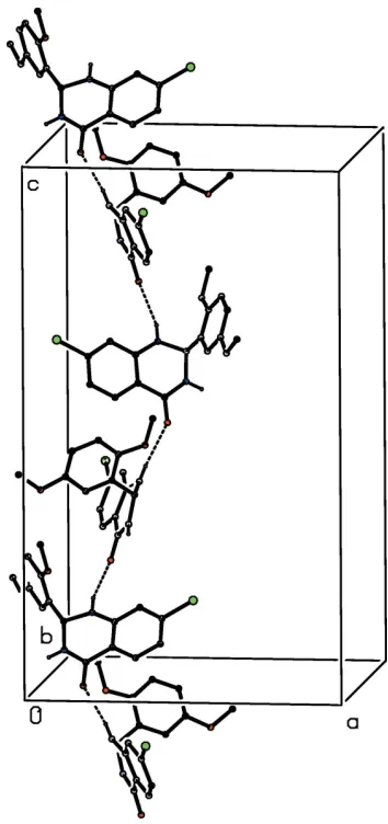
The structure of compound (I) contains just two N—H O hydrogen bonds (Table 1) but these are sufficient to link all of the molecules into a three-dimensional framework structure, whose formation is readily analysed in terms of the actions of the two individual hydrogen bonds. The hydrogen bond having atom N1 as the donor links molecules related by the 41
screw axis along (0.25, 0.5, z) into aC(6) chain (Etter, 1990;
Etteret al., 1990; Bernsteinet al., 1995) running parallel to the [001] direction (Fig. 2). Four chains of this type pass through each unit cell. The hydrogen bond having atom N3 as the donor links inversion-related pairs of molecules to form a cyclic dimer characterized by an R2
2(8) motif (Fig. 3). This
interaction directly links theC(6) chain around the 41screw
axis (1 4,
1
2, z) with four similar chains around the screw axes
along (3 4,
1 2,z), (
1 4,
1 2,z), (
1
4, 0,z) and ( 1
4, 1,z) (Fig. 4).
[image:2.610.47.296.71.263.2] [image:2.610.311.567.89.133.2]Propa-gation of these hydrogen bonds by the space-group symmetry operations links all of the C(6) chains, so linking all of the molecules into a very elegant three-dimensional structure generated by only two hydrogen bonds.
Table 1
Hydrogen-bond geometry (A˚ ,).
D—H A D—H H A D A D—H A
N1—H1 O4i 0.80 (3) 2.39 (3) 3.161 (3) 162 (2)
N3—H3 O4ii 0.83 (3) 2.04 (3) 2.854 (3) 166 (2)
Symmetry codes: (i)yþ3 4;xþ
1 4;zþ
1 4; (ii)xþ
1 2;yþ
1 2;zþ
1 2.
Figure 1
[image:2.610.352.529.327.703.2]The molecular structure of compound (I) showing the atom-labelling scheme. Displacement ellipsoids are drawn at the 30% probability level.
Figure 2
4. Database survey
It is of interest briefly to compare the molecular and supra-molecular structure of (I) reported here with those of some
related structures. In (RS )-2-(2-chlorophenyl)-2,3-dihydro-quinazolin-4(1H)-one (Li & Feng, 2009), the heterocyclic ring has a screw–boat conformation, as opposed to the envelope form in (I). As in (I), the structure contains two N—H O hydrogen bonds, and these were described in the original report as generating a polymer alongb, but without further specification. However, examination of the published atomic coordinates shows clearly that the molecules are linked into a chain of centrosymmetric, edge-fused rings running parallel to the [100] direction, in which R2
2(8) rings centred at (n, 1, 0)
alternate with R2
4(12) rings centred at (n + 1
2, 1, 0), where n
represents an integer in each case (Fig. 5).
In 5-chloro-3-hydroxy-2,2-dimethyl-2,3-dihydroquinazolin-4(1H)-one (Vembu et al., 2006), the heterocyclic ring again adopts the screw–boat conformation, and a combination of N—H O and O—H O hydrogen bonds links the mol-ecules into complex sheets, within which rings ofS(5),R2
[image:3.610.64.273.73.309.2]2(4)
Figure 4
A projection along [001] of part of the crystal structure of compound (I) showing the linking of theC(6) chains by theR2
[image:3.610.309.565.291.674.2]2(8) rings. Hydrogen bonds are drawn as dashed lines and, for the sake of clarity, only the heterocyclic ring, along with its hydrogen-bond acceptors and donors, is shown for each molecule.
Figure 5
Part of the crystal structure of (RS )-2-(2-chlorophenyl)-2,3-dihydro-quinazolin-4(1H)-one showing the formation of a hydrogen-bonded chain of edge-fused rings along [100]. The published atomic coordinates (Li & Feng, 2009) have been used. Hydrogen bonds are drawn as dashed lines and, for the sake of clarity, the H atoms bonded to C atoms have been omitted.
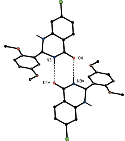
Figure 3
Part of the crystal structure of compound (I) showing the formation of a cyclic hydrogen-bonded dimer. Hydrogen bonds are drawn as dashed lines and, for the sake of clarity, the unit-cell outline and the H atoms bonded to C atoms have been omitted. The atoms marked with an asterisk (*) are at the symmetry position (1
2x, 1 2y,
[image:3.610.46.294.446.694.2]andR22(10) types can be identified. There is no carbonyl group
in (RS)-2-methyl-4-phenyl-3,4-dihydroquinazoline, and here molecules which are related by a 31screw axis are linked by an
N-–H N hydrogen bond to form aC(5) chain (Valkonenet al., 2011).
Finally, we note the structures of a number of 2,3-di-hydroquinazolin-4(1H)-ones in which there is a substituent at atom N3 (Butcheret al., 2007; Tozeet al., 2018; Zaytsevet al., 2018). In each of these examples, the molecules are linked by a single N—H O hydrogen bond to form a C(6) chain. However, when the substituent at atom N3 is an arylmethyl-amino group, the heterocyclic ring adopts a screw–boat conformation (Butcheret al., 2007), but in five examples where this substituent is either a benzyl group or a furanylmethyl unit, the heterocyclic ring adopts an envelope conformation, folded across the N N line (Tozeet al., 2018; Zaytsevet al. 2018).
5. Synthesis and crystallization
A sample of compound (I) was prepared using a recently published general procedure (Narasimhamurthyet al., 2014). Potassiumtert-butoxide (3.3 mmol) was added to a suspension of 2-(dibromomethyl)-1,4-dimethoxybenzene (3.3 mmol) and 2-amino-4-chlorobenzamide (3.5 mmol) in a pyridine-di-methylformamide mixture (3:1, v/v). The resulting mixture was heated at 313 K for 4 h, with TLC monitoring. When the reaction was judged to be complete, an excess of water was
added, followed by extraction with ethyl acetate (220 ml). The combined organic extract was washed with brine and then dried over anhydrous sodium sulfate. The solvent was removed under reduced pressure and the crude product was purified by column chromatography using silica gel mesh 60– 120, with 30% ethyl acetate in hexane as eluent, to give the product (I) in 79% yield. Crystals suitable for single crystal X-ray diffraction were grown by slow evaporation, at ambient temperature and in the presence of air, of a solution in di-methylsulfoxide: m.p. 481–483 K.
6. Refinement
Crystal data, data collection and structure refinement details are given in Table 2. In the setting of space groupI41/a, No. 88,
employed here the origin is located at a centre of inversion. All H atoms were located in difference maps. The H atoms bonded to C atoms were then treated as riding atoms in geometrically idealized position with C—H 0.93 A˚ (aromatic), 0.96 A˚ (CH3) or 0.98 A˚ (aliphatic C—H), and withUiso(H) =
kUeq(C), where k = 1.5 for the methyl groups, which were
permitted to rotate but not to tilt, and 1.2 for all other H atoms bonded to C atoms. For the H atoms bonded to N atoms, the atomic coordinates were refined with Uiso(H) = 1.2Ueq(N),
giving the N—H distances shown in Table 1.
Acknowledgements
KHN is grateful to UGC, RFSMS, Government of India for a Research Fellowship. BKS thanks the University of Mysore, for research facilities.
Funding information
KSR and HSY are grateful to UGC, New Delhi, for the award of a BSR Faculty Fellowship for a period of three years.
References
Badolato, M., Aiello, F. & Neamati, N. (2018).RSC Adv.8, 20894– 20921.
Bernstein, J., Davis, R. E., Shimoni, L. & Chang, N.-L. (1995).Angew. Chem. Int. Ed. Engl.34, 1555–1573.
Boeyens, J. C. A. (1978).J. Cryst. Mol. Struct.8, 317–320.
Bruker (2015). APEX2, SADABS and SAINT. Bruker AXS Inc., Madison, Wisconsin, USA.
Butcher, R. J., Jasinski, J. P., Narayana, B., Sunil, K. & Yathirajan, H. S. (2007).Acta Cryst.E63, o4025–o4026.
Etter, M. C. (1990).Acc. Chem. Res.23, 120–126.
Etter, M. C., MacDonald, J. C. & Bernstein, J. (1990).Acta Cryst.B46, 256–262.
Ferguson, G., Glidewell, C. & Patterson, I. L. J. (1996).Acta Cryst.
C52, 420–423.
Li, M.-J. & Feng, C.-J. (2009).Acta Cryst.E65, o2145.
Narasimhamurthy, K. H., Chandrappa, S., Sharath Kumar, K. S., Harsha, K. B., Ananda, H. & Rangappa, K. S. (2014).RSC Adv.4, 34479–34486.
Seip, H. M. & Seip, R. (1973).Acta Chem. Scand.27, 4024–4027. Sheldrick, G. M. (2008).Acta Cryst.A64, 112–122.
[image:4.610.43.292.89.367.2]Sheldrick, G. M. (2015).Acta Cryst.C71, 3–8. Spek, A. L. (2009).Acta Cryst.D65, 148–155. Table 2
Experimental details.
Crystal data
Chemical formula C16H15ClN2O3
Mr 318.75
Crystal system, space group Tetragonal,I41/a
Temperature (K) 296
a,c(A˚ ) 15.314 (7), 25.736 (12)
V(A˚3) 6036 (6)
Z 16
Radiation type MoK
(mm1) 0.27
Crystal size (mm) 0.260.220.18
Data collection
Diffractometer Bruker APEXII CCD
Absorption correction Multi-scan (SADABS; Bruker,
2015)
Tmin,Tmax 0.913, 0.953
No. of measured, independent and observed [I> 2(I)] reflections
42994, 3149, 1848
Rint 0.072
(sin/)max(A˚
1
) 0.629
Refinement
R[F2> 2(F2)],wR(F2),S 0.049, 0.128, 1.04
No. of reflections 3149
No. of parameters 207
H-atom treatment H atoms treated by a mixture of
independent and constrained refinement
max,min(e A˚
3
) 0.17,0.27
Computer programs:APEX2andSAINT(Bruker, 2015),SHELXS97(Sheldrick, 2008),
Toze, F. A. A., Zaytsev, V. P., Chervyakova, L. V., Kvyatkovskaya, E. A., Dorovatovskii, P. V. & Khrustalev, V. N. (2018).Acta Cryst.
E74, 10–14.
Valkonen, A., Kolehmainen, E., Zakrzewska, A., Skotnicka, A. & Gawinecki, R. (2011).Acta Cryst.E67, o923–o924.
Vembu, N., Spencer, E. C., Lee, J., Kelly, J. G., Nolan, K. B. & Devocelle, M. (2006).Acta Cryst.E62, o5003–o5005.
sup-1
Acta Cryst. (2019). E75, 843-847
supporting information
Acta Cryst. (2019). E75, 843-847 [https://doi.org/10.1107/S2056989019007023]
The crystal structure of (
RS
)-7-chloro-2-(2,5-dimethoxyphenyl)-2,3-dihydro-quinazolin-4(1
H
)-one: two hydrogen bonds generate an elegant
three-dimensional framework structure
Kereyagalahally H. Narasimhamurthy, Chandra, Belakavadi K. Sagar, Kanchugarakoppal S.
Rangappa, Hemmige S. Yathirajan and Christopher Glidewell
Computing details
Data collection: APEX2 (Bruker, 2015); cell refinement: SAINT (Bruker, 2015); data reduction: SAINT (Bruker, 2015);
program(s) used to solve structure: SHELXS97 (Sheldrick, 2008); program(s) used to refine structure: SHELXL2014
(Sheldrick, 2015); molecular graphics: PLATON (Spek, 2009); software used to prepare material for publication:
SHELXL2014 (Sheldrick, 2015) and PLATON (Spek, 2009).
(RS)-7-Chloro-2-(2,5-dimethoxyphenyl)-2,3-dihydroquinazolin-4(1H)-one
Crystal data
C16H15ClN2O3
Mr = 318.75
Tetragonal, I41/a
a = 15.314 (7) Å
c = 25.736 (12) Å
V = 6036 (6) Å3
Z = 16
F(000) = 2656
Dx = 1.403 Mg m−3
Mo Kα radiation, λ = 0.71073 Å Cell parameters from 3465 reflections
θ = 1.6–27.6°
µ = 0.27 mm−1
T = 296 K Block, colourless 0.26 × 0.22 × 0.18 mm
Data collection
Bruker APEXII CCD diffractometer
Radiation source: fine focus sealed tube Graphite monochromator
Detector resolution: 0.3333 pixels mm-1
φ and ω scans
Absorption correction: multi-scan (SADABS; Bruker, 2015)
Tmin = 0.913, Tmax = 0.953
42994 measured reflections 3149 independent reflections 1848 reflections with I > 2σ(I)
Rint = 0.072
θmax = 26.6°, θmin = 1.6°
h = −19→19
k = −19→19
l = −32→26
Refinement
Refinement on F2
Least-squares matrix: full
R[F2 > 2σ(F2)] = 0.049
wR(F2) = 0.128
S = 1.03 3149 reflections 207 parameters
0 restraints
Primary atom site location: difference Fourier map
Hydrogen site location: mixed
sup-2
Acta Cryst. (2019). E75, 843-847
w = 1/[σ2(F
o2) + (0.0505P)2 + 3.5139P]
where P = (Fo2 + 2Fc2)/3
(Δ/σ)max < 0.001
Δρmax = 0.17 e Å−3
Δρmin = −0.27 e Å−3
Special details
Geometry. All esds (except the esd in the dihedral angle between two l.s. planes) are estimated using the full covariance matrix. The cell esds are taken into account individually in the estimation of esds in distances, angles and torsion angles; correlations between esds in cell parameters are only used when they are defined by crystal symmetry. An approximate (isotropic) treatment of cell esds is used for estimating esds involving l.s. planes.
Fractional atomic coordinates and isotropic or equivalent isotropic displacement parameters (Å2)
x y z Uiso*/Ueq
N1 0.30909 (14) 0.40264 (14) 0.37953 (7) 0.0531 (6)
H1 0.3275 (17) 0.4092 (16) 0.4083 (10) 0.064*
C2 0.30616 (16) 0.31258 (15) 0.36210 (8) 0.0482 (6)
H2 0.3646 0.2879 0.3670 0.058*
N3 0.28752 (14) 0.31121 (14) 0.30684 (7) 0.0506 (5)
H3 0.2932 (16) 0.2628 (17) 0.2929 (9) 0.061*
C4 0.24435 (15) 0.37295 (16) 0.28060 (8) 0.0467 (6)
O4 0.22338 (12) 0.36121 (11) 0.23471 (6) 0.0603 (5)
C4A 0.22505 (14) 0.45429 (15) 0.30792 (8) 0.0434 (5)
C5 0.17657 (16) 0.51985 (16) 0.28452 (9) 0.0521 (6)
H5 0.1566 0.5118 0.2507 0.062*
C6 0.15743 (16) 0.59599 (17) 0.30984 (9) 0.0569 (7)
H6 0.1250 0.6397 0.2939 0.068*
C7 0.18794 (16) 0.60558 (16) 0.35987 (9) 0.0556 (6)
Cl7 0.16054 (6) 0.70075 (5) 0.39245 (3) 0.0873 (3)
C8 0.23775 (16) 0.54326 (16) 0.38428 (9) 0.0522 (6)
H8 0.2583 0.5527 0.4178 0.063*
C8A 0.25726 (14) 0.46570 (15) 0.35832 (8) 0.0424 (5)
C21 0.24297 (15) 0.25723 (14) 0.39324 (8) 0.0432 (5)
C22 0.26659 (17) 0.23605 (16) 0.44405 (8) 0.0522 (6)
C23 0.2110 (2) 0.18716 (17) 0.47413 (9) 0.0678 (8)
H23 0.2271 0.1735 0.5080 0.081*
C24 0.1321 (2) 0.15794 (18) 0.45543 (10) 0.0682 (8)
H24 0.0951 0.1257 0.4767 0.082*
C25 0.10822 (18) 0.17630 (16) 0.40582 (9) 0.0559 (7)
C26 0.16454 (16) 0.22579 (15) 0.37492 (8) 0.0488 (6)
H26 0.1485 0.2379 0.3408 0.059*
O221 0.34622 (12) 0.26875 (12) 0.45915 (6) 0.0718 (6)
C221 0.3739 (2) 0.2493 (2) 0.51083 (11) 0.0918 (11)
H22A 0.3823 0.1874 0.5143 0.138*
H22B 0.3302 0.2684 0.5350 0.138*
H22C 0.4279 0.2789 0.5179 0.138*
O251 0.03307 (13) 0.14750 (13) 0.38268 (8) 0.0767 (6)
C251 −0.0300 (2) 0.1052 (2) 0.41418 (14) 0.0939 (11)
H25A −0.0488 0.1442 0.4412 0.141*
sup-3
Acta Cryst. (2019). E75, 843-847
H25C −0.0793 0.0889 0.3932 0.141*
Atomic displacement parameters (Å2)
U11 U22 U33 U12 U13 U23
N1 0.0655 (14) 0.0565 (13) 0.0373 (10) −0.0097 (11) −0.0105 (10) −0.0042 (10)
C2 0.0520 (14) 0.0582 (15) 0.0343 (12) 0.0045 (12) −0.0054 (10) −0.0063 (10)
N3 0.0645 (14) 0.0545 (13) 0.0328 (10) 0.0040 (11) 0.0019 (9) −0.0061 (9)
C4 0.0508 (14) 0.0574 (15) 0.0321 (11) −0.0045 (12) 0.0049 (10) −0.0021 (11)
O4 0.0872 (13) 0.0620 (11) 0.0316 (8) 0.0061 (9) −0.0056 (8) −0.0085 (7)
C4A 0.0427 (13) 0.0524 (14) 0.0351 (11) −0.0070 (11) 0.0026 (10) −0.0054 (10)
C5 0.0539 (15) 0.0618 (16) 0.0404 (12) −0.0005 (13) −0.0050 (11) −0.0082 (11)
C6 0.0585 (16) 0.0595 (17) 0.0528 (14) 0.0039 (13) −0.0050 (12) −0.0054 (12)
C7 0.0568 (16) 0.0551 (16) 0.0548 (14) −0.0041 (13) 0.0012 (12) −0.0154 (12)
Cl7 0.1085 (7) 0.0736 (5) 0.0800 (5) 0.0165 (4) −0.0147 (4) −0.0338 (4)
C8 0.0580 (16) 0.0593 (16) 0.0395 (12) −0.0130 (13) −0.0033 (11) −0.0104 (11)
C8A 0.0404 (13) 0.0508 (14) 0.0359 (11) −0.0119 (11) 0.0002 (10) −0.0033 (10)
C21 0.0560 (15) 0.0410 (13) 0.0326 (11) 0.0068 (11) −0.0025 (10) −0.0045 (9)
C22 0.0692 (17) 0.0490 (15) 0.0384 (12) 0.0063 (13) −0.0121 (12) −0.0073 (11)
C23 0.109 (2) 0.0588 (17) 0.0354 (13) 0.0081 (17) −0.0032 (14) 0.0095 (12)
C24 0.088 (2) 0.0621 (18) 0.0540 (16) −0.0059 (16) 0.0068 (15) 0.0091 (13)
C25 0.0707 (18) 0.0458 (14) 0.0514 (14) 0.0010 (13) 0.0006 (13) −0.0025 (11)
C26 0.0612 (16) 0.0484 (14) 0.0369 (11) 0.0042 (12) −0.0039 (11) 0.0006 (10)
O221 0.0866 (14) 0.0768 (13) 0.0521 (11) 0.0029 (11) −0.0292 (10) −0.0026 (9)
C221 0.124 (3) 0.091 (2) 0.0607 (17) 0.024 (2) −0.0496 (18) −0.0082 (16)
O251 0.0707 (13) 0.0784 (14) 0.0810 (13) −0.0176 (11) −0.0024 (11) 0.0074 (11)
C251 0.072 (2) 0.082 (2) 0.128 (3) −0.0106 (17) 0.012 (2) 0.026 (2)
Geometric parameters (Å, º)
N1—C8A 1.364 (3) C21—C26 1.377 (3)
N1—C2 1.451 (3) C21—C22 1.395 (3)
N1—H1 0.80 (3) C22—C23 1.373 (4)
C2—N3 1.451 (3) C22—O221 1.374 (3)
C2—C21 1.516 (3) C23—C24 1.376 (4)
C2—H2 0.9800 C23—H23 0.9300
N3—C4 1.337 (3) C24—C25 1.357 (3)
N3—H3 0.83 (2) C24—H24 0.9300
C4—O4 1.237 (3) C25—O251 1.369 (3)
C4—C4A 1.461 (3) C25—C26 1.397 (3)
C4A—C5 1.386 (3) C26—H26 0.9300
C4A—C8A 1.399 (3) O221—C221 1.427 (3)
C5—C6 1.368 (3) C221—H22A 0.9600
C5—H5 0.9300 C221—H22B 0.9600
C6—C7 1.377 (3) C221—H22C 0.9600
C6—H6 0.9300 O251—C251 1.418 (3)
C7—C8 1.374 (3) C251—H25A 0.9600
sup-4
Acta Cryst. (2019). E75, 843-847
C8—C8A 1.395 (3) C251—H25C 0.9600
C8—H8 0.9300
C8A—N1—C2 122.09 (19) C26—C21—C22 117.8 (2)
C8A—N1—H1 119.2 (19) C26—C21—C2 124.84 (19)
C2—N1—H1 114.6 (19) C22—C21—C2 117.4 (2)
N3—C2—N1 108.84 (19) C23—C22—O221 126.1 (2)
N3—C2—C21 112.62 (19) C23—C22—C21 119.6 (2)
N1—C2—C21 112.82 (18) O221—C22—C21 114.2 (2)
N3—C2—H2 107.4 C22—C23—C24 121.7 (2)
N1—C2—H2 107.4 C22—C23—H23 119.2
C21—C2—H2 107.4 C24—C23—H23 119.2
C4—N3—C2 125.6 (2) C25—C24—C23 119.9 (3)
C4—N3—H3 117.8 (17) C25—C24—H24 120.1
C2—N3—H3 114.6 (17) C23—C24—H24 120.1
O4—C4—N3 120.5 (2) C24—C25—O251 124.7 (2)
O4—C4—C4A 122.1 (2) C24—C25—C26 118.8 (3)
N3—C4—C4A 117.39 (19) O251—C25—C26 116.5 (2)
C5—C4A—C8A 120.1 (2) C21—C26—C25 122.2 (2)
C5—C4A—C4 121.12 (19) C21—C26—H26 118.9
C8A—C4A—C4 118.8 (2) C25—C26—H26 118.9
C6—C5—C4A 121.7 (2) C22—O221—C221 116.8 (2)
C6—C5—H5 119.2 O221—C221—H22A 109.5
C4A—C5—H5 119.2 O221—C221—H22B 109.5
C5—C6—C7 117.6 (2) H22A—C221—H22B 109.5
C5—C6—H6 121.2 O221—C221—H22C 109.5
C7—C6—H6 121.2 H22A—C221—H22C 109.5
C8—C7—C6 122.8 (2) H22B—C221—H22C 109.5
C8—C7—Cl7 119.83 (19) C25—O251—C251 118.1 (2)
C6—C7—Cl7 117.4 (2) O251—C251—H25A 109.5
C7—C8—C8A 119.4 (2) O251—C251—H25B 109.5
C7—C8—H8 120.3 H25A—C251—H25B 109.5
C8A—C8—H8 120.3 O251—C251—H25C 109.5
N1—C8A—C8 122.4 (2) H25A—C251—H25C 109.5
N1—C8A—C4A 119.2 (2) H25B—C251—H25C 109.5
C8—C8A—C4A 118.4 (2)
C8A—N1—C2—N3 33.6 (3) C5—C4A—C8A—C8 1.3 (3)
C8A—N1—C2—C21 −92.2 (2) C4—C4A—C8A—C8 −179.4 (2)
N1—C2—N3—C4 −26.5 (3) N3—C2—C21—C26 −14.9 (3)
C21—C2—N3—C4 99.4 (3) N1—C2—C21—C26 108.8 (2)
C2—N3—C4—O4 −171.9 (2) N3—C2—C21—C22 164.5 (2)
C2—N3—C4—C4A 9.3 (3) N1—C2—C21—C22 −71.8 (3)
O4—C4—C4A—C5 4.7 (3) C26—C21—C22—C23 −1.5 (3)
N3—C4—C4A—C5 −176.6 (2) C2—C21—C22—C23 179.0 (2)
O4—C4—C4A—C8A −174.6 (2) C26—C21—C22—O221 179.1 (2)
N3—C4—C4A—C8A 4.1 (3) C2—C21—C22—O221 −0.4 (3)
sup-5
Acta Cryst. (2019). E75, 843-847
C4—C4A—C5—C6 179.4 (2) C21—C22—C23—C24 0.2 (4)
C4A—C5—C6—C7 −0.1 (4) C22—C23—C24—C25 1.0 (4)
C5—C6—C7—C8 1.5 (4) C23—C24—C25—O251 177.2 (2)
C5—C6—C7—Cl7 −178.02 (19) C23—C24—C25—C26 −0.8 (4)
C6—C7—C8—C8A −1.5 (4) C22—C21—C26—C25 1.8 (3)
Cl7—C7—C8—C8A 178.07 (18) C2—C21—C26—C25 −178.8 (2)
C2—N1—C8A—C8 158.6 (2) C24—C25—C26—C21 −0.6 (4)
C2—N1—C8A—C4A −24.0 (3) O251—C25—C26—C21 −178.8 (2)
C7—C8—C8A—N1 177.4 (2) C23—C22—O221—C221 0.6 (4)
C7—C8—C8A—C4A 0.0 (3) C21—C22—O221—C221 179.9 (2)
C5—C4A—C8A—N1 −176.2 (2) C24—C25—O251—C251 8.7 (4)
C4—C4A—C8A—N1 3.1 (3) C26—C25—O251—C251 −173.4 (2)
Hydrogen-bond geometry (Å, º)
D—H···A D—H H···A D···A D—H···A
N1—H1···O4i 0.80 (3) 2.39 (3) 3.161 (3) 162 (2)
N3—H3···O4ii 0.83 (3) 2.04 (3) 2.854 (3) 166 (2)