3-Oxo-
N
000,2-diphenyl-2,3-dihydro-1
H
-pyrazole-4-carbohydrazide
Joel T. Mague,aShaaban K. Mohamed,b,cMehmet Akkurt,d Eman A. Ahmedeand Mustafa R. Albayatif*
aDepartment of Chemistry, Tulane University, New Orleans, LA 70118, USA, bChemistry and Environmental Division, Manchester Metropolitan University, Manchester M1 5GD, England,cChemistry Department, Faculty of Science, Minia University, 61519 El-Minia, Egypt,dDepartment of Physics, Faculty of Sciences, Erciyes University, 38039 Kayseri, Turkey,eChemistry Department, Faculty of Science, Sohag University, 61519 Sohag, Egypt, andfKirkuk University, College of Science, Department of Chemistry, Kirkuk, Iraq
Correspondence e-mail: shaabankamel@yahoo.com
Received 27 May 2014; accepted 28 May 2014
Key indicators: single-crystal X-ray study;T= 150 K; mean(C–C) = 0.002 A˚; Rfactor = 0.043;wRfactor = 0.112; data-to-parameter ratio = 18.5.
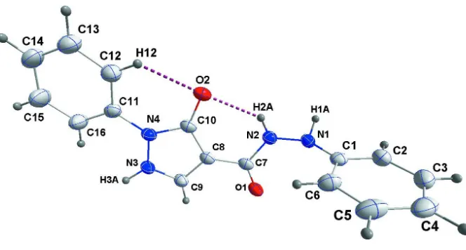
In the title compound, C16H14N4O2, the pyrazole ring makes a
dihedral angle of 10.49 (8) with its N-bound phenyl group,
while it is nearly perpendicular to the other phenyl ring [dihedral angle = 88.47 (5)]. The molecular conformation is
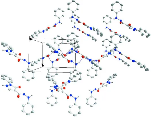
stabilized by intramolecular C—H O and N—H O hydrogen bonds. In the crystal, the packing involves sheets of molecules parallel to (100) linked by N—H O hydrogen bonds. A C—H O interaction is also observed.
Related literature
For the diverse biological activities of pyrazolone compounds, see: Guckianet al.(2010); Fanet al.(2006); Castagnoloet al.
(2008); Idreeset al.(2009); Abdel-Azizet al.(2009); Manoj-kumaret al.(2009); Sheteet al.(2011); Sujathaet al.(2009); El-Hawashet al.(2006); Kawaiet al.(1997); Wuet al.(20026). For industrial applications of pyrazolones, see: Basaifet al.(2007); Ho (2005); Kirschke et al.(1984); Chande et al. (1993); El-Saraf & El-Sayed (2003). For graph-set motif notation, see: Bernsteinet al.(1995).
Experimental
Crystal data
C16H14N4O2
Mr= 294.31 Monoclinic,P21=c
a= 8.4488 (12) A˚
b= 11.5605 (17) A˚
c= 14.642 (2) A˚ = 91.565 (2)
V= 1429.6 (4) A˚3
Z= 4
MoKradiation = 0.09 mm1
T= 150 K
0.260.200.07 mm
Data collection
Bruker SMART APEX CCD diffractometer
Absorption correction: multi-scan (SADABS; Bruker, 2013)
Tmin= 0.85,Tmax= 0.99
25571 measured reflections 3674 independent reflections 2895 reflections withI> 2(I)
Rint= 0.049
Refinement
R[F2> 2(F2)] = 0.043
wR(F2) = 0.112
S= 1.03 3674 reflections
199 parameters
H-atom parameters constrained
max= 0.25 e A˚
3
min=0.21 e A˚3
Table 1
Hydrogen-bond geometry (A˚ ,).
D—H A D—H H A D A D—H A
N1—H1A O2i
0.91 2.06 2.9244 (14) 158 N2—H2A O2 0.91 2.14 2.8597 (14) 136 N3—H3A O1ii
0.91 1.75 2.6527 (15) 169 C12—H12 O2 0.95 2.28 2.9133 (18) 124 C16—H16 O1ii
0.95 2.51 3.2745 (18) 137
Symmetry codes: (i)xþ1;yþ1;zþ1; (ii)xþ1;yþ1 2;zþ
3 2.
Data collection:APEX2(Bruker, 2013); cell refinement:SAINT
(Bruker, 2013); data reduction:SAINT; program(s) used to solve structure: SHELXTL (Sheldrick, 2008); program(s) used to refine structure: SHELXL2014 (Sheldrick, 2008); molecular graphics:
DIAMOND(Brandenburg & Putz, 2012); software used to prepare material for publication:SHELXTL.
JTM thanks Tulane University for support of the Tulane Crystallography Laboratory.
Supporting information for this paper is available from the IUCr electronic archives (Reference: BT6982).
References
Abdel-Aziz, M., El-Din, G., Abuo-Rahma, A. & Hassan, A. A. (2009).Eur. J. Med. Chem.44, 3480–3487.
Basaif, S. A., Hassan, M. A. & Gobouri, A. A. (2007).Dyes Pigm.72, 387–391. Bernstein, J., Davis, R. E., Shimoni, L. & Chang, N.-L. (1995).Angew. Chem.
Int. Ed. Engl.34, 1555–1573.
Brandenburg, K. & Putz, H. (2012).DIAMOND. Crystal Impact GbR, Bonn, Germany.
Bruker (2013).APEX2,SADABSandSAINT. Bruker AXS Inc., Madison, Wisconsin, USA.
Castagnolo, D., Logu, A. D., Radi, M., Bechi, B., Manetti, F., Magnani, M., Supino, S., Meleddu, R., Chisu, L. & Botta, M. (2008).Bioorg. Med. Chem. Lett.16, 8587–8591.
Chande, M. S., Bhandari, J. D., Joshi, V. R. & Joshi, V. R. (1993).Indian J. Chem. Sect. B,32, 1218–1228.
El-Hawash, S. A. M., Badawey, E. A. M. & El-Ashmawey, I. M. (2006).Eur. J. Med. Chem.41, 155–165.
El-Saraf, G. A. & El-Sayed, A. M. (2003).Heteroat. Chem.14, 211–217.
organic compounds
Acta Cryst.(2014). E70, o819–o820 doi:10.1107/S1600536814012392 Magueet al.
o819
Acta Crystallographica Section EStructure Reports Online
(2006).Bioorg. Med. Chem. Lett.16, 3224–3228.
Guckian, K., Carter, M. B., Lin, E. Y., Choi, M., Sun, L., Boriack-Sjodin, P. A., Chuaqui, C., Lane, B., Cheung, K., Ling, L. & Lee, W. C. (2010).Bioorg. Med. Chem. Lett.20, 326–329.
Ho, Y. W. (2005).Dyes Pigm.64, 223–230.
Idrees, G. A., Aly, O. M., El-Din, G., Abuo-Rahma, A. A. & Radwan, M. F. (2009).Eur. J. Med. Chem.44, 3973–3980.
Kawai, H., Nakai, H., Suga, M., Yuki, S., Watanabe, T. & Saito, K. I. (1997).J. Pharmacol. Exp. Ther.281, 921–927.
Manojkumar, P., Ravi and, T. K. & Gopalakrishnan, S. (2009).Eur. J. Med. Chem.44, 4690–4694.
Sheldrick, G. M. (2008).Acta Cryst.A64, 112–122.
Shete, R. B., Antre, R. V., Oswal, R. J., Kshirsagar, S. S., Jangam, S. S. & Nimje, H. M. (2011).Int. J. Drug Des. Discov.2, 648–660.
Sujatha, K., Shanthi, G., Selvam, N. P., Manoharan, S., Perumal, P. T. & Rajendran, M. (2009).Bioorg. Med. Chem. Lett.19, 4501–4503.
supporting information
sup-1 Acta Cryst. (2014). E70, o819–o820
supporting information
Acta Cryst. (2014). E70, o819–o820 [https://doi.org/10.1107/S1600536814012392]
3-Oxo-
N
′
,2-diphenyl-2,3-dihydro-1
H
-pyrazole-4-carbohydrazide
Joel T. Mague, Shaaban K. Mohamed, Mehmet Akkurt, Eman A. Ahmed and Mustafa R. Albayati
S1. Comment
Compounds containing a pyrazole core and related analogs have received signicant attention due to their chemical, medicinal, and pharmaceutical applications. Several reports showed the pyrazolone moiety to be one of most active pharmacophores and possesses anti-cancer (Guckian et al., 2010), anti-viral (Fan et al., 2006), anti-tubercular
(Castagnolo et al., 2008), anti-hyperlipedaemic (Idrees et al., 2009), anti-depressant, anti-convulsant (Abdel-Aziz et al., 2009), anti-oxidant, anti-bacterial (Manojkumar et al., 2009; Shete et al., 2011), anti-HIV (Sujatha et al., 2009), anti-inflammatory, analgesic and anti-pyretic (El-Hawash et al., 2006) activities. The pyrazolone-like edaravone has been developed as a drug for brain ischemia (Kawai et al., 1997) and has also been reported to be effective for myocardial ischemia (Wu et al., 2002). Additionally, pyrazolones have been reported to be the key starting materials for the synthesis of commercial aryl/heteroaropyrazolone dyes (Basaif et al., 2007; Ho, 2005). Halogenated pyrazolones are also useful synthetic intermediates for synthesis of diazo-dyes (Kirschke et al., 1984), fused (Chande et al., 1993) and spiro-heterocyclic compounds (El-Saraf & El-Sayed 2003). In this context we report the synthesis and crystal structure of the title compound.
The phenyl ring C1–C6 is nearly perpendicular to the 5-membered ring (dihedral angle = 88.47 (5)°) while the latter makes a dihedral angle of 10.49 (8)° with the phenyl ring C11–C16. The rotational orientation of the latter phenyl ring as well as that of the N2—H2a unit are determined by the intramolecular C16—H16···O2 and N2—H2a···O2 hydrogen bonds (Table 1, Fig. 1) forming S(6) ring motifs (Bernstein et al., 1995). The packing involves sheets of molecules parallel to the (100) plane formed by N3—H3a···O1 and pairwise N1—H1a···O2 intermolecular hydrogen bonds (Table 1, Figs. 2 and 3).
S2. Experimental
A mixture of 10 mmol (2.31 g) of 4-[(dimethylamino)methylene]-1-phenylpyrazolidine-3,5-dione and 10 mmol (1.08 g) of phenyl hydrazine in 15 ml dioxane was refluxed for 6 h. After cooling, the resulting solid was collected by filtration and recrystallized from ethanol. Colourless crystals, 84%, m.p. = 483 K.
S3. Refinement
Figure 1
The title molecule showing intramolecular hydrogen bonds as dotted lines. Displacement ellipsoids are drawn at the 50% probability level.
Figure 2
[image:4.610.137.444.297.487.2]supporting information
[image:5.610.134.399.72.283.2]sup-3 Acta Cryst. (2014). E70, o819–o820
Figure 3
Packing viewed down the c axis with intermolecular hydrogen bonds shown as dotted lines.
3-Oxo-N′,2-diphenyl-2,3-dihydro-1H-pyrazole-4-carbohydrazide
Crystal data
C16H14N4O2
Mr = 294.31
Monoclinic, P21/c
Hall symbol: -P 2ybc
a = 8.4488 (12) Å
b = 11.5605 (17) Å
c = 14.642 (2) Å
β = 91.565 (2)°
V = 1429.6 (4) Å3
Z = 4
F(000) = 616
Dx = 1.367 Mg m−3
Mo Kα radiation, λ = 0.71073 Å Cell parameters from 9891 reflections
θ = 2.2–28.6°
µ = 0.09 mm−1
T = 150 K Plate, colourless 0.26 × 0.20 × 0.07 mm
Data collection
Bruker SMART APEX CCD diffractometer
Radiation source: fine-focus sealed tube Graphite monochromator
Detector resolution: 8.3660 pixels mm-1
φ and ω scans
Absorption correction: multi-scan
(SADABS; Bruker, 2013)
Tmin = 0.85, Tmax = 0.99
25571 measured reflections 3674 independent reflections 2895 reflections with I > 2σ(I)
Rint = 0.049
θmax = 28.7°, θmin = 2.2°
h = −11→11
k = −15→15
l = −19→19
Refinement
Refinement on F2
Least-squares matrix: full
R[F2 > 2σ(F2)] = 0.043
wR(F2) = 0.112
S = 1.03 3674 reflections 199 parameters
0 restraints
Primary atom site location: structure-invariant direct methods
Secondary atom site location: difference Fourier map
where P = (Fo2 + 2Fc2)/3
(Δ/σ)max < 0.001
Δρmin = −0.21 e Å−3
Special details
Geometry. Bond distances, angles etc. have been calculated using the rounded fractional coordinates. All su's are estimated from the variances of the (full) variance-covariance matrix. The cell e.s.d.'s are taken into account in the estimation of distances, angles and torsion angles
Refinement. Refinement on F2 for ALL reflections except those flagged by the user for potential systematic errors.
Weighted R-factors wR and all goodnesses of fit S are based on F2, conventional R-factors R are based on F, with F set to
zero for negative F2. The observed criterion of F2 > σ(F2) is used only for calculating -R-factor-obs etc. and is not relevant
to the choice of reflections for refinement. R-factors based on F2 are statistically about twice as large as those based on F,
and R-factors based on ALL data will be even larger.
Fractional atomic coordinates and isotropic or equivalent isotropic displacement parameters (Å2)
x y z Uiso*/Ueq
supporting information
sup-5 Acta Cryst. (2014). E70, o819–o820
H13 0.28090 0.97360 0.29030 0.0560* H14 0.12040 1.12490 0.33540 0.0550* H15 0.08530 1.15780 0.49080 0.0610* H16 0.20200 1.03470 0.60010 0.0520*
Atomic displacement parameters (Å2)
U11 U22 U33 U12 U13 U23
O1 0.0487 (6) 0.0260 (5) 0.0230 (4) −0.0009 (4) 0.0018 (4) −0.0005 (3) O2 0.0371 (5) 0.0345 (5) 0.0239 (4) −0.0022 (4) 0.0012 (4) −0.0072 (4) N1 0.0270 (5) 0.0257 (5) 0.0286 (5) −0.0012 (4) 0.0029 (4) −0.0043 (4) N2 0.0299 (5) 0.0286 (5) 0.0229 (5) 0.0003 (4) 0.0018 (4) −0.0019 (4) N3 0.0432 (6) 0.0268 (5) 0.0212 (5) 0.0004 (5) 0.0013 (4) −0.0033 (4) N4 0.0355 (6) 0.0275 (5) 0.0209 (5) −0.0029 (4) 0.0012 (4) −0.0026 (4) C1 0.0268 (6) 0.0398 (7) 0.0186 (5) −0.0005 (5) 0.0015 (4) −0.0041 (5) C2 0.0369 (7) 0.0458 (8) 0.0354 (7) 0.0058 (6) −0.0009 (6) −0.0100 (6) C3 0.0378 (8) 0.0735 (12) 0.0386 (8) 0.0187 (8) −0.0043 (6) −0.0178 (8) C4 0.0271 (7) 0.0945 (14) 0.0300 (7) −0.0009 (8) −0.0005 (6) −0.0155 (8) C5 0.0343 (8) 0.0735 (12) 0.0326 (7) −0.0175 (8) −0.0005 (6) −0.0012 (7) C6 0.0314 (7) 0.0462 (8) 0.0294 (6) −0.0083 (6) −0.0002 (5) 0.0010 (6) C7 0.0286 (6) 0.0248 (6) 0.0243 (6) −0.0078 (5) 0.0027 (5) −0.0024 (5) C8 0.0301 (6) 0.0249 (6) 0.0239 (6) −0.0053 (5) 0.0027 (5) −0.0020 (4) C9 0.0371 (7) 0.0258 (6) 0.0227 (6) −0.0027 (5) 0.0013 (5) −0.0007 (5) C10 0.0278 (6) 0.0269 (6) 0.0246 (6) −0.0065 (5) 0.0028 (5) −0.0034 (5) C11 0.0323 (6) 0.0303 (6) 0.0254 (6) −0.0079 (5) −0.0029 (5) 0.0012 (5) C12 0.0551 (9) 0.0380 (8) 0.0274 (7) −0.0010 (7) 0.0031 (6) 0.0003 (6) C13 0.0633 (10) 0.0502 (9) 0.0272 (7) −0.0051 (8) −0.0014 (7) 0.0052 (6) C14 0.0510 (9) 0.0471 (9) 0.0378 (8) −0.0043 (7) −0.0130 (7) 0.0098 (7) C15 0.0572 (10) 0.0515 (10) 0.0427 (9) 0.0147 (8) −0.0115 (7) −0.0006 (7) C16 0.0503 (9) 0.0473 (9) 0.0307 (7) 0.0106 (7) −0.0064 (6) −0.0034 (6)
Geometric parameters (Å, º)
C4—C5 1.376 (3) C14—H14 0.9500 C5—C6 1.394 (2) C15—H15 0.9500 C7—C8 1.4493 (17) C16—H16 0.9500
N2—N1—C1 116.56 (10) N4—C11—C12 119.25 (12) N1—N2—C7 120.04 (10) N4—C11—C16 120.65 (11) N4—N3—C9 108.74 (10) C12—C11—C16 120.09 (13) N3—N4—C10 108.80 (10) C11—C12—C13 119.18 (14) N3—N4—C11 120.98 (10) C12—C13—C14 121.35 (14) C10—N4—C11 130.15 (10) C13—C14—C15 119.04 (15) N2—N1—H1A 110.00 C14—C15—C16 120.65 (16) C1—N1—H1A 113.00 C11—C16—C15 119.64 (14) C7—N2—H2A 119.00 C1—C2—H2 120.00 N1—N2—H2A 121.00 C3—C2—H2 120.00 N4—N3—H3A 121.00 C2—C3—H3 120.00 C9—N3—H3A 126.00 C4—C3—H3 120.00 C2—C1—C6 119.79 (12) C3—C4—H4 120.00 N1—C1—C2 117.86 (12) C5—C4—H4 120.00 N1—C1—C6 122.33 (12) C4—C5—H5 120.00 C1—C2—C3 119.71 (15) C6—C5—H5 120.00 C2—C3—C4 120.54 (17) C1—C6—H6 120.00 C3—C4—C5 119.57 (15) C5—C6—H6 120.00 C4—C5—C6 120.87 (16) N3—C9—H9 125.00 C1—C6—C5 119.50 (14) C8—C9—H9 125.00 N2—C7—C8 115.01 (10) C11—C12—H12 120.00 O1—C7—N2 123.43 (11) C13—C12—H12 120.00 O1—C7—C8 121.56 (11) C12—C13—H13 119.00 C7—C8—C10 127.58 (11) C14—C13—H13 119.00 C9—C8—C10 107.30 (11) C13—C14—H14 120.00 C7—C8—C9 125.12 (11) C15—C14—H14 120.00 N3—C9—C8 110.09 (11) C14—C15—H15 120.00 N4—C10—C8 104.97 (10) C16—C15—H15 120.00 O2—C10—C8 129.92 (12) C11—C16—H16 120.00 O2—C10—N4 125.08 (11) C15—C16—H16 120.00
supporting information
sup-7 Acta Cryst. (2014). E70, o819–o820
N3—N4—C11—C12 170.15 (13) C9—C8—C10—N4 1.08 (14) N3—N4—C11—C16 −8.37 (19) N4—C11—C12—C13 179.11 (14) C10—N4—C11—C12 −13.3 (2) C16—C11—C12—C13 −2.4 (2) C10—N4—C11—C16 168.14 (13) N4—C11—C16—C15 179.27 (14) N1—C1—C2—C3 −179.61 (13) C12—C11—C16—C15 0.8 (2) C6—C1—C2—C3 −1.4 (2) C11—C12—C13—C14 2.0 (2) N1—C1—C6—C5 179.80 (12) C12—C13—C14—C15 −0.1 (3) C2—C1—C6—C5 1.64 (19) C13—C14—C15—C16 −1.6 (3) C1—C2—C3—C4 0.1 (2) C14—C15—C16—C11 1.2 (3)
Hydrogen-bond geometry (Å, º)
D—H···A D—H H···A D···A D—H···A
N1—H1A···O2i 0.91 2.06 2.9244 (14) 158
N2—H2A···O2 0.91 2.14 2.8597 (14) 136 N3—H3A···O1ii 0.91 1.75 2.6527 (15) 169
C12—H12···O2 0.95 2.28 2.9133 (18) 124 C16—H16···O1ii 0.95 2.51 3.2745 (18) 137