Received 2 May 2018 Accepted 28 May 2018
Edited by V. Khrustalev, Russian Academy of Sciences, Russia
Keywords:crystal structure; quinoxalin; ene-di-thiocarbonate; carbonodithioate; hydrogen bonding interaction; molybdopterin.
CCDC reference:1845671
Supporting information:this article has supporting information at journals.iucr.org/e
Crystal structure of
1-ethyl-3-(2-oxo-1,3-dithiol-4-yl)quinoxalin-2(1
H
)-one
Nicolas Chrysochos and Carola Schulzke*
Institut fu¨r Biochemie, Ernst-Moritz-Arndt-Universita¨t Greifswald, 4 Felix-Hausdorff-Strasse, 17487 Greifswald, Germany. *Correspondence e-mail: carola.schulzke@uni-greifswald.de
The title compoundI, C13H10N2O2S2, crystallizes in the monoclinic space group C2/cwith eight molecules in the unit cell. Excluding for the ethyl substituent, the molecule of I adopts a nearly coplanar conformation (r.m.s. deviations is 0.058 A˚ ), which is supported by the intramolecular C—H O hydrogen-bonding interaction between the two ring systems [C O = 2.859 (3) A˚ ]. In the crystal, the molecules form dimeric associates via two bifurcated C—H O hydrogen-bonding interactions between an ene hydrogen atom and a carbonyl functional group of an adjacent molecule [C O = 3.133 (3) A˚ ] andvice versa. The crystal structure is further stabilized by a three-dimensional network of weak hydrogen bonds between one molecule and six adjacent molecules as well as offset–stacking. The combination of the quinoxaline 2(1H)-one moiety with the dithiocarbonate moiety extends the aromaticity of the quinoxaline scaffold towards the substituent as well as influencing the -system of the quinoxaline. The title compound is the direct precursor for a dithiolene ligand mimicking the natural cofactor ligand molybdopterin.
1. Chemical context
Non-innocent dithiolenes and their role as interesting ligand systems were discovered in the early 1960s. As a result of their unusual redox and structural characteristics and those of their metal complexes, they immediately attracted considerable scientific interest (Schrauzer & Mayweg, 1962). Initially, di-thiolene systems were studied predominantly in the context of electronic and photonic conductors (Wudlet al., 1972; Ferraris
et al., 1973). Later, metal dithiolene complexes found appli-cation in the purifiappli-cation and separation of olefins (Wang & Stiefel, 2001). In the early 1980s Rajagopalan and co-workers discovered and characterized the natural molybdopterin ligand (mpt) in the active sites of enzymes. Mpt is present in nearly all molybdenum enzymes and all tungsten enzymes and binds the respective central metal by a dithiolene moiety. As these enzymes are ubiquitous to all kingdoms of life, this brought dithiolene chemistry again to the focus of scientific attention (Johnsonet al., 1980; Johnson & Rajagopalan, 1982; Kramer et al., 1987). Quinoxaline constitutes a widely exploited platform in the development of pharmaceuticals (Shi et al., 2018). The title compound is a dithiolene ligand precursor, which can be used for the synthesis of molybdo-pterin cofactor model complexes bearing quinoxaline substituents. The target dithiolene ligand replicates the pyra-zine moiety of mpt in its half-reduced form and in addition contains an oxofunction in the position of the pyran ring of the natural product. By itself it is an interesting example of an
extended -system involving (by resonance) three different heteroatoms (N, O and S).
2. Structural commentary
The title moleculeIcrystallizes in the monoclinic space group
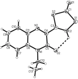
C2/cwithZ= 8. The quinoxaline ring system [C4–C11, N1, N2; largest deviation from plane = 0.041 (2) A˚ for C5] and the dithiolene ring [C1–C3, S1, S2; largest deviation from plane = 0.012 (1) A˚ for C3)], which are connected by the C3—C4 bond [length = 1.465 (3) A˚ ], are essentially coplanar, with an angle of only 4.89 (12) between the two planes (Fig. 1). This
planitarity is supported by intramolecular hydrogen bonding between the dithiolene hydrogen atom and the quinoxaline carbonyl oxygen atom [C2—H2 O2 with D A = 2.859 (3) A˚ ; Table 1]. Only the alkyl substituent C12—C13 subtends out of the planar geometry with an N1—C12—C13 torsion angle of 112.78 (18). While the N1—C12 bond [amine
nitrogen and ethyl substituent; 1.475 (3) A˚ ] is of explicit single-bond character, all other N—C distances are decidedly shorter, ranging from 1.296 (3) A˚ for the, according to the chemical structure, double bond of imine nitrogen (N2 C4) to 1.392 (3) A˚ for the amine nitrogen-to-benzene ring formal single bond (N1—C6). The longest C—C bond of the benzene ring is the one that is shared with the N-heterocycle [C6—C11, 1.411 (3) A˚ ]. This, together with the adjacent N—C bonds [N1—C6, 1.392 (3) A˚ and C11—N2, 1.374 (3) A˚ ] being significantly shorter than single bonds indicates resonance throughout the entire quinoxaline substituent. The C O bond [1.212 (3) A˚ ] of the carbonodithioate moiety is slightly shorter than the carbonyl C O bond [1.232 (3) A˚ ] of the quinoxaline substituent, suggesting that the latter might be involved to a small extent in resonance effects of the-system, whereas the former is not. The C2 C3 double bond of the ene-dithiocarbonate moiety is at 1.341 (3) A˚ slightly longer than the average value for C C double bonds of 1.331 (9) A˚ (Allenet al., 1995), which again may be due to participation in resonance effects throughout the entire molecule. The devia-tion from the average value of 1.751 (17) A˚ for S—Csp2bonds (Allen et al., 1995) of the S1—C2 bond [1.724 (2) A˚ ] is substantial enough to suggest that the resonance effects extend up to this bond to which partial double-bond character can be assigned. All other C—S bonds concur with typical single bonds.
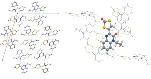
By bidirectional intermolecular hydrogen bonding, the title compound crystallizes as dimeric associate with the same donor and acceptor roles for both monomers [C2— H2 O2(x,y+ 2,z+ 1) and O2 H2—C2(x,y+ 2,
z+ 1);D A= 3.133 (3) A˚ ]. Here, the exact same atoms are involved as in the intramolecular hydrogen bond mentioned above. The respective hydrogen atom H2 is therefore bound to the ene carbon atom C2 and hydrogen bonded to the carbonyl oxygen (O2) of the quinoxaline moiety of the same molecule as well as that of the adjacent molecule (Fig. 2, left). There are only two crystal sructures of very closely related systems reported in the literature. In one (A), the quinoxalin substituent of the present molecule is replaced by a coumarine (Ghoshet al., 2016). In the other (B), the ene-dithiocarbonate is replaced by an aminothiazole (Mamedovet al., 2005). The metrical parameters in both structures are, as far as compar-able, very similar to the ones observed here. Notable differ-ences comprise (i) a slightly stronger resonance involvement
902
Chrysochos and Schulzke C [image:2.610.313.564.80.182.2]13H10N2O2S2 Acta Cryst.(2018). E74, 901–904
research communications
Figure 1
[image:2.610.94.247.100.239.2]The molecular structure of 1-ethyl-3-(2-oxo-1,3-dithiol-4yl-)quinoxalin-2(1H)-one showing the atom labelling, 50% probability displacement ellipsoids and the intramolecular non-classical hydrogen bond (dashed line).
Table 1
Hydrogen-bond geometry (A˚ ,).
D—H A D—H H A D A D—H A
C2—H2 O2 0.95 (2) 2.30 (3) 2.859 (3) 117.2 (19) C2—H2 O2i 0.95 (2) 2.44 (2) 3.133 (3) 129 (2) C7—H7 O1ii 0.95 2.51 3.272 (3) 138
C9—H9 S2iii 0.95 2.99 3.652 (3) 128 C12—H12A O2iv 0.99 2.69 3.367 (3) 126
Symmetry codes: (i) x;yþ2;zþ1; (ii) x;yþ1;zþ1 2; (iii)
xþ1 2;yþ
1
[image:2.610.316.566.445.699.2]of the thiazole in B compared to the ene-dithiocarbonate while the quinoxalin carbonyl and amine functions are embraced to a lesser extent and (ii) an overall weaker reso-nance inA, in which the benzene ring C—C distances are all very similar (i.e. strongly resonant) whereas all other distances are of more pronounced single- and double-bond character and of less aromatic character.
3. Supramolecular features
In the crystal, the associated dimers are linked by (partly rather weak) C—H O and C—H S hydrogen-bonding interactions, forming a three-dimensional network (Fig. 2, Table 1). In the three-dimensional network, each molecule forms hydrogen-bonding interactions to six surrounding molecules. These are donor interactions involving C2 [C2— H2 O2(x,y+ 2,z+ 1);D A= 3.133 (3) A˚ ], C7 [C7— H7 O1(x, y + 1,z + 1
2);D A= 3.272 (3) A˚ ], C9 [C9—
H9 S2(x+1 2,y+
1
2,z+ 1);D A= 3.652 (3) A˚ ], C12
[C12—H12A O2(x, y 1, z); D A = 3.367 (3) A˚ ] and acceptor interactions involving S2 [S2 H9—C9(x + 1
2,
y + 12, z + 1)], O2 [O2 H2—C2(x, y + 2, z + 1),
O2 H12A—C12(x,y–1,z)] and O1 [O1 H7—C7(x,y+ 1,
z1
2)]. Even though there are coplanar alignments of layers,
only offset–stacking was observed with centroid–centroid distances of 3.587 (3) A˚ between the benzene ring of one molecule and the pyrazine ring of a molecule in the layer above or below.
4. Synthesis and crystallization
The title compound, 1-ethyl-3-(2-oxo-1,3-dithiol-4yl-)quinox-alin-2(1H)-one was synthesized based on a reported literature procedure (Mamedov et al., 2005). The compound was synthesized in five steps starting from o-phenylenediamine. The last step in the synthetical pathway was carried outviaan acid-catalysed Tchugaeff ring closure reaction, which led to the formation of the dithiolene ring.
Synthesis of 1-ethyl-3-(2-oxo-1,3-dithiol-4yl-)quinoxalin-2(1H)-one: To a solution of S -2-(4-ethyl-3-oxo-4-dihydro-quinoxalin-2-yl)-2-oxo-ethyl o-isopropyl carbonodithioate (11.180 g, 31.9 mmol) in 250 ml DCM/Et2O 1:1 at ambient
temperature, H2SO4 (25.50 ml) was added. The reaction
mixture was stirred at room temperature for 2h. After that, the reaction was quenched by addition of 250 ml of ice and the mixture was stirred for 30 min. The organic phase was washed with brine and water 3250 ml. The solvent was reduced to 10 mlin vacuoand the greenish precipitate was filtered off and washed on the filter with cold acetone 3 50 ml. The title compound was obtained as a greenish-white powder. Single crystals suitable for X-ray analysis were obtained by slow diffusion of solvents with chloroform and Et2O Yield: 1.85g
(20%).
1
H NMR (300MHz, CD3Cl) 8.79 ppm (s, 1H), 7.87 ppm
(m, 1H), 7.61 ppm (m, 1H), 7.4 ppm (m, 1H), 4.39 ppm (q,J= 7.2Hz, 2H), 1.42 ppm (t,J= 7.3Hz, 3H).13C NMR (300MHz, CD3Cl) 152.54 ppm, 144.75 ppm, 133.25 ppm, 132.58 ppm,
[image:3.610.46.566.444.716.2]131.14 ppm, 130.32 ppm, 126.98 ppm, 124.00 ppm, 113.47 ppm, 37.47 ppm, 12.20 ppm. IR (KBr pellet): (cm1) = 3495 (br),
Figure 2
1734 (w), 1646 (sst), 1601 (st), 1579 (st), 1535 (st), 1463 (st), 1383 (w), 1280 (st), 1248 (w), 1216 (w), 1173 (st), 1128 (w), 1087 (w), 1045 (w), 950 (w), 892 (st), 868 (w), 825 (st), 785 (w), 758 (st), 631 (w), 554 (w), 529 (w), 467 (w), 432 (w). APCI–MS (m/s) = 291 (M++ H+). Analysis calculated for C13H10N2O2S2:
C, 53.78; H 3.47; N 9.65; S 22.09. Found: C, 53.41; H 3.25; N 9.86; S 22.32.
5. Refinement
Crystal data, data collection and structure refinement details are summarized in Table 2. The hydrogen atom of the di-thiolene unit (H2) was refined freely without any constraints or restraints. All other C-bound hydrogen atoms were attached in calculated positions and treated as riding: C—H =
0.98 A˚ withUiso(H) = 1.5Ueq(C) for the methyl group, C—H =
0.99 A˚ withUiso(H) = 1.2Ueq(C) for the methylene group and
C—H = 0.95 A˚ with Uiso(H) = 1.2Ueq(C) for the aromatic
atoms.
Acknowledgements
Generous financial support from the European Research Council (project MocoModels) is gratefully acknowledged.
Funding information
Funding for this research was provided by: FP7 Ideas: European Research Council (grant No. 281257 to Carola Schulzke).
References
Allen, F. H., Kennard, O., Watson, D. G., Brammer, L., Orpen, A. G., Taylor, R. & Wilson, A. J. C. (1995). International Tables for Crystallography, Vol. C. Boston: Kluwer Academic Publishers. Altomare, A., Cascarano, G., Giacovazzo, C., Guagliardi, A., Burla,
M. C., Polidori, G. & Camalli, M. (1994).J. Appl. Cryst.27, 435. Ferraris, J., Cowan, D. O., Walatka, V. & Perlstein, J. H. (1973).J. Am.
Chem. Soc.95, 948–949.
Ghosh, A. C., Weisz, K. & Schulzke, C. (2016).Eur. J. Inorg. Chem. pp. 208–218.
Johnson, J. L., Hainline, B. E. & Rajagopalan, K. V. (1980).J. Biol. Chem.255, 1783–1786.
Johnson, J. L. & Rajagopalan, K. V. (1982). Proc. Natl Acad. Sci. USA,79, 6856–6860.
Kramer, S. P., Johnson, J. L., Ribeiro, A. A., Millington, D. S. & Rajagopalan, K. V. (1987).J. Biol. Chem.262, 16357–16363. Macrae, C. F., Edgington, P. R., McCabe, P., Pidcock, E., Shields, G. P.,
Taylor, R., Towler, M. & van de Streek, J. (2006).J. Appl. Cryst.39, 453–457.
Mamedov, V. A., Kalinin, A. A., Gubaidullin, A. T., Isaikina, O. G. & Litvinov, I. A. (2005).Russ. J. Org. Chem.41, 599–606.
Schrauzer, G. N. & Mayweg, V. (1962).J. Am. Chem. Soc.84, 3221– 3221.
Sheldrick, G. M. (2008).Acta Cryst.A64, 112–122.
Sheldrick, G. (2013).CIFTAB. University of Go¨ttingen, Germany. Sheldrick, G. M. (2015).Acta Cryst.C71, 3–8.
Shi, L. L., Hu, W., Wu, J. F., Zhou, H. Y., Zhou, H. & Li, X. (2018). Mini Rev. Med. Chem.18, 392–413.
Stoe & Cie (2010).X-AREA,X-RED32andX-SHAPE. Stoe & Cie, Darmstadt, Germany.
Wang, K. & Stiefel, E. I. (2001).Science,291, 106–109.
Wudl, F., Wobschall, D. & Hufnagel, E. J. (1972).J. Am. Chem. Soc. 94, 670–672.
904
Chrysochos and Schulzke C [image:4.610.44.294.89.376.2]13H10N2O2S2 Acta Cryst.(2018). E74, 901–904
research communications
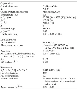
Table 2
Experimental details.
Crystal data
Chemical formula C13H10N2O2S2
Mr 290.35
Crystal system, space group Monoclinic,C2/c
Temperature (K) 170
a,b,c(A˚ ) 25.531 (6), 4.8522 (10), 20.861 (4)
() 107.81 (3)
V(A˚3) 2460.4 (10)
Z 8
Radiation type MoK
(mm1) 0.43
Crystal size (mm) 0.480.460.04
Data collection
Diffractometer Stoe IPDS 2T
Absorption correction Numerical (X-RED32and X-SHAPE; Stoe & Cie, 2010) Tmin,Tmax 0.680, 0.887
No. of measured, independent and observed [I> 2(I)] reflections
10078, 2599, 2052
Rint 0.107
(sin /)max(A˚
1) 0.633
Refinement
R[F2> 2(F2)],wR(F2),S 0.043, 0.113, 1.03 No. of reflections 2599
No. of parameters 177
H-atom treatment H atoms treated by a mixture of independent and constrained refinement
max,min(e A˚
3) 0.39,0.44
Computer programs:X-AREA (Stoe & Cie, 2010), SIR92(Altomare et al., 1994),
SHELXL2016/6(Sheldrick, 2015),XPinSHELXTL(Sheldrick, 2008),Mercury(Macrae
sup-1 Acta Cryst. (2018). E74, 901-904
supporting information
Acta Cryst. (2018). E74, 901-904 [https://doi.org/10.1107/S2056989018007892]
Crystal structure of 1-ethyl-3-(2-oxo-1,3-dithiol-4-yl)quinoxalin-2(1
H
)-one
Nicolas Chrysochos and Carola Schulzke
Computing details
Data collection: X-AREA (Stoe & Cie, 2010); cell refinement: X-AREA (Stoe & Cie, 2010); data reduction: X-AREA (Stoe & Cie, 2010); program(s) used to solve structure: SIR92 (Altomare et al., 1994); program(s) used to refine structure:
SHELXL2016/6 (Sheldrick, 2015); molecular graphics: XP in SHELXTL (Sheldrick, 2008) and Mercury (Macrae et al., 2006); software used to prepare material for publication: CIFTAB (Sheldrick, 2013) and Mercury (Macrae et al.,, 2006).
1-Ethyl-3-(2-oxo-1,3-dithiol-4-yl)quinoxalin-2(1H)-one
Crystal data
C13H10N2O2S2 Mr = 290.35
Monoclinic, C2/c a = 25.531 (6) Å
b = 4.8522 (10) Å
c = 20.861 (4) Å
β = 107.81 (3)°
V = 2460.4 (10) Å3 Z = 8
F(000) = 1200
Dx = 1.568 Mg m−3
Mo Kα radiation, λ = 0.71073 Å Cell parameters from 10798 reflections
θ = 3.4–53.5°
µ = 0.43 mm−1 T = 170 K Platelet, green
0.48 × 0.46 × 0.04 mm
Data collection
Stoe IPDS2T diffractometer
Radiation source: fine-focus sealed tube Detector resolution: 6.67 pixels mm-1 ω scans
Absorption correction: numerical
(X-Red32 and X-Shape; Stoe & Cie, 2010)
Tmin = 0.680, Tmax = 0.887
10078 measured reflections 2599 independent reflections 2052 reflections with I > 2σ(I)
Rint = 0.107
θmax = 26.7°, θmin = 1.7° h = −32→32
k = −6→6
l = −26→23
Refinement
Refinement on F2
Least-squares matrix: full
R[F2 > 2σ(F2)] = 0.043 wR(F2) = 0.113 S = 1.03 2599 reflections 177 parameters 0 restraints
Hydrogen site location: mixed
H atoms treated by a mixture of independent and constrained refinement
w = 1/[σ2(F
o2) + (0.0557P)2 + 1.3022P]
where P = (Fo2 + 2Fc2)/3
(Δ/σ)max < 0.001
Δρmax = 0.39 e Å−3
supporting information
sup-2 Acta Cryst. (2018). E74, 901-904
Special details
Geometry. All esds (except the esd in the dihedral angle between two l.s. planes) are estimated using the full covariance matrix. The cell esds are taken into account individually in the estimation of esds in distances, angles and torsion angles; correlations between esds in cell parameters are only used when they are defined by crystal symmetry. An approximate (isotropic) treatment of cell esds is used for estimating esds involving l.s. planes.
Fractional atomic coordinates and isotropic or equivalent isotropic displacement parameters (Å2)
x y z Uiso*/Ueq
S1 0.06586 (2) 1.17332 (13) 0.38431 (3) 0.02924 (17) S2 0.16189 (2) 0.80790 (12) 0.43550 (3) 0.02882 (17) O1 0.14629 (7) 1.1274 (4) 0.32955 (8) 0.0379 (4) O2 0.04198 (6) 0.7134 (3) 0.56260 (8) 0.0296 (4) N1 0.09576 (7) 0.3591 (4) 0.61708 (9) 0.0233 (4) N2 0.16734 (7) 0.4593 (4) 0.54239 (9) 0.0242 (4) C1 0.12850 (9) 1.0512 (5) 0.37418 (11) 0.0290 (5) C2 0.06873 (9) 0.9742 (5) 0.45374 (11) 0.0258 (5) C3 0.11163 (9) 0.8032 (5) 0.47709 (11) 0.0237 (4) C4 0.12261 (8) 0.6022 (5) 0.53199 (10) 0.0229 (4) C5 0.08324 (8) 0.5692 (5) 0.57104 (10) 0.0236 (4) C6 0.14444 (8) 0.2091 (5) 0.63101 (11) 0.0231 (4) C7 0.15922 (9) 0.0081 (5) 0.68143 (11) 0.0263 (5) H7 0.135972 −0.028724 0.708326 0.032* C8 0.20757 (9) −0.1364 (5) 0.69197 (11) 0.0278 (5) H8 0.217320 −0.273223 0.726147 0.033* C9 0.24248 (9) −0.0852 (5) 0.65328 (12) 0.0296 (5) H9 0.275622 −0.187017 0.661099 0.036* C10 0.22885 (9) 0.1124 (5) 0.60399 (11) 0.0273 (5) H10 0.252595 0.147249 0.577626 0.033* C11 0.17997 (9) 0.2633 (5) 0.59235 (11) 0.0237 (4) C12 0.05590 (9) 0.2972 (5) 0.65349 (12) 0.0269 (5) H12A 0.054369 0.095209 0.659446 0.032* H12B 0.018905 0.359079 0.625995 0.032* C13 0.07039 (10) 0.4352 (6) 0.72202 (12) 0.0353 (6) H13A 0.108738 0.394349 0.747326 0.053* H13B 0.046176 0.365385 0.746832 0.053* H13C 0.065600 0.634974 0.716145 0.053* H2 0.0387 (10) 0.987 (5) 0.4716 (12) 0.028 (6)*
Atomic displacement parameters (Å2)
U11 U22 U33 U12 U13 U23
sup-3 Acta Cryst. (2018). E74, 901-904
C1 0.0287 (11) 0.0313 (12) 0.0272 (11) 0.0004 (10) 0.0089 (9) 0.0011 (10) C2 0.0228 (10) 0.0296 (12) 0.0260 (10) −0.0002 (9) 0.0087 (8) 0.0003 (10) C3 0.0221 (10) 0.0253 (11) 0.0245 (10) −0.0006 (8) 0.0085 (8) −0.0029 (9) C4 0.0220 (10) 0.0239 (10) 0.0237 (10) −0.0003 (8) 0.0083 (8) −0.0029 (9) C5 0.0202 (10) 0.0262 (11) 0.0251 (10) −0.0015 (9) 0.0081 (8) −0.0037 (9) C6 0.0200 (10) 0.0228 (10) 0.0260 (10) −0.0005 (8) 0.0065 (8) −0.0038 (9) C7 0.0268 (10) 0.0262 (11) 0.0269 (11) −0.0030 (9) 0.0096 (8) −0.0009 (9) C8 0.0293 (11) 0.0238 (11) 0.0277 (11) 0.0007 (9) 0.0048 (9) 0.0018 (9) C9 0.0255 (11) 0.0278 (12) 0.0340 (12) 0.0065 (9) 0.0067 (9) −0.0016 (10) C10 0.0223 (10) 0.0315 (12) 0.0287 (11) 0.0016 (9) 0.0088 (8) −0.0022 (10) C11 0.0226 (10) 0.0243 (10) 0.0247 (10) −0.0009 (8) 0.0077 (8) −0.0021 (9) C12 0.0226 (10) 0.0282 (11) 0.0357 (12) −0.0010 (9) 0.0173 (9) 0.0010 (10) C13 0.0362 (12) 0.0401 (14) 0.0366 (13) −0.0039 (11) 0.0215 (10) −0.0032 (11)
Geometric parameters (Å, º)
S1—C2 1.724 (2) C6—C11 1.411 (3) S1—C1 1.778 (2) C7—C8 1.378 (3) S2—C3 1.755 (2) C7—H7 0.9500 S2—C1 1.758 (2) C8—C9 1.395 (3) O1—C1 1.212 (3) C8—H8 0.9500 O2—C5 1.232 (3) C9—C10 1.371 (3) N1—C5 1.370 (3) C9—H9 0.9500 N1—C6 1.392 (3) C10—C11 1.403 (3) N1—C12 1.475 (3) C10—H10 0.9500 N2—C4 1.296 (3) C12—C13 1.519 (3) N2—C11 1.374 (3) C12—H12A 0.9900 C2—C3 1.341 (3) C12—H12B 0.9900 C2—H2 0.95 (2) C13—H13A 0.9800 C3—C4 1.465 (3) C13—H13B 0.9800 C4—C5 1.483 (3) C13—H13C 0.9800 C6—C7 1.399 (3)
supporting information
sup-4 Acta Cryst. (2018). E74, 901-904
N2—C4—C3 115.80 (19) C13—C12—H12A 109.0 N2—C4—C5 124.1 (2) N1—C12—H12B 109.0 C3—C4—C5 120.11 (19) C13—C12—H12B 109.0 O2—C5—N1 121.94 (19) H12A—C12—H12B 107.8 O2—C5—C4 123.6 (2) C12—C13—H13A 109.5 N1—C5—C4 114.42 (18) C12—C13—H13B 109.5 N1—C6—C7 122.65 (19) H13A—C13—H13B 109.5 N1—C6—C11 118.2 (2) C12—C13—H13C 109.5 C7—C6—C11 119.16 (19) H13A—C13—H13C 109.5 C8—C7—C6 119.8 (2) H13B—C13—H13C 109.5 C8—C7—H7 120.1
C3—S2—C1—O1 −178.3 (2) N2—C4—C5—N1 −4.3 (3) C3—S2—C1—S1 1.57 (15) C3—C4—C5—N1 174.71 (18) C2—S1—C1—O1 179.1 (2) C5—N1—C6—C7 175.2 (2) C2—S1—C1—S2 −0.82 (15) C12—N1—C6—C7 −3.6 (3) C1—S1—C2—C3 −0.6 (2) C5—N1—C6—C11 −4.9 (3) S1—C2—C3—C4 −176.09 (18) C12—N1—C6—C11 176.29 (19) S1—C2—C3—S2 1.9 (3) N1—C6—C7—C8 178.9 (2) C1—S2—C3—C2 −2.1 (2) C11—C6—C7—C8 −0.9 (3) C1—S2—C3—C4 176.21 (16) C6—C7—C8—C9 0.3 (3) C11—N2—C4—C3 −178.92 (19) C7—C8—C9—C10 0.2 (4) C11—N2—C4—C5 0.1 (3) C8—C9—C10—C11 0.0 (3) C2—C3—C4—N2 178.4 (2) C4—N2—C11—C10 −179.0 (2) S2—C3—C4—N2 0.4 (3) C4—N2—C11—C6 1.9 (3) C2—C3—C4—C5 −0.7 (4) C9—C10—C11—N2 −179.8 (2) S2—C3—C4—C5 −178.67 (16) C9—C10—C11—C6 −0.7 (3) C6—N1—C5—O2 −174.7 (2) N1—C6—C11—N2 0.3 (3) C12—N1—C5—O2 4.1 (3) C7—C6—C11—N2 −179.8 (2) C6—N1—C5—C4 6.6 (3) N1—C6—C11—C10 −178.7 (2) C12—N1—C5—C4 −174.59 (18) C7—C6—C11—C10 1.2 (3) N2—C4—C5—O2 177.0 (2) C5—N1—C12—C13 −96.3 (2) C3—C4—C5—O2 −4.0 (3) C6—N1—C12—C13 82.5 (3)
Hydrogen-bond geometry (Å, º)
D—H···A D—H H···A D···A D—H···A
C2—H2···O2 0.95 (2) 2.30 (3) 2.859 (3) 117.2 (19) C2—H2···O2i 0.95 (2) 2.44 (2) 3.133 (3) 129 (2)
C7—H7···O1ii 0.95 2.51 3.272 (3) 138
C9—H9···S2iii 0.95 2.99 3.652 (3) 128
C12—H12A···O2iv 0.99 2.69 3.367 (3) 126