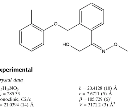
2-Methoxyimino-2-{2-[(2-methylphen-oxy)methyl]phenyl}ethanol
Rajni Kant,aVivek K. Gupta,aKamini Kapoor,aChetan S. Shripanavarband Kaushik Banerjeeb*
aX-ray Crystallography Laboratory, Post-Graduate Department of Physics &
Electronics, University of Jammu, Jammu Tawi 180 006, India, andbNational
Research Centre for Grapes, Pune 412 307, India Correspondence e-mail: [email protected]
Received 25 July 2012; accepted 8 August 2012
Key indicators: single-crystal X-ray study;T= 293 K; mean(C–C) = 0.005 A˚; disorder in main residue;Rfactor = 0.066;wRfactor = 0.212; data-to-parameter ratio = 13.7.
In the title compound, C17H19NO3, the dihedral angle between
the benzene rings is 68.0 (1). The C—O—C—C torsion angle
of the atoms joining these rings is 179.7 (2). The atoms of the
methanol group were refined as disordered over two sets of sites with fixed occupancies of 0.86 and 0.14. The H atoms of the hydroxy group in the major component are disordered over a further two sets of sites with equal occupancies. This is a necessary arrangement to allow for hydrogen bonding without unrealistic H H contacts. In the crystal, O—H N and O— H O hydrogen bonds connect molecules into chains along [001].
Related literature
The title compound was derived from kresoxim-methyl. For the biological activity of kresoxim-methyl, see: Anke et al.
(1977); Clinton et al. (2011); Balba (2007); Sudisha et al.
(2005). For related structures, see: Chopraet al.(2004); Kantet al.(2012a,b).
Experimental
Crystal data
C17H19NO3 Mr= 285.33
Monoclinic,C2=c ˚
b= 20.4128 (10) A˚ c= 7.6711 (5) A˚
= 105.729 (6) ˚
Z= 8
MoKradiation
= 0.08 mm1
T= 293 K 0.30.20.1 mm
Data collection
Oxford Diffraction Xcalibur Sapphire3 diffractometer Absorption correction: multi-scan
(CrysAlis PRO; Oxford Diffraction, 2010) Tmin= 0.790,Tmax= 1.000
11340 measured reflections 2788 independent reflections 1497 reflections withI> 2(I) Rint= 0.052
Refinement
R[F2> 2(F2)] = 0.066 wR(F2) = 0.212 S= 1.08 2788 reflections 204 parameters
2 restraints
H-atom parameters constrained max= 0.39 e A˚3
[image:1.610.52.259.572.747.2]min=0.22 e A˚3
Table 1
Hydrogen-bond geometry (A˚ ,).
D—H A D—H H A D A D—H A
O11A—H11Y O11Ai
0.84 1.77 2.614 (8) 178
O11A—H11Z O11Aii
0.84 2.11 2.950 (14) 178
O11B—H11X N3iii 0.84 2.21 3.046 (18) 177
Symmetry codes: (i)xþ1;y;zþ2; (ii)xþ1;y;zþ3
2; (iii)x;y;zþ12. Data collection: CrysAlis PRO(Oxford Diffraction, 2010); cell refinement: CrysAlis PRO; data reduction: CrysAlis PRO; program(s) used to solve structure: SHELXS97 (Sheldrick, 2008); program(s) used to refine structure:SHELXL97(Sheldrick, 2008); molecular graphics: ORTEP-3 (Farrugia, 1997) and SHELXTL
(Sheldrick, 2008); software used to prepare material for publication:
PLATON(Spek, 2009).
RK acknowledges the Department of Science & Tech-nology for access to the single-crystal X-ray diffractometer sanctioned as a National Facility under project No. SR/S2/ CMP-47/2003.
Supplementary data and figures for this paper are available from the IUCr electronic archives (Reference: LH5508).
References
Anke, T., Oberwinkler, F., Steglich, W. & Schramm, G. (1977).J. Antibiot.30, 806–810.
Balba, H. (2007).J. Environ. Sci. Health B,42, 441–451.
Chopra, D., Mohan, T. P., Rao, K. S. & Guru Row, T. N. (2004).Acta Cryst.E60, o2421–o2423.
Clinton, B., Warden, A. C., Haboury, S., Easton, C. J., Kotsonis, S., Taylor, M. C., Oakeshott, J. G., Russell, R. J. & &Scott, C. (2011).Biocatal. Biotransfor. 28, 119–129.
Farrugia, L. J. (1997).J. Appl. Cryst.30, 565.
Kant, R., Gupta, V. K., Kapoor, K., Shripanavar, C. S. & Banerjee, K. (2012a). Acta Cryst.E68, o2425.
Kant, R., Gupta, V. K., Kapoor, K., Shripanavar, C. S. & Banerjee, K. (2012b). Acta Cryst.E68, o2426.
Oxford Diffraction (2010).CrysAlis PRO. Oxford Diffraction Ltd, Yarnton, England.
Sheldrick, G. M. (2008).Acta Cryst.A64, 112–122. Spek, A. L. (2009).Acta Cryst.D65, 148–155.
Sudisha, J., Amruthesh, K. N., Deepak, S. A., Shetty, N. P., Sarosh, B. R. & Shekar Shetty, H. (2005).Pest. Biochem. Physiol.81, 188–197.
Acta Crystallographica Section E
Structure Reports
Online
supporting information
Acta Cryst. (2012). E68, o2697 [doi:10.1107/S160053681203499X]
2-Methoxyimino-2-{2-[(2-methylphenoxy)methyl]phenyl}ethanol
Rajni Kant, Vivek K. Gupta, Kamini Kapoor, Chetan S. Shripanavar and Kaushik Banerjee
S1. Comment
Kresoxim-methyl is a widely used agricultural fungicide of the strobilurin group (Anke et al., 1977; Clinton et al., 2011;
Balba, 2007). It is a broad-spectrum systemic compound with novel mode of action (Sudisha et al., 2005). While
exploring its fate in the environment, we have derived a new compound by the process of reduction. This may contribute
to the understanding of the metabolic and environmental fate of this compound. The crystal structure of the title
compound (I) is presented herein.
In (I)(Fig. 1), all bond lengths and angles are normal and correspond to those observed in the related structures (Chopra
et al., 2004; Kant et al., 2012a,b). The dihedral angle between the two benzene rings is 68.0 (1)°. The C—O—C—C
torsion angle of the atoms joining these rings is 179.7 (2) °. The atoms of the methanol group were refined as disordered
over two sets of sites with fixed occupancies of 0.86 and 0.14. The H atoms of the hydroxy group in the major component
are disordered over a further two sets of sites with equal occupancies. This is a necessary arrangement to allow for
hydrogen bonding without unrealistic H···H contacts. The O—H···O hydrogen bond motif of one the O—H disorder
components is shown in Fig. 2. For the other disorder component in the O—H···O hydrogen bonds, the acceptors become
donors and vice-versa. In the crystal, O—H···N and O—H···O hydrogen bonds connect molecules to form chains along
[001].
S2. Experimental
Finely powdered sodium borohydride (6 eq., 0.06 mol) was suspended in tetrahydrofuran in presence of kresoxim-methyl
(3.13 g m, 0.01 mol) under reflux (343 K) with stirring for 1 h. Then methanol (8 ml) was slowly added drop wise.
Stirring and refluxing were maintained until the reaction was completed as monitored by TLC. After the end of the
reaction, the reaction mixture was cooled to room temperature and quenched with a saturated solution of ammonium
chloride (15 ml) for further period of 1.5 h. The product was separated by extraction with ethyl acetate (2x25 ml). The
organic extracts were combined and dried over sodium sulfate and concentrated under low pressure to yield the final
product. The synthesized compound was dissolved in methanol and subjected to slow evaporation to produce colourless
crystals.
S3. Refinement
All H atoms were positioned geometrically and were treated as riding on their parent atoms, with O—H distance of 0.84
Å and C—H distances of 0.93–0.97 Å and with Uiso(H) = 1.2Ueq(C) or 1.5Ueq(methyl C,O). The disordered H atoms of the
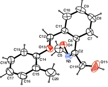
Figure 1
The molecular structure of the title compound with ellipsoids drawn at the 40% probability level.
Figure 2
Part of the crystal structure showing the hydrogen bonds along [001] as dashed lines. For the other disorder component in
[image:3.610.129.481.396.660.2]2-Methoxyimino-2-{2-[(2-methylphenoxy)methyl]phenyl}ethanol
Crystal data
C17H19NO3 Mr = 285.33 Monoclinic, C2/c
Hall symbol: -C 2yc
a = 21.0394 (14) Å
b = 20.4128 (10) Å
c = 7.6711 (5) Å
β = 105.729 (6)°
V = 3171.2 (3) Å3 Z = 8
F(000) = 1216
Dx = 1.195 Mg m−3
Mo Kα radiation, λ = 0.71073 Å Cell parameters from 2909 reflections
θ = 3.6–29.0°
µ = 0.08 mm−1 T = 293 K Block, colourless 0.3 × 0.2 × 0.1 mm
Data collection
Oxford Diffraction Xcalibur Sapphire3 diffractometer
Radiation source: fine-focus sealed tube Graphite monochromator
Detector resolution: 16.1049 pixels mm-1 ω scans
Absorption correction: multi-scan
(CrysAlis PRO; Oxford Diffraction, 2010)
Tmin = 0.790, Tmax = 1.000
11340 measured reflections 2788 independent reflections 1497 reflections with I > 2σ(I)
Rint = 0.052
θmax = 25.0°, θmin = 3.6°
h = −23→24
k = −23→24
l = −9→9
Refinement
Refinement on F2
Least-squares matrix: full
R[F2 > 2σ(F2)] = 0.066 wR(F2) = 0.212 S = 1.08 2788 reflections 204 parameters 2 restraints
Primary atom site location: structure-invariant direct methods
Secondary atom site location: difference Fourier map
Hydrogen site location: inferred from neighbouring sites
H-atom parameters constrained
w = 1/[σ2(Fo2) + (0.0921P)2]
where P = (Fo2 + 2Fc2)/3
(Δ/σ)max = 0.002
Δρmax = 0.39 e Å−3
Δρmin = −0.22 e Å−3
Special details
Experimental. CrysAlis PRO, Oxford Diffraction Ltd., Version 1.171.34.40 (release 27–08-2010 CrysAlis171. NET) (compiled Aug 27 2010,11:50:40) Empirical absorption correction using spherical harmonics, implemented in SCALE3 ABSPACK scaling algorithm.
Geometry. All e.s.d.'s (except the e.s.d. in the dihedral angle between two l.s. planes) are estimated using the full covariance matrix. The cell e.s.d.'s are taken into account individually in the estimation of e.s.d.'s in distances, angles and torsion angles; correlations between e.s.d.'s in cell parameters are only used when they are defined by crystal symmetry. An approximate (isotropic) treatment of cell e.s.d.'s is used for estimating e.s.d.'s involving l.s. planes.
Refinement. Refinement of F2 against ALL reflections. The weighted R-factor wR and goodness of fit S are based on F2,
conventional R-factors R are based on F, with F set to zero for negative F2. The threshold expression of F2 > σ(F2) is used
only for calculating R-factors(gt) etc. and is not relevant to the choice of reflections for refinement. R-factors based on F2
are statistically about twice as large as those based on F, and R- factors based on ALL data will be even larger.
Fractional atomic coordinates and isotropic or equivalent isotropic displacement parameters (Å2)
x y z Uiso*/Ueq Occ. (<1)
O13 0.24680 (10) 0.14485 (9) 0.7894 (3) 0.0689 (7)
N3 0.37326 (13) 0.10321 (12) 0.5462 (4) 0.0709 (8)
C2 0.38302 (15) 0.11036 (14) 0.7167 (4) 0.0638 (8)
C5 0.3638 (2) 0.1523 (2) 0.2722 (5) 0.1041 (13)
H5A 0.3630 0.1934 0.2110 0.156*
H5B 0.3242 0.1283 0.2174 0.156*
H5C 0.4014 0.1273 0.2629 0.156*
C6 0.39179 (15) 0.17396 (14) 0.8143 (3) 0.0544 (8)
C7 0.45455 (16) 0.19128 (17) 0.9162 (4) 0.0736 (9)
H7A 0.4898 0.1630 0.9228 0.088*
C8 0.4652 (2) 0.2500 (2) 1.0079 (5) 0.0912 (12)
H8A 0.5077 0.2616 1.0736 0.109*
C9 0.4141 (3) 0.2911 (2) 1.0029 (5) 0.0943 (12)
H9A 0.4216 0.3307 1.0653 0.113*
C10 0.3504 (2) 0.27400 (16) 0.9042 (5) 0.0785 (10)
H10A 0.3155 0.3022 0.9023 0.094*
C11 0.33831 (16) 0.21524 (14) 0.8083 (4) 0.0577 (8)
C12 0.27043 (14) 0.19845 (14) 0.7035 (4) 0.0635 (9)
H12A 0.2418 0.2361 0.6974 0.076*
H12B 0.2701 0.1864 0.5810 0.076*
C14 0.18363 (16) 0.12314 (14) 0.7087 (4) 0.0596 (8)
C15 0.16296 (18) 0.06974 (15) 0.7930 (5) 0.0722 (9)
C16 0.0989 (2) 0.04775 (18) 0.7189 (6) 0.0901 (12)
H16A 0.0832 0.0129 0.7735 0.108*
C17 0.0578 (2) 0.0762 (2) 0.5666 (7) 0.0991 (13)
H17A 0.0152 0.0604 0.5195 0.119*
C18 0.07975 (19) 0.1270 (2) 0.4858 (5) 0.0902 (12)
H18A 0.0523 0.1457 0.3820 0.108*
C19 0.14248 (17) 0.15098 (15) 0.5562 (5) 0.0737 (9)
H19A 0.1572 0.1862 0.5006 0.088*
C20 0.2088 (2) 0.03868 (18) 0.9558 (5) 0.1147 (15)
H20A 0.2484 0.0250 0.9269 0.172*
H20B 0.1877 0.0013 0.9919 0.172*
H20C 0.2197 0.0698 1.0532 0.172*
O11A 0.4567 (3) 0.0312 (2) 0.8725 (8) 0.253 (3) 0.86
H11Y 0.4837 0.0106 0.9553 0.380* 0.86
H11Z 0.4806 0.0312 0.8007 0.380* 0.86
C11A 0.3946 (2) 0.0467 (2) 0.8269 (7) 0.0952 (17) 0.86
H11A 0.3796 0.0522 0.9351 0.114* 0.43
H11B 0.3691 0.0116 0.7556 0.114* 0.43
O11B 0.3776 (10) −0.0022 (7) 0.757 (2) 0.107 (7) 0.14
H11X 0.3746 −0.0298 0.8361 0.161* 0.14
C11B 0.3637 (19) 0.0532 (6) 0.822 (4) 0.0952 (17) 0.14
H11C 0.3875 0.0566 0.9491 0.114* 0.14
Atomic displacement parameters (Å2)
U11 U22 U33 U12 U13 U23
O4 0.1028 (18) 0.0737 (14) 0.0500 (13) −0.0026 (12) 0.0234 (11) 0.0048 (10)
O13 0.0712 (15) 0.0688 (13) 0.0615 (13) −0.0153 (11) 0.0090 (11) 0.0183 (10)
N3 0.091 (2) 0.0592 (15) 0.0623 (18) 0.0014 (13) 0.0212 (14) −0.0015 (13)
C2 0.080 (2) 0.0564 (18) 0.0553 (19) −0.0021 (16) 0.0193 (15) 0.0060 (15)
C5 0.141 (4) 0.120 (3) 0.050 (2) −0.007 (3) 0.023 (2) −0.008 (2)
C6 0.067 (2) 0.0615 (18) 0.0362 (16) −0.0124 (16) 0.0163 (14) 0.0013 (12)
C7 0.074 (2) 0.095 (2) 0.054 (2) −0.0094 (19) 0.0206 (17) −0.0034 (18)
C8 0.088 (3) 0.118 (3) 0.070 (2) −0.043 (3) 0.026 (2) −0.019 (2)
C9 0.128 (4) 0.081 (3) 0.083 (3) −0.043 (3) 0.045 (3) −0.030 (2)
C10 0.110 (3) 0.063 (2) 0.075 (2) −0.004 (2) 0.046 (2) −0.0010 (17)
C11 0.071 (2) 0.0549 (17) 0.0504 (17) −0.0103 (17) 0.0220 (15) 0.0061 (13)
C12 0.073 (2) 0.0567 (18) 0.0632 (19) −0.0037 (15) 0.0227 (16) 0.0184 (15)
C14 0.067 (2) 0.0572 (18) 0.0570 (19) −0.0061 (16) 0.0201 (16) −0.0041 (14)
C15 0.082 (3) 0.0623 (19) 0.078 (2) −0.0110 (19) 0.0319 (19) −0.0012 (17)
C16 0.094 (3) 0.073 (2) 0.115 (3) −0.025 (2) 0.050 (3) −0.016 (2)
C17 0.070 (3) 0.096 (3) 0.130 (4) −0.011 (2) 0.026 (3) −0.035 (3)
C18 0.071 (3) 0.092 (3) 0.101 (3) 0.004 (2) 0.011 (2) −0.012 (2)
C19 0.068 (2) 0.075 (2) 0.076 (2) 0.0006 (19) 0.0161 (18) 0.0016 (17)
C20 0.145 (4) 0.092 (3) 0.110 (3) −0.029 (2) 0.038 (3) 0.036 (2)
O11A 0.166 (5) 0.175 (4) 0.355 (9) 0.019 (4) −0.040 (4) 0.159 (5)
C11A 0.089 (5) 0.075 (3) 0.113 (4) 0.026 (3) 0.012 (3) 0.024 (3)
O11B 0.20 (2) 0.031 (8) 0.083 (12) 0.001 (11) 0.026 (12) −0.011 (8)
C11B 0.089 (5) 0.075 (3) 0.113 (4) 0.026 (3) 0.012 (3) 0.024 (3)
Geometric parameters (Å, º)
O4—N3 1.403 (3) C14—C19 1.375 (4)
O4—C5 1.412 (4) C14—C15 1.396 (4)
O13—C14 1.378 (3) C15—C16 1.388 (5)
O13—C12 1.434 (3) C15—C20 1.497 (5)
N3—C2 1.277 (3) C16—C17 1.379 (5)
C2—C6 1.485 (4) C16—H16A 0.9300
C2—C11A 1.533 (5) C17—C18 1.352 (5)
C2—C11B 1.535 (7) C17—H17A 0.9300
C5—H5A 0.9600 C18—C19 1.374 (4)
C5—H5B 0.9600 C18—H18A 0.9300
C5—H5C 0.9600 C19—H19A 0.9300
C6—C7 1.386 (4) C20—H20A 0.9600
C6—C11 1.396 (4) C20—H20B 0.9600
C7—C8 1.378 (5) C20—H20C 0.9600
C7—H7A 0.9300 O11A—C11A 1.298 (6)
C8—C9 1.355 (5) O11A—H11Y 0.8400
C8—H8A 0.9300 O11A—H11Z 0.8399
C9—C10 1.394 (5) C11A—H11A 0.9700
C10—C11 1.394 (4) O11B—C11B 1.300 (8)
C10—H10A 0.9300 O11B—H11X 0.8400
C11—C12 1.477 (4) C11B—H11C 0.9700
C12—H12A 0.9700 C11B—H11D 0.9700
C12—H12B 0.9700
N3—O4—C5 108.7 (2) C19—C14—C15 120.9 (3)
C14—O13—C12 116.8 (2) O13—C14—C15 115.2 (3)
C2—N3—O4 111.8 (2) C16—C15—C14 116.9 (3)
N3—C2—C6 125.5 (3) C16—C15—C20 122.6 (3)
N3—C2—C11A 115.2 (3) C14—C15—C20 120.4 (3)
C6—C2—C11A 118.9 (3) C17—C16—C15 121.8 (4)
N3—C2—C11B 117.4 (12) C17—C16—H16A 119.1
C6—C2—C11B 114.4 (9) C15—C16—H16A 119.1
O4—C5—H5A 109.5 C18—C17—C16 119.9 (4)
O4—C5—H5B 109.5 C18—C17—H17A 120.1
H5A—C5—H5B 109.5 C16—C17—H17A 120.1
O4—C5—H5C 109.5 C17—C18—C19 120.3 (4)
H5A—C5—H5C 109.5 C17—C18—H18A 119.9
H5B—C5—H5C 109.5 C19—C18—H18A 119.9
C7—C6—C11 120.1 (3) C18—C19—C14 120.2 (3)
C7—C6—C2 118.5 (3) C18—C19—H19A 119.9
C11—C6—C2 121.5 (3) C14—C19—H19A 119.9
C8—C7—C6 120.5 (3) C15—C20—H20A 109.5
C8—C7—H7A 119.7 C15—C20—H20B 109.5
C6—C7—H7A 119.7 H20A—C20—H20B 109.5
C9—C8—C7 120.4 (4) C15—C20—H20C 109.5
C9—C8—H8A 119.8 H20A—C20—H20C 109.5
C7—C8—H8A 119.8 H20B—C20—H20C 109.5
C8—C9—C10 119.9 (3) C11A—O11A—H11Y 138.5
C8—C9—H9A 120.0 C11A—O11A—H11Z 124.1
C10—C9—H9A 120.0 H11Y—O11A—H11Z 95.5
C9—C10—C11 120.9 (3) O11A—C11A—C2 110.6 (4)
C9—C10—H10A 119.6 O11A—C11A—H11A 109.5
C11—C10—H10A 119.6 C2—C11A—H11A 109.5
C10—C11—C6 118.1 (3) O11A—C11A—H11B 109.5
C10—C11—C12 119.9 (3) C2—C11A—H11B 109.5
C6—C11—C12 122.0 (3) H11A—C11A—H11B 108.1
O13—C12—C11 109.4 (2) C11B—O11B—H11X 104.0
O13—C12—H12A 109.8 O11B—C11B—C2 109.9 (12)
C11—C12—H12A 109.8 O11B—C11B—H11C 109.7
O13—C12—H12B 109.8 C2—C11B—H11C 109.7
C11—C12—H12B 109.8 O11B—C11B—H11D 109.7
H12A—C12—H12B 108.2 C2—C11B—H11D 109.7
C19—C14—O13 123.9 (3) H11C—C11B—H11D 108.2
C5—O4—N3—C2 174.3 (3) C10—C11—C12—O13 −110.4 (3)
O4—N3—C2—C11A −175.6 (3) C12—O13—C14—C19 −1.9 (4)
O4—N3—C2—C11B 157.0 (14) C12—O13—C14—C15 178.2 (2)
N3—C2—C6—C7 −105.8 (4) C19—C14—C15—C16 −1.8 (5)
C11A—C2—C6—C7 66.3 (4) O13—C14—C15—C16 178.0 (3)
C11B—C2—C6—C7 93.5 (16) C19—C14—C15—C20 178.3 (3)
N3—C2—C6—C11 75.6 (4) O13—C14—C15—C20 −1.8 (4)
C11A—C2—C6—C11 −112.3 (3) C14—C15—C16—C17 1.5 (5)
C11B—C2—C6—C11 −85.1 (16) C20—C15—C16—C17 −178.6 (4)
C11—C6—C7—C8 −1.9 (4) C15—C16—C17—C18 −0.2 (6)
C2—C6—C7—C8 179.5 (3) C16—C17—C18—C19 −0.9 (6)
C6—C7—C8—C9 1.5 (5) C17—C18—C19—C14 0.5 (5)
C7—C8—C9—C10 −0.1 (5) O13—C14—C19—C18 −179.0 (3)
C8—C9—C10—C11 −0.7 (5) C15—C14—C19—C18 0.9 (5)
C9—C10—C11—C6 0.2 (4) N3—C2—C11A—O11A 87.9 (5)
C9—C10—C11—C12 −179.5 (3) C6—C2—C11A—O11A −85.0 (5)
C7—C6—C11—C10 1.1 (4) C11B—C2—C11A—O11A −171 (3)
C2—C6—C11—C10 179.6 (3) N3—C2—C11B—O11B 38 (3)
C7—C6—C11—C12 −179.2 (2) C6—C2—C11B—O11B −160 (2)
C2—C6—C11—C12 −0.6 (4) C11A—C2—C11B—O11B −53.1 (15)
C14—O13—C12—C11 179.7 (2)
Hydrogen-bond geometry (Å, º)
D—H···A D—H H···A D···A D—H···A
O11A—H11Y···O11Ai 0.84 1.77 2.614 (8) 178
O11A—H11Z···O11Aii 0.84 2.11 2.950 (14) 178
O11B—H11X···N3iii 0.84 2.21 3.046 (18) 177