research communications
Acta Cryst.(2014).E70, 415–417 doi:10.1107/S1600536814023216
415
Received 9 October 2014 Accepted 21 October 2014
Edited by H. Stoeckli-Evans, University of Neuchaˆtel, Switzerland
†
Keywords:crystal structure; acenaphthylene; hydrazinecarbothioamide; thiosemicarbazones; hydrogen bonding; C—H interactions
CCDC reference:1030348
Supporting information:this article has supporting information at journals.iucr.org/e
Crystal structure of (2
E
)-
N
-methyl-2-(2-oxo-1,2-di-
hydroacenaphthylen-1-ylidene)hydrazinecarbo-thioamide
G. Vimala,aJ. Govindaraj,bJ. Haribabu,cR. Karvembucand A. SubbiahPandia,c*
a
Department of Physics, Presidency College (Autonomous), Chennai 600 005, India,bDepartment of Physics, Pachaiyappa’s College for Men, Kancheepuram 631 501, India, andcDepartment of Chemistry, National Institute of Technology, Trichy 620 015, India. *Correspondence e-mail: aspandian59@gmail.com
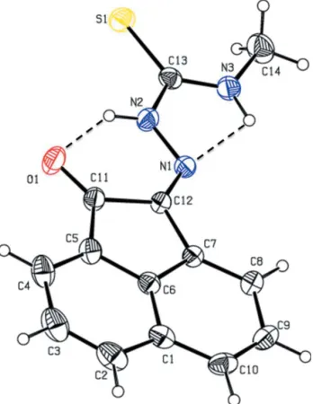
In the title compound, C14H11N3OS, the acenaphthylene ring system and
hydrazinecarbothioamide unit (=N—NH—C=S—NH–) are essentially coplanar [with maximum deviations from their mean planes of 0.009 (2) and 0.033 (2) A˚ , respectively], and make a dihedral angle of 1.59 (9). The molecular
conformation is stabilized by two weak intramolecular hydrogen bonds (N— H O and N—H N), which generateS(6) andS(5) ring motifs. In the crystal, molecules are linked by N—H S hydrogen bonds, forming chains along [010]. The chains are linkedviapairs of C—H O hydrogen bonds, enclosingR22(10)
ring motifs, and C—H interactions, forming a three-dimensional framework. The absolute structure of the title compound was determined by resonant scattering.
1. Chemical context
The design and synthesis of thiosemicarbazones are of considerable interest because of their versatile chemistry and various biological activities, such as antitumor, antibacterial, antiviral, antiamoebic and antimalarial (Kelly et al., 1996). They comprise an intriguing class of chelating molecules, which possess a wide range of beneficial medicinal properties (Prabhakaranet al.2008). Thiosemicarbazones are a versatile class of ligands that have been studied for their biological activity (Chellanet al., 2010), their interesting binding motifs (Lobanaet al., 2009) and their use as ligands in catalysis (Xieet al., 2010). In view of their biological importance, the crystal structure of the title compound has been determined and the results are presented herein.
2. Structural commentary
The molecular structure of the title compound is illustrated in Fig. 1. The atoms of both the acenaphthylene ring system and the =N—NH—C=S—NH– segment are essentially coplanar, the maximum deviations from their mean planes being
tively. The dihedral angle between the benzene and cyclo-pentane rings of the acenapthalene unit is 1.59 (9). The
molecular structure is stabilized by N—H O and N—H N hydrogen bonds, forming S(6) andS(5) ring motifs, respec-tively (Table 1 and Fig. 1).
3. Supramolecular features
In the crystal, molecules are linked by N—H S hydrogen bonds (Table 1 and Fig. 2), forming chains along [010]. The chains are linked via pairs of C—H O hydrogen bonds, enclosing R2
2(10) ring motifs, and C—H interactions,
forming a three-dimensional framework (Table 1 and Fig. 2).
4. Database survey
A search of the Cambridge Structural Database (Version 5.35, last update May 2014; Groom & Allen, 2014) for the substructure 2-(imino)acenaphthylen-1(2H)-one gave 13 hits, including that of the ethyl analogue of the title compound,
acenaphthylene-1,2-dione 4-ethylthiosemicarbazone (GUR-HAD; Pascu et al., 2010). The two molecules differ in the dihedral angle between the mean planes of the acenaphthyl-ene ring system and hydrazinecarbothioamide unit (=N— NH—C=S—NH–) which is 1.59 (9)in the title compound but
9.14 (6)in the ethyl analogue (GURHAD; Pascuet al., 2010). In the crystals of both compounds, molecules are linkedvia N—H S hydrogen bonds, forming chains along [010].
5. Synthesis and crystallization
An ethanolic solution of N-methylhydrazinecarbothioamide (0.01 mol) was added to an ethanolic solution (50 ml) containing acenaphthylene-1,2-dione (0.01 mol). The mixture was refluxed for 2 h during which time a yellow precipitate separated out. The reaction mixture was then cooled to room temperature and the precipitate was filtered off. It was then washed with ethanol and dried under vacuum. The yield of the isolated product was 89%. Single crystals suitable for X-ray diffraction were obtained by slow evaporation of a solution of the title compound in ethanol at room temperature.
6. Refinement
Crystal data, data collection and structure refinement details are summarized in Table 2. All H atoms were fixed
geom-416
Vimalaet al. C14H11N3OS Acta Cryst.(2014).E70, 415–417
[image:2.610.345.533.71.382.2]research communications
Figure 1
[image:2.610.79.253.74.299.2]The molecular structure of the title compound, with the atom labelling. Displacement ellipsoids are drawn at the 30% probability level. Hydrogen bonds are shown as dashed lines (see Table 1 for details).
Table 1
Hydrogen-bond geometry (A˚ ,).
Cgis the centroid of ring C1/C6–C10.
D—H A D—H H A D A D—H A
N2—H2 O1 0.86 2.03 2.7178 (19) 136 N3—H3 N1 0.86 2.26 2.6437 (19) 107 N3—H3 S1i 0.86 2.64 3.4407 (15) 156 C4—H4 O1ii 0.93 2.47 3.246 (2) 141 C2—H2A Cgiii 0.93 2.76 3.502 (2) 137
Symmetry codes: (i) xþ1;y1 2;zþ
1
2; (ii) xþ
1 2;yþ
1
2;z; (iii)
xþ1 2;y
1 2;z.
Figure 2
[image:2.610.45.296.661.727.2]etrically and allowed to ride on their parent atoms: N—H = 0.86 and C—H = 0.93–0.97 A˚ and withUiso(H) = 1.5Ueq(C) for
methyl H atoms and = 1.2Ueq(C) for other H atoms. The
absolute structure of the title compound was determined by resonant scattering, with a Flack parameter of 0.02 (8).
Acknowledgements
The authors thank Dr Babu Varghese, SAIF, IIT, Chennai, India for the data collection.
References
Bruker (2008). APEX2, SAINT and SADABS. Bruker AXS Inc., Madison, Wisconsin, USA.
Chellan, P., Shunmoogam-Gounden, N., Hendricks, D. T., Gut, J., Rosenthal, P. J., Lategan, C., Smith, P. J., Chibale, K. & Smith, G. S. (2010).Eur. J. Inorg. Chem.pp, 3520–3528.
Farrugia, L. J. (2012).J. Appl. Cryst.45, 849–854. Flack, H. D. (1983).Acta Cryst.A39, 876–881.
Groom, C. R. & Allen, F. H. (2014).Angew. Chem. Int. Ed.53, 662– 671.
Kelly, P. F., Slawin, A. M. Z. & Soriano-Rama, A. (1996).J. Chem. Soc. Dalton Trans.pp. 53–59.
Lobana, T. S., Sharma, R., Bawa, G. & Khanna, S. (2009).Coord. Chem. Rev.253, 977–1055.
Pascu, S. I., Waghorn, P. A., Kennedy, B. W. C., Arrowsmith, R. L., Bayly, S. R., Dilworth, J. R., Christlieb, M., Tyrrell, R. M., Zhong, J., Kowalczyk, R. M., Collison, D., Aley, P. K., Churchill, G. C. & Aigbirhio, F. I. (2010).Chem. Asian J.5, 506–519.
Prabhakaran, R., Huang, R., Renukadevi, S. V., Karvembu, R., Zeller, M. & Natarajan, K. (2008).Inorg. Chim. Acta,361, 2547– 2552.
Sheldrick, G. M. (2008).Acta Cryst.A64, 112–122. Spek, A. L. (2009).Acta Cryst.D65, 148–155.
Xie, G., Chellan, P., Mao, J., Chibale, K. & Smith, G. S. (2010).Adv. Synth. Catal.352, 1641–1647.
research communications
Acta Cryst.(2014).E70, 415–417 Vimalaet al. C
[image:3.610.43.291.87.366.2]14H11N3OS
417
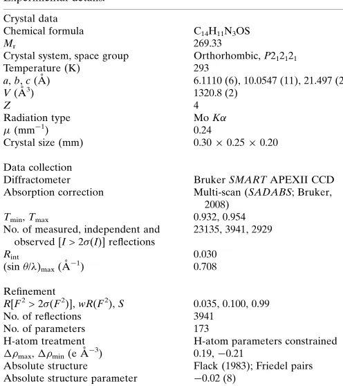
Table 2
Experimental details.
Crystal data
Chemical formula C14H11N3OS
Mr 269.33
Crystal system, space group Orthorhombic,P212121 Temperature (K) 293
a,b,c(A˚ ) 6.1110 (6), 10.0547 (11), 21.497 (2)
V(A˚3) 1320.8 (2)
Z 4
Radiation type MoK
(mm1) 0.24
Crystal size (mm) 0.300.250.20
Data collection
Diffractometer BrukerSMARTAPEXII CCD Absorption correction Multi-scan (SADABS; Bruker,
2008)
Tmin,Tmax 0.932, 0.954 No. of measured, independent and
observed [I> 2(I)] reflections
23135, 3941, 2929
Rint 0.030
(sin/)max(A˚
1
) 0.708
Refinement
R[F2> 2(F2)],wR(F2),S 0.035, 0.100, 0.99 No. of reflections 3941
No. of parameters 173
H-atom treatment H-atom parameters constrained
max,min(e A˚
3
) 0.19,0.21
Absolute structure Flack (1983); Friedel pairs Absolute structure parameter 0.02 (8)
supporting information
sup-1
Acta Cryst. (2014). E70, 415-417supporting information
Acta Cryst. (2014). E70, 415-417 [doi:10.1107/S1600536814023216]
Crystal structure of (2
E
)-
N
-methyl-2-(2-oxo-1,2-dihydroacenaphthylen-1-yl-idene)hydrazinecarbothioamide
G. Vimala, J. Govindaraj, J. Haribabu, R. Karvembu and A. SubbiahPandi
Computing details
Data collection: APEX2 (Bruker, 2008); cell refinement: SAINT (Bruker, 2008); data reduction: SAINT (Bruker, 2008); program(s) used to solve structure: SHELXS97 (Sheldrick, 2008); program(s) used to refine structure: SHELXL97
(Sheldrick, 2008); molecular graphics: ORTEP-3 for Windows (Farrugia, 2012) and PLATON (Spek, 2009); software used to prepare material for publication: SHELXL97 (Sheldrick, 2008) and PLATON (Spek, 2009).
(2E)-N-Methyl-2-(2-oxo-1,2-dihydroacenaphthylen-1-ylidene)hydrazinecarbothioamide
Crystal data
C14H11N3OS
Mr = 269.33
Orthorhombic, P212121
Hall symbol: P 2ac 2ab
a = 6.1110 (6) Å
b = 10.0547 (11) Å
c = 21.497 (2) Å
V = 1320.8 (2) Å3
Z = 4
F(000) = 560
Dx = 1.354 Mg m−3
Mo Kα radiation, λ = 0.71073 Å
µ = 0.24 mm−1
T = 293 K Block, yellow
0.30 × 0.25 × 0.20 mm
Data collection
Bruker SMART APEXII CCD diffractometer
Radiation source: fine-focus sealed tube Graphite monochromator
ω and φ scans
Absorption correction: multi-scan (SADABS; Bruker, 2008)
Tmin = 0.932, Tmax = 0.954
23135 measured reflections 3941 independent reflections 2929 reflections with I > 2σ(I)
Rint = 0.030
θmax = 30.2°, θmin = 2.2°
h = −8→8
k = −13→14
l = −29→29
Refinement
Refinement on F2
Least-squares matrix: full
R[F2 > 2σ(F2)] = 0.035
wR(F2) = 0.100
S = 0.99 3941 reflections 173 parameters 0 restraints
Primary atom site location: structure-invariant direct methods
Secondary atom site location: difference Fourier map
Hydrogen site location: inferred from neighbouring sites
H-atom parameters constrained
w = 1/[σ2(F
o2) + (0.0532P)2 + 0.2048P]
where P = (Fo2 + 2Fc2)/3
(Δ/σ)max = 0.001
Δρmax = 0.19 e Å−3
supporting information
sup-2
Acta Cryst. (2014). E70, 415-417Absolute structure: Flack (1983); Friedel pairs Absolute structure parameter: −0.02 (8)
Special details
Geometry. All e.s.d.'s (except the e.s.d. in the dihedral angle between two l.s. planes) are estimated using the full covariance matrix. The cell e.s.d.'s are taken into account individually in the estimation of e.s.d.'s in distances, angles and torsion angles; correlations between e.s.d.'s in cell parameters are only used when they are defined by crystal symmetry. An approximate (isotropic) treatment of cell e.s.d.'s is used for estimating e.s.d.'s involving l.s. planes.
Refinement. Refinement of F2 against ALL reflections. The weighted R-factor wR and goodness of fit S are based on F2,
conventional R-factors R are based on F, with F set to zero for negative F2. The threshold expression of F2 > σ(F2) is used
only for calculating R-factors(gt) etc. and is not relevant to the choice of reflections for refinement. R-factors based on F2
are statistically about twice as large as those based on F, and R-factors based on ALL data will be even larger.
Fractional atomic coordinates and isotropic or equivalent isotropic displacement parameters (Å2)
x y z Uiso*/Ueq
supporting information
sup-3
Acta Cryst. (2014). E70, 415-417Atomic displacement parameters (Å2)
U11 U22 U33 U12 U13 U23
S1 0.0421 (2) 0.0413 (2) 0.0654 (3) 0.0080 (2) 0.0033 (2) −0.01024 (19) N1 0.0345 (6) 0.0331 (6) 0.0420 (7) 0.0008 (6) 0.0020 (7) −0.0007 (5) C6 0.0373 (9) 0.0370 (8) 0.0369 (8) −0.0031 (7) −0.0004 (7) −0.0047 (6) O1 0.0621 (8) 0.0423 (7) 0.0605 (8) 0.0123 (7) 0.0046 (7) 0.0140 (6) N2 0.0397 (7) 0.0361 (7) 0.0488 (8) 0.0054 (6) 0.0055 (7) 0.0043 (6) C7 0.0353 (8) 0.0338 (8) 0.0336 (7) 0.0004 (6) −0.0001 (6) −0.0013 (6) C13 0.0337 (8) 0.0365 (8) 0.0438 (8) −0.0028 (6) 0.0001 (7) −0.0089 (7) N3 0.0401 (8) 0.0406 (7) 0.0528 (8) 0.0015 (6) 0.0077 (7) −0.0009 (7) C12 0.0356 (8) 0.0326 (7) 0.0368 (7) 0.0007 (6) −0.0014 (7) 0.0021 (6) C5 0.0498 (10) 0.0395 (9) 0.0381 (8) −0.0029 (8) 0.0047 (8) −0.0009 (7) C11 0.0436 (9) 0.0357 (8) 0.0415 (8) −0.0005 (7) −0.0003 (7) 0.0040 (7) C8 0.0454 (10) 0.0386 (9) 0.0407 (9) 0.0029 (8) 0.0000 (8) 0.0019 (7) C9 0.0523 (10) 0.0432 (9) 0.0521 (10) 0.0129 (9) −0.0088 (8) −0.0013 (8) C14 0.0583 (12) 0.0610 (12) 0.0707 (13) −0.0037 (11) 0.0226 (12) 0.0021 (9) C1 0.0388 (9) 0.0476 (9) 0.0441 (9) −0.0044 (8) 0.0042 (7) −0.0129 (8) C2 0.0513 (12) 0.0702 (14) 0.0596 (12) −0.0090 (10) 0.0196 (10) −0.0180 (10) C4 0.0742 (14) 0.0496 (10) 0.0495 (10) −0.0103 (11) 0.0164 (11) 0.0068 (8) C3 0.0747 (16) 0.0684 (15) 0.0581 (12) −0.0179 (12) 0.0285 (12) −0.0009 (10) C10 0.0397 (10) 0.0547 (11) 0.0572 (11) 0.0100 (8) −0.0044 (8) −0.0157 (9)
Geometric parameters (Å, º)
S1—C13 1.6744 (17) C5—C11 1.472 (3) N1—C12 1.283 (2) C8—C9 1.412 (2) N1—N2 1.3528 (19) C8—H8 0.9300 C6—C1 1.394 (2) C9—C10 1.367 (3)
C6—C5 1.405 (2) C9—H9 0.9300
C6—C7 1.410 (2) C14—H14A 0.9600 O1—C11 1.213 (2) C14—H14B 0.9600 N2—C13 1.362 (2) C14—H14C 0.9600
N2—H2 0.8600 C1—C2 1.409 (3)
C7—C8 1.363 (2) C1—C10 1.411 (3) C7—C12 1.461 (2) C2—C3 1.367 (3) C13—N3 1.312 (2) C2—H2A 0.9300 N3—C14 1.449 (2) C4—C3 1.403 (3)
N3—H3 0.8600 C4—H4 0.9300
C12—C11 1.515 (2) C3—H3A 0.9300 C5—C4 1.364 (3) C10—H10 0.9300
supporting information
sup-4
Acta Cryst. (2014). E70, 415-417C13—N2—H2 119.6 N3—C14—H14B 109.5 C8—C7—C6 118.78 (16) H14A—C14—H14B 109.5 C8—C7—C12 134.48 (16) N3—C14—H14C 109.5 C6—C7—C12 106.73 (13) H14A—C14—H14C 109.5 N3—C13—N2 116.67 (15) H14B—C14—H14C 109.5 N3—C13—S1 125.49 (13) C6—C1—C2 115.65 (19) N2—C13—S1 117.84 (13) C6—C1—C10 115.92 (16) C13—N3—C14 124.05 (16) C2—C1—C10 128.43 (18) C13—N3—H3 118.0 C3—C2—C1 121.1 (2) C14—N3—H3 118.0 C3—C2—H2A 119.4 N1—C12—C7 125.20 (14) C1—C2—H2A 119.4 N1—C12—C11 127.56 (15) C5—C4—C3 117.9 (2) C7—C12—C11 107.22 (14) C5—C4—H4 121.1 C4—C5—C6 119.92 (18) C3—C4—H4 121.1 C4—C5—C11 132.73 (18) C2—C3—C4 122.4 (2) C6—C5—C11 107.34 (15) C2—C3—H3A 118.8 O1—C11—C5 129.05 (16) C4—C3—H3A 118.8 O1—C11—C12 125.21 (16) C9—C10—C1 120.19 (17) C5—C11—C12 105.74 (14) C9—C10—H10 119.9 C7—C8—C9 118.19 (17) C1—C10—H10 119.9
supporting information
sup-5
Acta Cryst. (2014). E70, 415-417Hydrogen-bond geometry (Å, º)
Cg is the centroid of ring C1/C6–C10.
D—H···A D—H H···A D···A D—H···A
N2—H2···O1 0.86 2.03 2.7178 (19) 136 N3—H3···N1 0.86 2.26 2.6437 (19) 107 N3—H3···S1i 0.86 2.64 3.4407 (15) 156
C4—H4···O1ii 0.93 2.47 3.246 (2) 141
C2—H2A···Cgiii 0.93 2.76 3.502 (2) 137