Received 22 July 2017 Accepted 4 August 2017
Edited by W. T. A. Harrison, University of Aberdeen, Scotland
Keywords:crystal structure; redetermination; hydrogen bonding; symmetric hydrogen bonds; refinement constraints; refinement restraints; Cambridge Structural Database.
CCDC reference:1566932
Supporting information:this article has supporting information at journals.iucr.org/e
Institute of Physics of the Czech Academy of Sciences, Na Slovance 2, 182 21 Praha 8, Czech Republic. *Correspondence e-mail: fabry@fzu.cz
The structure of the title molecular salt, C10H13N4 +
N3
, has been redetermined from the data published by Qian & Huang [Acta Cryst. (2010), E66, o3086;
refcode WACMIY (Groom et al., 2016)]. The improvement of the present
redetermination consists in a correction of the site-occupancy parameter of the bridging H atom between the pyridine rings, as well as of its position. The present study has shown that the bridging H atom (site symmetry 2) is involved in a symmetric N H N hydrogen bond, which is one of the longest ever observed [N N = 2.678 (3) A˚ ]. In addition, there are also present weaker
Nam—H Naz hydrogen bonds (am = amine and az = azide) of moderate
strength and-electron pyridine -electron interactions in the structure. All the azide N atoms also lie on a twofold axis.
1. Chemical context
Structures that contain hydroxyl and secondary and primary amine groups are sometimes determined incorrectly because of an assumed geometry of these groups from which the applied constraints or restraints were inferred. In such cases, the correct geometry is missed as it is not verified by inspec-tion of the difference electron-density maps. Thus, a consid-erable number of structures could have been determined more accurately – cf. Figs. 1 and 2 in Fa´bry et al. (2014). The inclusion of such erroneous structures causes bias in crystal-lographic databases such as the Cambridge Structural Data-base (Groomet al., 2016).
In the course of recalculation of suspect structures that were retrieved from the Cambridge Structural Database (Groomet al., 2016), the structure determination of the title structure by Qian & Huang (2010) with the pertinent CSD refcode WACMIY became a candidate for a checking recalculation. The reason was that both the primary and secondary amine groups were constrained with distance constraints equal to 0.86 A˚ , with planar conformation andUiso(H) = 1.2Ueq(N).
related by a crystallographic twofold axis, was modelled by two (undisordered) H atoms both with occupational para-meters equal to 1: such a structural motif is impossible. The present article describes the redetermination of bis(4-amino-pyridinium)(1+) azide(1), which was reported by Qian & Huang (2010).
2. Structural commentary
The components of the title molecular salt are shown in Fig. 1. It is seen that the bridging hydrogen atom (H2a) interconnects symmmetry-related 4-aminopyridine molecules; the symmetry operation for atoms with the suffix ‘a’ is the same as symmetry
code (i) in Table 1 and Fig. 2, viz. x + 1, y, z + 1 2. The
interplanar angle between the pyridine rings N2/C1–C5 and N2i/C1i–C5iis 87.90 (7).
Table 1 lists the hydrogen bonds in the structure. The packing of the ions in the unit cell is shown in Fig. 2. Fig. 3 shows the difference electron-density map calculated without
the bridging hydrogen atom H2a in the region
N2 (H2a) N2i. A well-defined, single peak in this map indicates that H2a is situated on a twofold axis, i.e. it is involved in a symmetric hydrogen bond while not being disordered. This hydrogen bond is the strongest hydrogen bond in the structure and is one of the family of long symmetric hydrogen bonds N H N as listed in Table 1. As Tables 1 and 2 show, the title structure contains the second
longest known truly symmetric N H N hydrogen bond
after CAFHAT01.
The remaining N—Ham Naz (am = primary amine, az =
azide) hydrogen bonds are considerably weaker, though still
research communications
Acta Cryst.(2017). E73, 1344–1347 Jan Fa´bry C
[image:2.610.45.294.72.214.2]10H13N4+N3
1345
Table 1
Hydrogen-bond geometry (A˚ ,).
D—H A D—H H A D A D—H A
N2—H2a N2i 1.3391 (16) 1.3391 (16) 2.678 (3) 178 (2)
N1—H1a N3ii 0.927 (14) 2.067 (14) 2.990 (2) 173.6 (13) N1—H1b N5iii 0.857 (16) 2.154 (16) 3.010 (2) 177.9 (14)
Symmetry codes: (i)xþ1;y;zþ1
2; (ii)xþ1;y;z; (iii)xþ 1 2;y
[image:2.610.314.565.413.673.2]1 2;z.
Figure 2
A view of the title structure along the unit-cell axisa. Symmetry codes: (i)
x+ 1,y,z+1
2; (ii)x+ 1,y,z; (iii)x+ 1 2,y
1
2,z. Applied colours for atoms: grey = C and H, blue = N; applied colours for bonds: black = covalent bonds, dashed orange = H hydrogen bonds acceptor (Brandenburg & Putz, 2005).
Figure 1
View of the constituent molecules of the title structure after the improved refinement. The displacement ellipsoids are depicted at the 30% probability level (Spek, 2009).
Figure 3
A section of the difference electron-density map for the present redetermined title structure, which shows the build up of the electron density between the atoms N and Ni[symmetry code: (i)x+ 1,y,z+1
[image:2.610.46.297.446.690.2]of moderate strength (Gilli & Gilli, 2009). Atom H1aforms a link to the terminal azide nitrogen atom N3 while H1bbonds to the other terminal azide atom N5. The graph-set motif is described in the Supramolecular featuressection. In addition to the hydrogen-bonding interactions, there are also -elec-tron ring -electron pyridine interactions in the structure. The distance between the ring centroids N2/C1–C5 and N2iv/C1iv–C5ivis 3.7145 (17) A˚ [symmetry code: (iv) x + 1,
y+ 1,z+ 1].
The primary amine group centered on N1 is almost planar
[C3—N1—H1a= 120.0 (9), C3—N1—H1b= 119.1 (9), H1a—
N1—H1b = 120.6 (13)] despite the somewhat lengthened
C3—N1 bond [1.345 (2) A˚ ]. The reason may be found in the
hydrogen bonds formed by the group with N—H N bond
angles being close to 180.
Once again, the present redetermination emphasizes the importance of careful examination of the difference electron-density maps during a structure determination.
3. Supramolecular features
In addition to the above-mentioned symmetric hydrogen bond N2 H2a N2i [symmetry code: (i) x + 1,y, z + 1
2] for
which the graph-set motif notation is missing (the donors act simultaneously as acceptors in the title structure; Etteret al., 1990) the principal graph-set motif in which the primary amine group as well the azide atoms are involved isR64(20).
In a detail, the atoms involved in this graph-set motif are as follows (Fig. 2): N3v–H1avi–N1vi–H1bvi–N5ii–N4ii–N3ii–H1a– N1–H1b–N5iii–H1bvii–N1vii–H1avii–N3–N4–N5–H1bviii–N1viii– H1aviii[symmetry codes: (ii)x+ 1,y,z; (iii)x+1
2,y 1 2,z; (v)
x+1 2,y+
1
2,z; (vi)x+ 3 2,y+
1 2,z+
3
2; (vii)x+ 1,y,z+ 3 2;
(viii)x1 2,y+
1 2,z].
The hydrogen bonds in this graph set motif are directed along the unit-cell parameterb.
4. Synthesis and crystallization
The preparation of the title compound was described by Qian & Huanget al. (2010) in the supporting information of their article.
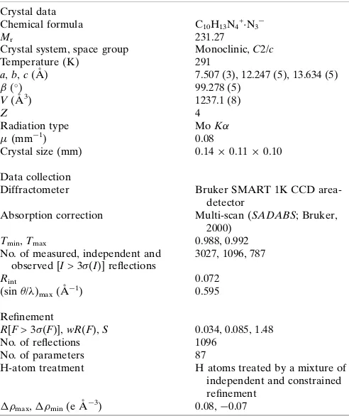
Radiation type MoK
(mm1) 0.08
Crystal size (mm) 0.140.110.10
Data collection
Diffractometer Bruker SMART 1K CCD area-detector
Absorption correction Multi-scan (SADABS; Bruker, 2000)
Tmin,Tmax 0.988, 0.992
No. of measured, independent and observed [I> 3(I)] reflections
3027, 1096, 787
Rint 0.072
(sin/)max(A˚
1
) 0.595
Refinement
R[F> 3(F)],wR(F),S 0.034, 0.085, 1.48 No. of reflections 1096
No. of parameters 87
H-atom treatment H atoms treated by a mixture of independent and constrained refinement
max, min(e A˚
3
) 0.08,0.07
Computer programs:SMARTandSAINT(Bruker, 2000),SHELXTL(Sheldrick, 2008),
PLATON (Spek, 2009), DIAMOND (Brandenburg & Putz, 2005) andJANA2006
(Petrˇı´cˇeket al., 2014).
CAFHAT01 1.34 1.37 2.7018 169.8 CAFHAT01b
1.35 1.35 2.7009 175.3 COFMUF10c 1.35 (10) 1.50 (10) 2.844 (7) 171 (11) DAHGUO01d
1.33 (6) 1.38 (6) 2.690 (8) 168 (6) EFAZOBe 1.32 (5) 1.38 (5) 2.692 (5) 176 (4) EPIWUXf
1.33 (3) 1.33 (2) 2.657 (9) 172 (8) FISROPg
1.45 (4) 1.51 (4) 2.963 (3) 173 (2) FOGKAPh
1.31 (4) 1.34 (4) 2.652 (5) 175 (4) HUJNUWi
1.341 (15) 1.414 (16) 2.68 (2) 152.7 (8) IYEVOXj 1.33 (7) 1.37 (7) 2.691 (6) 174 (6) MIJMUNk
1.27 (7) 1.56 (7) 2.812 (7) 165 (5) MIJMUNk 1.34 (9) 1.52 (10) 2.808 (7) 159 (8) OBUCOEl
1.33 (3) 1.43 (3) 2.736 (2) 165 (3) QUHFEGm 1.39 (4) 1.40 (4) 2.792 (10) 176 (5) SIZSUQn
1.317 (14) 1.319 (14) 2.63 (2) 176.8 (9) WOFGIIo 1.33 (4) 1.39 (4) 2.706 (4) 167 (3) XICRIMp
1.31 (4) 1.52 (4) 2.826 (3) 164 (3) ZEYLIAq
1.32 (4) 1.51 (4) 2.833 (4) 175 (3)
Notes: (a) 2-(1,3-Benzoxazol-2-yl)-1-phenylvinyl benzoate (Orozcoet al., 2009); (b) hydrogen bis[bis(2-{[(imidazol-4-yl)methylene]amino}ethyl){2-[(imidazolato)methyl-ene]amino}ethyl)amine]cobalt(III) triperchlorate heptahydrate (Marsh & Clemente, 2007); (c) 2,1,3-benzoselenadiazole 2,1,3-benzoselenadiazolium pentaiodide (Gierenet al., 1985); (d) bis{[1,4-diazoniabicyclo(2.2.2)octane][1-aza-4-azoniabicyclo(2.2.2)octane]} tetrakis(tribromide) dibromide (Heraviet al., 2005); (e) bis[(3,5-dimethylpyrazole)(3,5-dimethylpyrazolyl)]platinum(II) (Umakoshiet al., 2008); (f) 4-{2-(pyridin-4-yl)oxy]-1,2-bis(2,3,5,6-tetrafluoro-4-iodophenyl)ethoxy}pyridin-1-ium iodide bis(nitrobenzene) (Martı´-Rujas et al., 2012); (g) 5,6:14,15-dibenzo-1,4-dioxa-8-azonia-12-azacyclopenta-deca-5,14-diene 5,6:14,15-dibenzo-1,4-dioxa-8,12-diazacyclopentadeca-5,14-diene per-chlorate (Tusˇek-Bozˇic´et al., 2005); (h) dioxidotetrakis(methylpyridine)rhenium(V) 4-methylpyridinium 4-methylpyridine diodide (Krawczyk et al., 2014); (i) 4-methyl-pyridinium trans-bis(-picoline)tetrakis(thiocyanato)molybdenum 4-methylpyridine (Kitanovski et al., 2009); (j) bis(4,40-bipyridinium) hexakis(
2
-sulfido)tetra-germaniumtetrasulfide 4,40-bipyridine heptahydrate (Wang et al., 2003); (k) 4,40 -bipyridinium 4-(pyrid-4-yl)pyridinium 4,40-bipyridine hexakis(isothiocyanato-N)-iron (Weiet al., 2002); (l) tris(2-benzimidazolylmethyl)ammonium dinitrobenzoate 3,5-dinitrobenzoic acid clathrate (Ji et al., 2004); (m) (2R,4S,5R)-9-(hydroxyimino)-60 -methoxycinchonan-1-ium (2R,4S,5R)-N-hydroxy-60-methoxycinchonan-9-imine chloride methanol solvate (Zohri et al., 2015); (n) catena-[bis(2-aqua)-(5-cyano-2H
[image:3.610.314.560.92.385.2] [image:3.610.44.295.144.337.2]5. Database survey
The structure determination by Qian & Huang (2010) has been included into the Cambridge Structural Database (Groomet al., 2016) under the refcode WACMIY.
6. Refinement
Table 3 lists the details regarding the crystal data, data collection and the refinement. The starting structural model was taken from the determination by Qian & Huang (2010). All hydrogen atoms were discernible in the difference elec-tron-density map. The aryl hydrogen atoms were constrained by Caryl—Haryl = 0.93 A˚ and Uiso(Haryl) = 1.2Ueq(Caryl). The
positional parameters of the primary amine hydrogen atoms were refined freely while their displacement parameters were constrained by Uiso(HN2) = 1.2Ueq(N2). The bridging
hydrogen atom H2ainvolved in the symmetric hydrogen bond
N2 H2a N2i was refined freely. Refinements using
JANA2006 andSHELXL(Sheldrick, 2008) with the threshold for observed diffractionsI= 2(I) led to the same result of the bridging hydrogen atom being located on the twofold axis.
Acknowledgements
The support by the grant of the Czech Science Foundation 15– 12653S is gratefully acknowledged.
References
Al-Azmi, A., George, P. & El-Dusouqui, O. M. E. (2007). Heterocycles,71, 2183–2201.
Braga, D., Giaffreda, S. L., Grepioni, F., Palladino, G. & Polito, M. (2008).New J. Chem.32, 820–828.
Brandenburg, K. & Putz, H. (2005). DIAMOND. Crystal Impact GbR, Bonn, Germany.
Bruker (2000).SMART, SAINTand SADABS. Bruker AXS Inc., Madison, Wisconsin. USA.
Cherukuvada, S., Bolla, G., Sikligar, K. & Nangia, A. (2013).Cryst. Growth Des.13, 1551–1557.
Etter, M. C., MacDonald, J. C. & Bernstein, J. (1990).Acta Cryst.B46, 256–262.
Fa´bry, J., Dusˇek, M., Vaneˇk, P., Rafalovskyi, I., Hlinka, J. & Urban, J. (2014).Acta Cryst.C70, 1153–1160.
Gieren, A., Hubner, T., Lamm, V., Neidlein, R. & Droste, D. (1985). Z. Anorg. Allg. Chem.523, 33–44.
Gilli, G. & Gilli, P. (2009).The Nature of the Hydrogen Bond, p. 61. New York: Oxford University Press Inc.
Groom, C. R., Bruno, I. J., Lightfoot, M. P. & Ward, S. C. (2016).Acta Cryst. B72, 171–179.
Heravi, M. M., Derikvand, F., Ghassemzadeh, M. & Neumu¨ller, B. (2005).Tetrahedron Lett.46, 6243–6245.
Ji, B., Jian, F., Xiao, H. & Du, V. (2004).Anal. Sci. X-ray Struct. Anal. Online,20, x101–x102.
Kitanovski, N., Golobic, A. & Ceh, B. (2009).Croat. Chem. Acta,82, 567–571.
Krawczyk, M. K., Krawczyk, M. S., Siczek, M. & Lis, T. (2014).Inorg. Chim. Acta,418, 84–92.
Marsh, R. E. & Clemente, D. A. (2007).Inorg. Chim. Acta,360, 4017– 4024.
Martı´-Rujas, J., Colombo, L., Lu, J., Dey, A., Terraneo, G., Metrangolo, P., Pilati, T. & Resnati, G. (2012).Chem. Commun.
48, 8207–8209.
Orozco, F., Insuasty, B., Cobo, J. & Glidewell, C. (2009).Acta Cryst. C65, o257–o260.
Perumalla, S. R., Pedireddi, V. R. & Sun, C. C. (2013).Cryst. Growth Des.13, 429–432.
Petrˇı´cˇek, V., Dusˇek, M. & Palatinus, L. (2014).Z. Kristallogr.229, 345–352.
Qian, H.-F. & Huang, W. (2010).Acta Cryst.E66, o3086. Sheldrick, G. M. (2008).Acta Cryst.A64, 112–122. Spek, A. L. (2009).Acta Cryst.D65, 148–155.
Tusˇek-Bozˇic´, L., Visˇnjevac, A., Marotta, E. & Kojic´-Prodic´, B. (2005). Polyhedron,24, 97–111.
Umakoshi, K., Kojima, T., Saito, K., Akatsu, S., Onishi, M., Ishizaka, S., Kitamura, N., Nakao, Y., Sakaki, S. & Ozawa, Y. (2008).Inorg. Chem.47, 5033–5035.
Wang, M.-S., Chen, W.-T., Cai, L.-Z., Zhou, G.-W., Guo, G.-C. & Huang, J.-S. (2003).J. Cluster Sci.14, 495–504.
Wei, Y., Zhu, Y., Song, Y., Hou, H. & Fan, Y. (2002).Inorg. Chem. Commun.5, 166–170.
Zohri, M., Wartchow, R. & Hoffmann, H. M. R. (2015). Private communication. CCDC, Cambridge, England.
research communications
Acta Cryst.(2017). E73, 1344–1347 Jan Fa´bry C
sup-1 Acta Cryst. (2017). E73, 1344-1347
azide(1
−
): redetermination from the original data
Jan F
á
bry
Computing details
Data collection: SMART (Bruker, 2000); cell refinement: SAINT (Bruker, 2000); data reduction: SAINT (Bruker, 2000);
program(s) used to solve structure: SHELXTL (Sheldrick, 2008); program(s) used to refine structure: JANA2006 (Petříček
et al., 2014); molecular graphics: PLATON (Spek, 2009), DIAMOND (Brandenburg & Putz, 2005) and JANA2006
(Petříček et al., 2014); software used to prepare material for publication: JANA2006 (Petříček et al., 2014).
µ-Hydrido-bis(4-aminopyridinium) azide
Crystal data
C10H13N4+·N3−
Mr = 231.27
Monoclinic, C2/c
Hall symbol: -C 2yc
a = 7.507 (3) Å
b = 12.247 (5) Å
c = 13.634 (5) Å
β = 99.278 (5)°
V = 1237.1 (8) Å3
Z = 4
F(000) = 488
Dx = 1.242 Mg m−3
Mo Kα radiation, λ = 0.71073 Å
Cell parameters from 1359 reflections
θ = 3.0–25.4°
µ = 0.08 mm−1
T = 291 K
Block, colourless 0.14 × 0.11 × 0.10 mm
Data collection
Bruker SMART 1K CCD area-detector diffractometer
Radiation source: fine-focus sealed tube Graphite monochromator
φ and ω scans
Absorption correction: multi-scan (SADABS; Bruker, 2000)
Tmin = 0.988, Tmax = 0.992
3027 measured reflections 1096 independent reflections 787 reflections with I > 3σ(I)
Rint = 0.072
θmax = 25.0°, θmin = 3.0°
h = −8→8
k = −12→14
l = −16→15
Refinement
Refinement on F2
R[F > 3σ(F)] = 0.034
wR(F) = 0.085
S = 1.48
1096 reflections 87 parameters 0 restraints 18 constraints
H atoms treated by a mixture of independent and constrained refinement
Weighting scheme based on measured s.u.'s w =
1/(σ2(I) + 0.0004I2)
(Δ/σ)max = 0.004
Δρmax = 0.08 e Å−3
supporting information
sup-2 Acta Cryst. (2017). E73, 1344-1347
Special details
Experimental. The structure was solved by direct methods (Bruker, 2000) and successive difference Fourier syntheses.
Fractional atomic coordinates and isotropic or equivalent isotropic displacement parameters (Å2)
x y z Uiso*/Ueq
C1 0.6719 (2) 0.44584 (12) 0.40174 (12) 0.0889 (6)
H1 0.732036 0.496631 0.368331 0.1067*
C2 0.72245 (17) 0.43524 (10) 0.50154 (11) 0.0778 (5)
H2 0.814514 0.478469 0.535055 0.0934*
C3 0.63529 (16) 0.35896 (9) 0.55351 (10) 0.0702 (5)
C4 0.49858 (17) 0.29730 (11) 0.49826 (11) 0.0793 (5)
H4 0.436698 0.245345 0.529429 0.0952*
C5 0.4560 (2) 0.31347 (12) 0.39842 (12) 0.0936 (6)
H5 0.364461 0.271568 0.362714 0.1123*
N1 0.68183 (18) 0.34619 (10) 0.65226 (9) 0.0859 (5)
H1a 0.776 (2) 0.3866 (12) 0.6868 (10) 0.1031*
H1b 0.631 (2) 0.2960 (12) 0.6815 (11) 0.1031*
N2 0.54018 (19) 0.38706 (11) 0.34930 (8) 0.0943 (5)
N3 0 0.47492 (16) 0.75 0.1020 (8)
N4 0 0.57130 (18) 0.75 0.0768 (6)
N5 0 0.66663 (17) 0.75 0.1074 (8)
H2a 0.5 0.389 (2) 0.25 0.160 (9)*
Atomic displacement parameters (Å2)
U11 U22 U33 U12 U13 U23
C1 0.0946 (10) 0.0866 (10) 0.0930 (11) 0.0183 (8) 0.0376 (9) 0.0120 (8)
C2 0.0752 (8) 0.0760 (8) 0.0853 (10) 0.0109 (6) 0.0224 (7) 0.0054 (7)
C3 0.0685 (7) 0.0688 (7) 0.0762 (9) 0.0166 (6) 0.0205 (6) 0.0039 (6)
C4 0.0762 (8) 0.0795 (8) 0.0847 (10) 0.0065 (6) 0.0205 (7) 0.0007 (7)
C5 0.0948 (10) 0.0999 (10) 0.0854 (11) 0.0105 (8) 0.0125 (8) −0.0094 (8)
N1 0.0909 (8) 0.0865 (8) 0.0804 (9) −0.0012 (5) 0.0144 (6) 0.0093 (6)
N2 0.1093 (9) 0.1039 (9) 0.0725 (8) 0.0233 (7) 0.0229 (7) 0.0027 (7)
N3 0.0983 (12) 0.0857 (11) 0.1212 (15) 0 0.0154 (10) 0
N4 0.0625 (8) 0.1024 (13) 0.0667 (9) 0 0.0143 (6) 0
N5 0.1140 (14) 0.0918 (12) 0.1263 (15) 0 0.0495 (12) 0
Geometric parameters (Å, º)
C1—H1 0.93 C5—H5 0.93
C1—C2 1.359 (2) C5—N2 1.340 (2)
C1—N2 1.334 (2) N1—H1a 0.927 (14)
C2—H2 0.93 N1—H1b 0.857 (16)
C2—C3 1.397 (2) H1a—H1b 1.55 (2)
C3—C4 1.3935 (19) N2—H2a 1.3391 (16)
C3—N1 1.345 (2) N3—N4 1.180 (3)
sup-3 Acta Cryst. (2017). E73, 1344-1347
C2—C3—C4 116.91 (12) C1—N2—C5 117.61 (13)
C2—C3—N1 121.21 (11) C1—N2—H2a 123.9 (8)
C4—C3—N1 121.88 (12) C5—N2—H2a 118.2 (9)
C3—C4—H4 120.16 N3—N4—N5 180.0 (5)
C3—C4—C5 119.68 (13) N2—H2a—N2i 178 (2)
H4—C4—C5 120.16
Symmetry code: (i) −x+1, y, −z+1/2.
Hydrogen-bond geometry (Å, º)
D—H···A D—H H···A D···A D—H···A
N2—H2a···N2i 1.3391 (16) 1.3391 (16) 2.678 (3) 178 (2)
N1—H1a···N3ii 0.927 (14) 2.067 (14) 2.990 (2) 173.6 (13)
N1—H1b···N5iii 0.857 (16) 2.154 (16) 3.010 (2) 177.9 (14)