798
https://doi.org/10.1107/S2056989017006387 Acta Cryst.(2017). E73, 798–803research communications
Received 11 April 2017 Accepted 27 April 2017
Edited by T. J. Prior, University of Hull, England
Keywords:crystal structure; carboxamide; pyrazine; pyridine; silver(I); Ag—Ag bond; three-dimensional coordination polymer; hydrogen bonding.
CCDC references:1546645; 1546644
Supporting information:this article has supporting information at journals.iucr.org/e
Crystal structure of a pyrazine-2,3-dicarboxamide
ligand and of its silver(I) nitrate complex, a
three-dimensional coordination polymer
Dilovan S. Catiaand Helen Stoeckli-Evansb*
a
Debiopharm International S.A., Chemin Messidor 5-7, CP 5911, CH-1002 Lausanne, Switzerland, andbInstitute of Physics, University of Neuchaˆtel, rue Emile-Argand 11, CH-2000 Neuchaˆtel, Switzerland. *Correspondence e-mail: [email protected]
The title ligand, C18H16N6O22H2O (L1) [N 2
,N3 -bis(pyridin-4-ylmethyl)-pyrazine-2,3-dicarboxamide], crystallized as a dihydrate. The molecule is U-shaped with the carboxamide groups beingcisto one another, making a dihedral angle of 81.6 (5). The terminal pyridine rings are inclined to one another by
58.5 (4). There is an intramolecular N—H Npyrazinehydrogen bond present,
forming anS(5) ring motif. In the crystal, adjacent molecules are linked by N— H Ocarboxamide hydrogen bonds, forming a chain along [001]. A chain of
hydrogen-bonded water molecules is linked to the chain of (L1) molecules by O—H N hydrogen bonds, forming columns propagating along thecaxis. The columns are linked by C—H O and C—H N hydrogen bonds, forming a three-dimensional supramolecular structure. The reaction of ligand (L1) with silver(I) nitrate led to the formation of a new three-dimensional coordination polymer, {[Ag(C18H16N6O2)]NO3}n, poly[[[4-N
2
,N3 -bis(pyridin-4-ylmeth-yl)pyrazine-2,3-dicarboxamide]silver(I)] nitrate] (I). The asymmetric unit is composed of half of one silver ion, located on a twofold rotation axis, half a ligand molecule and half a positionally disordered nitrate anion located about a twofold rotation axis. The full molecule of the ligand is generated by twofold rotational symmetry, with this twofold axis bisecting the Car—Carbonds of the
pyrazine ring and the Ag—Ag bond. The carboxamide groups are nowtransto one another, making a dihedral angle of 65.8 (4). The two terminal pyridine rings are inclined to one another by 6.6 (3). Two ligands wrap around an Ag—
Ag bond of 3.1638 (11) A˚ , forming a figure-of-eight-shaped complex molecule. Each silver ion is coordinated by two pyridine N atoms and by two carboxamide O atoms of neighbouring molecules, hence forming a three-dimensional framework. The nitrate anion is linked to the framework by N—H O and C—H O hydrogen bonds.
1. Chemical context
The title ligand, N2,N3 -bis(pyridin-4-ylmethyl)pyrazine-2,3-dicarboxamide (L1), is one of a series of ligands synthesized in
order to study the superexchange in supramolecular
complexes formed using pyrazine carboxamide derivatives and first row transition metal ions (Cati, 2002; Catiet al., 2004). To the best of our knowledge, neither the synthesis nor the crystal structure of (L1) have been described previously. It is very similar to the ligand N2,N3 -bis(pyridin-2-ylmethyl)-pyrazine-2,3-dicarboxamide (L2), for which a number of transition metal complexes have been described, including some interesting tetranuclear 22 grid-like and square complexes (Hausmannet al., 2003; Klingeleet al., 2007). Two such complexes, {[Cu4(L2)4](ClO4)4}5CH3OH4H2O and
{[Ni4(L2)4]Cl4}5CH3CN13H2O (Cati et al., 2004), exhibit
anion encapsulation, and magnetic susceptibility measure-ments indicate that they are weakly anti-ferromagnetic, withJ values of5.87 and2.64 cm1, respectively.
A search of the Cambridge Structural Database (CSD; Groomet al., 2016), indicated that silver nitrate is an excellent metal salt for the formation of multi-dimensional coordination polymers. The silver ion can have multiple coordination geometries and modes, and the nitrate anion has been shown to coordinate to metal ions in a number of different modes, many of which involve bridging metal ions. The properties of the complexes formed are extremely varied. For example, with the tetradendate ligand 1,6-bis(2H-1,2,3-triazol-2-yl)hexane, Huoet al. (2016) synthesized the three-dimensional coordin-ation polymer, catena-[[-2,20-(butane-1,4-diyl)bis(2H
-1,2,3-triazole)]bis(-nitrato)disilver]. They showed that it exhibits highly selective and sensitive luminescence sensing of Cr2O7
2
ions in aqueous solution. With the rigid tripodal arene-core-based nitrogen ligand, 1,3,5-tris(pyrazol-1-yl)benzene, Shu et al. (2006) formed a porous metal–organic framework, viz. catena-[bis(3-nitrato-O,O0,O00)bis(3
-1,3,5-tris(pyrazol-1-yl)-benzene-N,N0,N00)trisilver(I) nitrate]. The nitrate counter-anions located in the cationic framework can be exchanged reversibly without destruction of the structure. Hence, this compound can act as a zeolite-like porous material for anion exchange.
The title ligand has potentially two bidentate (N,N) and two monodentate (Npyridine) coordination sites. It is therefore an
interesting ligand to study its coordination behaviour with silver nitrate, and herein, we describe the solid state structures of ligand (L1), and the new three-dimensional coordination polymer, poly[[[4-N
2
,N3 -bis(pyridin-4-ylmethyl)pyrazine-2,3-dicarboxamide]silver(I)]nitrate] (I).
2. Structural commentary
The title ligand (L1) crystallized as a dihydrate, and its mol-ecular structure is illustrated in Fig. 1. The molecule is
U-shaped with the carboxamide groups (C6/N3/C5/O1) beingcis to one another, making a dihedral angle of 81.6 (5). The
terminal pyridine rings (N4/C7–C11) are inclined to one another by 58.5 (4). There is an intramolecular N—H N
hydrogen bond present, forming anS(5) ring motif (Fig. 1 and Table 1).
[image:2.610.53.289.376.591.2]The reaction of the ligand with silver(I) nitrate led to the formation of a three-dimensional coordination polymer (I). The coordination of the ligand to the silver ions is illustrated in Fig. 2. Selected bond lengths and angles in (I) are given in Table 2. The asymmetric unit is composed of a silver ion, located on a twofold rotation axis, half a ligand molecule and half a nitrate anion. The full molecule of the ligand is gener-ated by twofold rotational symmetry, with this twofold axis bisecting the C4—C4i bonds of the pyrazine ring and the Ag1—Ag1ibond (Table 2). The carboxamide groups (C6/N3/ C5/O1) are nowtransto one another, making a dihedral angle of 65.8 (4). The terminal pyridine rings (N4/C7–C11) are
inclined to one another by 6.6 (3). Two ligands effectively
wrap around a Ag—Ag bond of 3.1638 (11) A˚ , forming a figure-of-eight-shaped molecule, with each silver ion being coordinated by two pyridine N atoms. The silver ions are each further coordinated by the carboxamide O atom, O1, of neighbouring molecules, hence forming a three-dimensional
research communications
Acta Cryst.(2017). E73, 798–803 Cati and Stoeckli-Evans C
framework, illustrated in Fig. 3. If one considers that the silver ion, Ag1, is fivefold coordinate (N2O2Agi) then its
coordina-tion sphere can be described as distorted trigonal–bipyr-amidal, with a 5 value of 0.8 (5 = 1 for perfect trigonal–
pyramidal geometry and 0 for perfect square-pyramidal geometry; Addisonet al., 1984). However, if one considers the Ag1 ion to be fourfold coordinate, N2O2, with a4value of
0.55, its coordination sphere can be described as intermediate between trigonal–pyramidal and seesaw (4= 1 for a perfect
tetrahedral geometry and 0 for a perfect square-planar geometry. For intermediate structures, including trigonal– pyramidal and seesaw,4falls within the range of 0 to 1; Yang
et al., 2007). The nitrate anion that does not coordinate to the silver(I) ion is positionally disordered, and also located about a twofold rotation axis.
3. Supramolecular features
In the crystal of ligand (L1), molecules are linked by N— H O(water) hydrogen bonds forming chains propagating along thec-axis direction (Table 1 and Fig. 4). Parallel to this chain of molecules is a chain of hydrogen-bonded water molecules (Table 1 and Fig. 4), which is linked to the chain of (L1) molecules by O—H N hydrogen bonds, forming columns propagating along thecaxis. The columns are linked
by C—H O and C—H N hydrogen bonds, forming a
three-dimensional supramolecular structure (Table 1 and Fig. 5).
In (I), the nitrate anion is situated in the cavities of the three-dimensional framework and is linked to the framework by N—H O and C—H O hydrogen bonds (Table 3 and
800
Cati and Stoeckli-Evans C18H16N6O22H2O and [Ag(C18H16N6O2)]NO3 Acta Cryst.(2017). E73, 798–803
[image:3.610.48.283.70.306.2]research communications
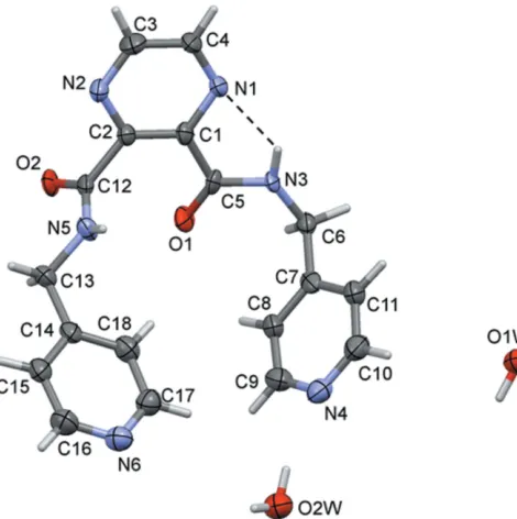
Figure 1
[image:3.610.315.565.263.324.2]A view of the molecular structure of ligand (L1), with atom labelling. Displacement ellipsoids are drawn at the 50% probability level. The intramolecular N—H N contact is shown as a dashed line (see Table 2).
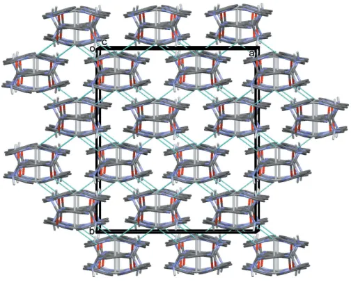
Figure 2
A view of the figure-eight arrangement of the title complex (I), with atom labelling for the asymmetric unit and some symmetry-related atoms (see Table 1 for details). The unlabelled atoms of the ligand on the left-hand-side of the figure are related to the labelled atoms by twofold rotational symmetry (symmetry operation:x+7
4,y+ 7
4,z). The nitrate anions have been omitted for clarity. Table 1
Hydrogen-bond geometry (A˚ ,) for (L1).
D—H A D—H H A D A D—H A
N3—H3N N1 0.88 (3) 2.08 (5) 2.718 (7) 129 (5) N5—H5N O1i 0.88 (3) 2.05 (4) 2.858 (7) 151 (6) O1W—H1WA O2Wii 0.86 1.91 2.762 (7) 177
O1W—H1WB N6iii 0.94 1.97 2.886 (7) 164 O2W—H2WA O1Wiv 0.86 1.91 2.765 (8) 172
O2W—H2WB N4 0.85 2.06 2.888 (7) 164 C3—H3 O1Wv 0.95 2.38 3.273 (8) 156
C4—H4 O2Wvi 0.95 2.58 3.253 (8) 128
C6—H6A N2vii 0.99 2.57 3.515 (8) 159
C16—H16 N4iv 0.95 2.60 3.451 (9) 149
Symmetry codes: (i) x1;y;z; (ii) x;yþ1;zþ1
2; (iii) xþ1;yþ1;zþ 1 2; (iv)
x1;yþ1;z1
2; (v)x1;yþ2;z 1
2; (vi)x;yþ1;z; (vii)xþ1;yþ2;zþ 1 2.
Table 2
Selected geometric parameters (A˚ ,) for (I).
Ag1—Ag1i 3.1638 (11) Ag1—O1ii 2.814 (5)
Ag1—N4 2.109 (5)
N4—Ag1—N4iii 173.0 (2) O1ii—Ag1—N4 97.80 (15) O1ii—Ag1—O1iv 109.48 (14) O1iv—Ag1—N4 86.26 (15)
Ag1i—Ag1—N4 86.51 (14) Ag1i—Ag1—O1ii 125.26 (10)
Symmetry codes: (i)x;yþ7 4;zþ
3 4; (ii)x
1 4;yþ
1 4;zþ
1 2; (iii)xþ
7 4;y;zþ
3 4; (iv) xþ2;yþ1
[image:3.610.116.487.567.713.2]Fig. 6). The nitrate anion in (I) is not essential for forming the three-dimensional structure, although it may act as a template for the formation of the framework (Battenet al., 2009). This is in contrast to the MOF catena-[bis(3-nitrato-O,O0,O00
)-bis(3-1,3,5-tris(pyrazol-1-yl)benzene-N,N0,N00)trisilver(I)
nitrate] mentioned above (Shuet al., 2006), in which there are nitrate anions coordinating the silver ions in a3fashion and
present also in the framework cavities. There are, of course, other examples reported in the Cambridge Structural Data-base (Groomet al., 2016).
In describing compound (I) as a three-dimensional coor-dination polymer, we make here the distinction between a coordination polymer and a metal–organic framework. Both have a three-dimensional framework but there are no cavities, even small ones, in the structure of (I). Hence, it should be classed as a three-dimensional coordination polymer according to the IUPAC recommendations on the ‘Termi-nology of metal–organic frameworks and coordination poly-mers’ (Battenet al., 2013).
4. Database survey
A search of the Cambridge Structural Database (Version 5.38, update February 2017; Groom et al., 2016) for Ag–Ag complexes, excluding silver ion clusters of any kind, gave 321 hits. Limiting the search to Ag–Ag complexes with each silver ion coordinated by two pyridine N atoms, gave 95 hits. The Ag—Ag distances vary betweenca2.6–3.6 A˚ . One compound, bis[2-2,7-di-tert-butyl-9,9-dimethyl-N,N0
-bis[(3-pyridyl)-methyl]xanthene-4,5-dicarboxamide]disilver bis(trifluoro-methanesulfonate) chloroform solvate (HIFKUD; Yueet al., 2007), is particularly interesting because it too involves a
research communications
Acta Cryst.(2017). E73, 798–803 Cati and Stoeckli-Evans C
18H16N6O22H2O and [Ag(C18H16N6O2)]NO3
801
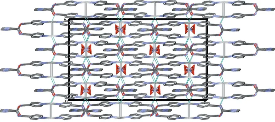
Figure 3
A view along thecaxis of the three-dimensional framework of complex (I), showing the Ag O bonds as dashed lines (see Table 2). The nitrate anions and the C-bound H atoms have been omitted for clarity.
Figure 4
[image:4.610.45.297.70.273.2] [image:4.610.312.567.94.169.2]A partial view along direction [111] of the crystal packing of ligand (L1). The hydrogen bonds are shown as dashed lines (see Table 1)
Table 3
Hydrogen-bond geometry (A˚ ,) for (I).
D—H A D—H H A D A D—H A
N3—H3N O11v 0.88 1.86 2.744 (14) 178
N3—H3N O13v 0.88 2.26 2.875 (13) 127 C4—H4 O11vi 0.95 2.45 3.378 (13) 165
C4—H4 O14vii 0.95 2.40 3.33 (2) 168 C9—H9 O13viii 0.95 2.50 3.224 (14) 133
C11—H11 O13v 0.95 2.51 3.154 (13) 126
Symmetry codes: (v)x;yþ1;z; (vi)xþ2;yþ1;z; (vii)xþ2;yþ3 4;z
1 4; (viii)
xþ1 4;yþ
3 4;zþ
[image:4.610.48.319.470.716.2] [image:4.610.125.553.477.703.2]1 2.
Figure 5
[image:4.610.312.564.487.688.2]dicarboxamide ligand, viz. N,N0 -bis[(3-pyridyl)methy]xanth-ene-4,5-dicarboxamide), that wraps around an Ag—Ag bond forming a similar figure-of-eight-shaped complex. Here the Ag—Ag bond length is 3.134 (1) A˚ , slightly shorter than the value of 3.1638 (11) A˚ observed in (I); Table 2. A search for the benzene analogue of ligand (L1), N -(4-pyridylmethyl)-carbamoyl)benzene, gave only two hits. Both of them are mercury(II) complexes,viz.the binuclear complex bis{2
-1,2-bis[N -(4-pyridylmethyl)carbamoyl]benzene}tetrakis(trifluoro-acetato)dimercury(II) methanol solvate (XAHSIJ; Burchell et al., 2004) and the two-dimensional networkcatena-[bis{2
-1,2-bis[N -(4-pyridylmethyl)carbamoyl]benzene}dichlorido-mercury(II) 1,2-dichloroethane solvate] (XAHSOP; Burchell et al., 2004). A search for the benzene analogue of ligand (L2), [N-(2-pyridylmethyl)carbamoyl]benzene, gave zero hits, while that for [N-(3-pyridylmethyl)carbamoyl]benzene gave eight hits. The latter includes the crystal structure of the dihydrate of the ligand itself (PANROM; Ge et al., 2005) and the structures of seven first-row transition metal one-, two- and three-dimensional coordination polymers.
5. Synthesis and crystallization
Ligand (L1) was prepared using the same procedure as for ligand (L2) (Cati et al., 2004). Dimethyl pyrazine-2,3-di-carboxylate (1.96 g, 10 mmol; Alvarez-Ibarraet al., 1994) and an excess of 4-(aminomethyl)pyridine (3.24 g, 30 mmol) in 35 ml of methanol were heated to reflux and heating was continued for 72 h in a two-necked flask (100 ml). The brown solution that formed was concentrated and 15 ml of water were added, which precipitated quantitatively ligand (L1). The solid was collected by filtration, washed with 10 ml of water and dried in air. Recrystallization in ethanol gave colourless plate-like crystals (yield is quantitative; m.p. 474 K). Spec-troscopic data:1H NMR (400 MHz, DMSO-d6): 9.33 (t, 1H,
Jhg= 6.1, Hh); 8.86 (s, 1H, Hn= Hm); 8.49 (dd, 2H,Jba= 4.5,
Jbe= 1.5, Hb= Hd); 7.39 (dd, 2H,Jab= 4.5,Jeb= 1.5, Ha= He);
4.52 (d, 2H,Jgh= 6.1, Hg). 13
C NMR (400 MHz, DMSO-d6): 165.8, 150.3, 148.9, 147.6, 145.6, 123.0, 42.2. IR (KBr pellet, cm1): 3273 (s), 3031 (s), 1675 (vs), 1602 (vs), 1564 (vs), 1520 (vs), 1416 (vs), 1364 (s), 1311 (s), 1292 (s), 1220 (s), 1185 (m), 1164 (m), 1124 (s), 1069 (m), 995 (s), 871 (w), 830 (m), 787 (m), 745 (m), 715 (m), 611 (m), 575 (w), 504 (m), 495 (m), 475 (m). Elemental analysis for [C18H16N6O2]H2O (Mr =
366.39 g mol1): calculated: C: 59.01 H: 4.95 N: 22.94%; found: C: 59.10 H: 5.05 N: 23.10%.
Complex (I): A solution of (L1) (46 mg; 0.126 mmol) in 6 ml CHCl3 was introduced into a 13 mm diameter glass tube. It
was layered with methanol (ca2 ml) used as a buffer zone. A solution of AgNO3(21 mg, 0.126 mmol) in MeOH (6 ml) was
then added gently to avoid possible mixing. The glass tube was sealed with a perforated parafilm and left at room tempera-ture. Colourless block-like crystals were obtained after a few
days (yield 60 mg, 92%). Elemental analysis for
AgC18H16N7O5: (Mr= 518.25 g mol
1
): calculated: C: 41.72 H: 3.11 N: 18.92%; found: C: 41.65 H: 3.09 N: 18.85%.
6. Refinement
Crystal data, data collection and structure refinement details are summarized in Table 4. For the ligand (L1), the NH and water H atoms were located in difference-Fourier maps and refined with distance restraints: O—H = 0.85 (2) A˚ , N—H = 0.88 (2) A˚ withUiso(H) = 1.5Ueq(O) and 1.2Ueq(N). In the final
cycles of refinement, the water H atoms were treated as riding atoms. For complex (I), the NH H atoms were included in calculated positions and treated as riding: N—H = 0.88 A˚ with Uiso(H) = 1.2Ueq(N). For both compounds, the C-bound H
atoms were included in calculated positions and refined as riding: C—H = 0.95–0.99 A˚ with Uiso(H) = 1.2Ueq(C). The
802
Cati and Stoeckli-Evans C18H16N6O22H2O and [Ag(C18H16N6O2)]NO3 Acta Cryst.(2017). E73, 798–803
[image:5.610.80.527.74.272.2]research communications
Figure 6
nitrate anion is positionally disordered about a twofold rota-tion axis and was refined with fixed occupancies (N10Aand N10B= 0.5, O11 and O13 = 0.5, O12 and O14 = 0.25), and all their ADP’s were made equal to that of atom O11. Using a one-circle image-plate diffraction system it is not possible to measure 100% of the Ewald sphere, particularly for triclinic or monoclinic systems. This is the case for ligand (L1), which crystallized in the monoclinic space group Pcand for which only 94.7% of the Ewald sphere was accessible.
Funding information
Funding for this research was provided by: Swiss National Science Foundation; University of Neuchaˆtel..
References
Addison, A. W., Rao, T. N., Reedijk, J., van Rijn, J. & Verschoor, G. C. (1984).J. Chem. Soc. Dalton Trans.pp. 1349–1356.
Alvarez-Ibarra, C., Cuervo-Rodrı´guez, R., Ferna´ndez-Monreal, M. C. & Ruiz, M. P. (1994).J. Org. Chem.59, 7284–7291.
Batten, S. R., Champness, N. R., Chen, X. M., Garcia-Martinez, J., Kitagawa, S., O¨ hrstro¨m, L., O’Keeffe, M., Suh, M. P. & Reedijk, J. (2013).Pure Appl. Chem.5, 1715–1724.
Batten, S. R., Neville, S. M. & Turner, D. R. (2009).Coordination Polymers, Design, Analysis and Applications. Cambridge: Royal Society of Chemistry.
Burchell, T. J., Eisler, D. J. & Puddephatt, R. J. (2004).Inorg. Chem.
43, 5550–5557.
Cati, D. (2002). PhD thesis, University of Neuchaˆtel, Switzerland. Cati, D. S., Ribas, J., Ribas-Arin˜o, J. & Stoeckli-Evans, H. (2004).
Inorg. Chem.43, 1021–1030.
Ge, C.-H., Kou, H.-Z., Wang, R.-J., Jiang, Y.-B. & Cui, A.-L. (2005).
Acta Cryst.E61, o2024–o2026.
Groom, C. R., Bruno, I. J., Lightfoot, M. P. & Ward, S. C. (2016).Acta Cryst.B72, 171–179.
Hausmann, J., Jameson, G. B. & Brooker, S. (2003).Chem. Commun.
pp. 2992–2993.
Huo, J. Z., Su, X. M., Wu, X. X., Liu, Y. Y. & Ding, B. (2016).
CrystEngComm,18, 6640–6652.
Klingele (ne´e Hausmann), J., Boas, J. F., Pilbrow, J. R., Moubaraki, B., Murray, K. S., Berry, K. J., Hunter, K. A., Jameson, G. B., Boyd, P. D. W. & Brooker, S. (2007).Dalton Trans.pp. 633–645. Macrae, C. F., Bruno, I. J., Chisholm, J. A., Edgington, P. R., McCabe,
P., Pidcock, E., Rodriguez-Monge, L., Taylor, R., van de Streek, J. & Wood, P. A. (2008).J. Appl. Cryst.41, 466–470.
Sheldrick, G. M. (2008).Acta Cryst.A64, 112–122. Sheldrick, G. M. (2015).Acta Cryst.C71, 3–8.
Shu, M., Tu, C., Xu, W., Jin, H. & Sun, J. (2006).Cryst. Growth Des.6, 1890–1896.
Spek, A. L. (2009).Acta Cryst.D65, 148–155.
Stoe & Cie (2004).IPDSI Bedienungshandbuch. Stoe & Cie GmbH, Darmstadt, Germany.
Westrip, S. P. (2010).J. Appl. Cryst.43, 920–925.
Yang, L., Powell, D. R. & Houser, R. P. (2007).Dalton Trans.pp. 955– 964.
Yue, N. L. S., Jennings, M. C. & Puddephatt, R. J. (2007).Eur. J. Inorg. Chem.pp. 1690–1697.
research communications
Acta Cryst.(2017). E73, 798–803 Cati and Stoeckli-Evans C
[image:6.610.47.565.93.384.2]18H16N6O22H2O and [Ag(C18H16N6O2)]NO3
803
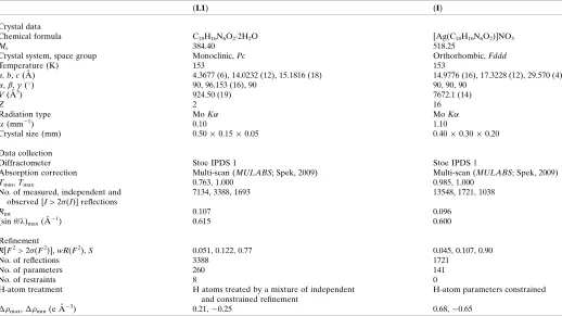
Table 4
Experimental details.
(L1) (I)
Crystal data
Chemical formula C18H16N6O22H2O [Ag(C18H16N6O2)]NO3
Mr 384.40 518.25
Crystal system, space group Monoclinic,Pc Orthorhombic,Fddd
Temperature (K) 153 153
a,b,c(A˚ ) 4.3677 (6), 14.0232 (12), 15.1816 (18) 14.9776 (16), 17.3228 (12), 29.570 (4)
,,(
) 90, 96.153 (16), 90 90, 90, 90
V(A˚3) 924.50 (19) 7672.1 (14)
Z 2 16
Radiation type MoK MoK
(mm1) 0.10 1.10
Crystal size (mm) 0.500.150.05 0.400.300.20
Data collection
Diffractometer Stoe IPDS 1 Stoe IPDS 1
Absorption correction Multi-scan (MULABS; Spek, 2009) Multi-scan (MULABS; Spek, 2009)
Tmin,Tmax 0.763, 1.000 0.985, 1.000
No. of measured, independent and observed [I> 2(I)] reflections
7134, 3388, 1693 13548, 1721, 1038
Rint 0.107 0.096
(sin/ )max(A˚
1) 0.615 0.600
Refinement
R[F2> 2(F2)],wR(F2),S 0.051, 0.122, 0.77 0.045, 0.107, 0.90
No. of reflections 3388 1721
No. of parameters 260 141
No. of restraints 8 0
H-atom treatment H atoms treated by a mixture of independent and constrained refinement
H-atom parameters constrained
max,min(e A˚3) 0.21,0.25 0.68,0.65
Computer programs:EXPOSE,CELLandINTEGRATEinIPDS-I(Stoe & Cie, 2004),SHELXS2014(Sheldrick, 2008),Mercury(Macraeet al., 2008),SHELXL2014(Sheldrick, 2015),
supporting information
sup-1
Acta Cryst. (2017). E73, 798-803
supporting information
Acta Cryst. (2017). E73, 798-803 [https://doi.org/10.1107/S2056989017006387]
Crystal structure of a pyrazine-2,3-dicarboxamide ligand and of its silver(I)
nitrate complex, a three-dimensional coordination polymer
Dilovan S. Cati and Helen Stoeckli-Evans
Computing details
For both compounds, data collection: EXPOSE in IPDS-I (Stoe & Cie, 2004); cell refinement: CELL in IPDS-I (Stoe &
Cie, 2004); data reduction: INTEGRATE in IPDS-I (Stoe & Cie, 2004); program(s) used to solve structure: SHELXS2014
(Sheldrick, 2008); program(s) used to refine structure: SHELXL2014 (Sheldrick, 2015); molecular graphics: Mercury
(Macrae et al., 2008); software used to prepare material for publication: SHELXL2014 (Sheldrick, 2015), PLATON (Spek,
2009) and publCIF (Westrip, 2010).
(L1) N2,N3-Bis(pyridin-4-ylmethyl)pyrazine-2,3-dicarboxamide
Crystal data
C18H16N6O2·2H2O Mr = 384.40 Monoclinic, Pc a = 4.3677 (6) Å
b = 14.0232 (12) Å
c = 15.1816 (18) Å
β = 96.153 (16)°
V = 924.50 (19) Å3 Z = 2
F(000) = 404
Dx = 1.381 Mg m−3
Mo Kα radiation, λ = 0.71073 Å Cell parameters from 3264 reflections
θ = 2.0–25.9°
µ = 0.10 mm−1 T = 153 K Plate, colourless 0.50 × 0.15 × 0.05 mm
Data collection
Stoe IPDS 1 diffractometer
Radiation source: fine-focus sealed tube Plane graphite monochromator
φ rotation scans
Absorption correction: multi-scan (MULABS; Spek, 2009)
Tmin = 0.763, Tmax = 1.000
7134 measured reflections 3388 independent reflections 1693 reflections with I > 2σ(I)
Rint = 0.107
θmax = 25.9°, θmin = 2.0° h = −5→5
k = −17→16
l = −18→18
Refinement
Refinement on F2
Least-squares matrix: full
R[F2 > 2σ(F2)] = 0.051 wR(F2) = 0.122 S = 0.77 3388 reflections 260 parameters 8 restraints
Primary atom site location: structure-invariant direct methods
Secondary atom site location: difference Fourier map
Hydrogen site location: mixed
H atoms treated by a mixture of independent and constrained refinement
w = 1/[σ2(F
o2) + (0.0453P)2]
supporting information
sup-2
Acta Cryst. (2017). E73, 798-803 (Δ/σ)max < 0.001
Δρmax = 0.21 e Å−3
Δρmin = −0.25 e Å−3
Extinction correction: (SHELXL2016; Sheldrick, 2015),
Fc*=kFc[1+0.001xFc2λ3/sin(2θ)]-1/4
Extinction coefficient: 0.036 (6)
Special details
Geometry. All esds (except the esd in the dihedral angle between two l.s. planes) are estimated using the full covariance
matrix. The cell esds are taken into account individually in the estimation of esds in distances, angles and torsion angles; correlations between esds in cell parameters are only used when they are defined by crystal symmetry. An approximate (isotropic) treatment of cell esds is used for estimating esds involving l.s. planes.
Fractional atomic coordinates and isotropic or equivalent isotropic displacement parameters (Å2)
x y z Uiso*/Ueq
O1 0.7750 (11) 0.9108 (3) 0.4491 (3) 0.0358 (13)
O2 0.5478 (11) 0.9994 (3) 0.2578 (3) 0.0351 (12)
N1 0.4263 (14) 1.1135 (3) 0.5363 (4) 0.0321 (15)
N2 0.1716 (14) 1.1331 (4) 0.3588 (4) 0.0330 (15)
N3 0.7147 (13) 0.9491 (3) 0.5921 (3) 0.0258 (14)
H3N 0.612 (13) 0.998 (3) 0.609 (4) 0.031*
N4 0.4923 (14) 0.5966 (4) 0.6225 (4) 0.0403 (16)
N5 0.1699 (14) 0.9119 (4) 0.3107 (4) 0.0313 (14)
H5N 0.002 (11) 0.906 (5) 0.337 (4) 0.038*
N6 −0.0999 (16) 0.5615 (4) 0.3180 (4) 0.0436 (17)
C1 0.4668 (15) 1.0457 (4) 0.4743 (4) 0.0267 (17)
C2 0.3401 (16) 1.0564 (4) 0.3878 (4) 0.0258 (16)
C3 0.1396 (19) 1.1999 (5) 0.4209 (5) 0.043 (2)
H3 0.030625 1.256737 0.403978 0.051*
C4 0.2586 (18) 1.1888 (4) 0.5079 (5) 0.0360 (19)
H4 0.219526 1.236935 0.549295 0.043*
C5 0.6613 (15) 0.9624 (4) 0.5039 (4) 0.0280 (16)
C6 0.9192 (16) 0.8748 (4) 0.6312 (5) 0.0290 (17)
H6A 0.985533 0.891453 0.693707 0.035*
H6B 1.105193 0.872221 0.599314 0.035*
C7 0.7688 (16) 0.7778 (4) 0.6278 (5) 0.0307 (17)
C8 0.8016 (18) 0.7151 (4) 0.5602 (5) 0.0376 (19)
H8 0.917205 0.733033 0.513344 0.045*
C9 0.669 (2) 0.6270 (5) 0.5600 (5) 0.046 (2)
H9 0.702062 0.584555 0.513239 0.056*
C10 0.4698 (19) 0.6573 (5) 0.6889 (5) 0.044 (2)
H10 0.357252 0.636975 0.735668 0.053*
C11 0.5967 (17) 0.7473 (5) 0.6950 (5) 0.0374 (19)
H11 0.568042 0.787610 0.743743 0.045*
C12 0.3639 (17) 0.9848 (4) 0.3138 (4) 0.0297 (17)
C13 0.1367 (17) 0.8444 (4) 0.2372 (5) 0.0337 (18)
H13A 0.332934 0.841179 0.210178 0.040*
H13B −0.024426 0.867753 0.191430 0.040*
C14 0.0529 (17) 0.7469 (4) 0.2655 (4) 0.0303 (17)
supporting information
sup-3
Acta Cryst. (2017). E73, 798-803
H15 −0.242090 0.714271 0.158778 0.043*
C16 −0.224 (2) 0.6001 (5) 0.2413 (5) 0.049 (2)
H16 −0.368187 0.563552 0.204091 0.058*
C17 0.1048 (19) 0.6151 (5) 0.3680 (6) 0.042 (2)
H17 0.200289 0.589113 0.421904 0.050*
C18 0.1843 (19) 0.7075 (5) 0.3446 (5) 0.0389 (18)
H18 0.327922 0.743333 0.382699 0.047*
O1W 0.7645 (12) 0.5986 (3) 0.9268 (3) 0.0503 (15)
H1WA 0.624209 0.595327 0.962023 0.075*
H1WB 0.768245 0.545186 0.889768 0.075*
O2W 0.3065 (12) 0.4183 (3) 0.5379 (3) 0.0478 (14)
H2WA 0.133161 0.418283 0.504471 0.072*
H2WB 0.327355 0.474826 0.557396 0.072*
Atomic displacement parameters (Å2)
U11 U22 U33 U12 U13 U23
O1 0.051 (3) 0.039 (3) 0.021 (3) 0.009 (2) 0.019 (3) 0.000 (2)
O2 0.052 (3) 0.040 (2) 0.015 (3) −0.005 (2) 0.015 (2) −0.0003 (19)
N1 0.055 (4) 0.024 (3) 0.018 (3) 0.001 (3) 0.007 (3) −0.002 (2)
N2 0.050 (4) 0.030 (3) 0.019 (3) 0.006 (3) 0.004 (3) 0.003 (2)
N3 0.041 (4) 0.025 (3) 0.012 (3) 0.004 (3) 0.009 (3) −0.001 (2)
N4 0.047 (4) 0.032 (3) 0.041 (4) 0.001 (3) 0.001 (4) 0.007 (3)
N5 0.038 (4) 0.034 (3) 0.023 (3) −0.010 (3) 0.006 (3) −0.006 (2)
N6 0.054 (4) 0.033 (3) 0.044 (4) −0.005 (3) 0.007 (4) −0.003 (3)
C1 0.035 (5) 0.029 (3) 0.018 (4) −0.002 (3) 0.009 (3) 0.001 (3)
C2 0.034 (4) 0.025 (3) 0.020 (4) 0.000 (3) 0.008 (3) 0.000 (3)
C3 0.061 (6) 0.027 (4) 0.040 (5) 0.011 (4) 0.003 (4) 0.003 (3)
C4 0.060 (5) 0.025 (4) 0.023 (4) 0.007 (4) 0.009 (4) −0.003 (3)
C5 0.037 (4) 0.031 (4) 0.017 (4) −0.001 (3) 0.006 (3) 0.008 (3)
C6 0.036 (4) 0.031 (4) 0.020 (4) 0.006 (3) 0.001 (3) 0.000 (3)
C7 0.032 (5) 0.030 (4) 0.027 (4) 0.013 (3) −0.007 (4) −0.002 (3)
C8 0.056 (5) 0.034 (4) 0.022 (5) 0.001 (4) 0.001 (4) −0.001 (3)
C9 0.066 (6) 0.031 (4) 0.040 (5) 0.005 (4) 0.001 (5) −0.005 (3)
C10 0.057 (6) 0.037 (4) 0.039 (5) 0.007 (4) 0.009 (4) 0.007 (4)
C11 0.053 (5) 0.035 (4) 0.024 (4) 0.002 (4) 0.006 (4) −0.001 (3)
C12 0.047 (5) 0.027 (4) 0.015 (4) 0.007 (3) −0.002 (4) 0.001 (3)
C13 0.047 (5) 0.029 (3) 0.025 (4) −0.003 (3) 0.003 (4) −0.007 (3)
C14 0.038 (5) 0.029 (3) 0.025 (5) 0.005 (3) 0.006 (4) −0.005 (3)
C15 0.047 (5) 0.035 (4) 0.028 (4) 0.005 (4) 0.007 (4) −0.003 (3)
C16 0.063 (6) 0.040 (4) 0.042 (5) −0.008 (4) 0.000 (5) −0.004 (4)
C17 0.052 (5) 0.038 (4) 0.038 (5) 0.001 (4) 0.012 (4) 0.003 (4)
C18 0.041 (5) 0.040 (4) 0.036 (5) −0.003 (4) 0.002 (4) −0.006 (3)
O1W 0.066 (4) 0.036 (3) 0.051 (4) −0.006 (3) 0.013 (3) −0.001 (2)
supporting information
sup-4
Acta Cryst. (2017). E73, 798-803 Geometric parameters (Å, º)
O1—C5 1.245 (7) C6—H6B 0.9900
O2—C12 1.248 (7) C7—C8 1.370 (9)
N1—C4 1.331 (8) C7—C11 1.398 (9)
N1—C1 1.364 (8) C8—C9 1.364 (10)
N2—C3 1.346 (9) C8—H8 0.9500
N2—C2 1.351 (8) C9—H9 0.9500
N3—C5 1.347 (8) C10—C11 1.378 (10)
N3—C6 1.456 (8) C10—H10 0.9500
N3—H3N 0.88 (3) C11—H11 0.9500
N4—C10 1.330 (9) C13—C14 1.491 (8)
N4—C9 1.354 (10) C13—H13A 0.9900
N5—C12 1.325 (8) C13—H13B 0.9900
N5—C13 1.458 (8) C14—C15 1.366 (10)
N5—H5N 0.88 (3) C14—C18 1.389 (10)
N6—C17 1.340 (10) C15—C16 1.386 (9)
N6—C16 1.344 (10) C15—H15 0.9500
C1—C2 1.377 (9) C16—H16 0.9500
C1—C5 1.485 (8) C17—C18 1.397 (10)
C2—C12 1.518 (9) C17—H17 0.9500
C3—C4 1.375 (10) C18—H18 0.9500
C3—H3 0.9500 O1W—H1WA 0.8569
C4—H4 0.9500 O1W—H1WB 0.9371
C6—C7 1.510 (9) O2W—H2WA 0.8649
C6—H6A 0.9900 O2W—H2WB 0.8483
C4—N1—C1 115.9 (6) C7—C8—H8 119.9
C3—N2—C2 114.8 (6) N4—C9—C8 123.9 (7)
C5—N3—C6 122.5 (5) N4—C9—H9 118.1
C5—N3—H3N 99 (4) C8—C9—H9 118.1
C6—N3—H3N 139 (5) N4—C10—C11 125.2 (7)
C10—N4—C9 115.0 (6) N4—C10—H10 117.4
C12—N5—C13 122.6 (6) C11—C10—H10 117.4
C12—N5—H5N 128 (4) C10—C11—C7 118.3 (6)
C13—N5—H5N 106 (4) C10—C11—H11 120.8
C17—N6—C16 116.6 (6) C7—C11—H11 120.8
N1—C1—C2 120.9 (6) O2—C12—N5 123.9 (6)
N1—C1—C5 116.8 (6) O2—C12—C2 119.7 (6)
C2—C1—C5 122.2 (5) N5—C12—C2 116.2 (6)
N2—C2—C1 123.1 (5) N5—C13—C14 112.5 (6)
N2—C2—C12 111.4 (6) N5—C13—H13A 109.1
C1—C2—C12 125.5 (6) C14—C13—H13A 109.1
N2—C3—C4 122.5 (7) N5—C13—H13B 109.1
N2—C3—H3 118.7 C14—C13—H13B 109.1
C4—C3—H3 118.7 H13A—C13—H13B 107.8
N1—C4—C3 122.6 (6) C15—C14—C18 116.6 (6)
supporting information
sup-5
Acta Cryst. (2017). E73, 798-803
C3—C4—H4 118.7 C18—C14—C13 121.5 (6)
O1—C5—N3 123.0 (6) C14—C15—C16 121.0 (7)
O1—C5—C1 120.7 (6) C14—C15—H15 119.5
N3—C5—C1 116.3 (5) C16—C15—H15 119.5
N3—C6—C7 112.7 (6) N6—C16—C15 122.9 (7)
N3—C6—H6A 109.1 N6—C16—H16 118.6
C7—C6—H6A 109.1 C15—C16—H16 118.6
N3—C6—H6B 109.1 N6—C17—C18 123.0 (8)
C7—C6—H6B 109.1 N6—C17—H17 118.5
H6A—C6—H6B 107.8 C18—C17—H17 118.5
C8—C7—C11 117.3 (6) C14—C18—C17 119.9 (7)
C8—C7—C6 121.6 (6) C14—C18—H18 120.1
C11—C7—C6 121.1 (6) C17—C18—H18 120.1
C9—C8—C7 120.3 (7) H1WA—O1W—H1WB 113.1
C9—C8—H8 119.9 H2WA—O2W—H2WB 105.0
C4—N1—C1—C2 0.2 (9) C7—C8—C9—N4 2.1 (12)
C4—N1—C1—C5 178.1 (6) C9—N4—C10—C11 3.4 (12)
C3—N2—C2—C1 0.9 (10) N4—C10—C11—C7 −1.4 (12)
C3—N2—C2—C12 179.9 (6) C8—C7—C11—C10 −0.5 (11)
N1—C1—C2—N2 0.3 (10) C6—C7—C11—C10 −178.9 (6)
C5—C1—C2—N2 −177.6 (6) C13—N5—C12—O2 −4.5 (10)
N1—C1—C2—C12 −178.5 (6) C13—N5—C12—C2 171.7 (6)
C5—C1—C2—C12 3.6 (10) N2—C2—C12—O2 78.2 (8)
C2—N2—C3—C4 −2.5 (11) C1—C2—C12—O2 −102.8 (8)
C1—N1—C4—C3 −1.8 (10) N2—C2—C12—N5 −98.1 (7)
N2—C3—C4—N1 3.2 (12) C1—C2—C12—N5 80.8 (9)
C6—N3—C5—O1 2.1 (10) C12—N5—C13—C14 148.9 (6)
C6—N3—C5—C1 −175.4 (5) N5—C13—C14—C15 141.5 (7)
N1—C1—C5—O1 −161.3 (6) N5—C13—C14—C18 −40.3 (9)
C2—C1—C5—O1 16.6 (9) C18—C14—C15—C16 1.4 (10)
N1—C1—C5—N3 16.2 (8) C13—C14—C15—C16 179.6 (6)
C2—C1—C5—N3 −165.8 (6) C17—N6—C16—C15 −0.6 (11)
C5—N3—C6—C7 −79.3 (7) C14—C15—C16—N6 −0.9 (12)
N3—C6—C7—C8 94.4 (7) C16—N6—C17—C18 1.6 (11)
N3—C6—C7—C11 −87.3 (8) C15—C14—C18—C17 −0.4 (10)
C11—C7—C8—C9 0.1 (11) C13—C14—C18—C17 −178.7 (6)
C6—C7—C8—C9 178.5 (7) N6—C17—C18—C14 −1.2 (11)
C10—N4—C9—C8 −3.8 (11)
Hydrogen-bond geometry (Å, º)
D—H···A D—H H···A D···A D—H···A
N3—H3N···N1 0.88 (3) 2.08 (5) 2.718 (7) 129 (5)
N5—H5N···O1i 0.88 (3) 2.05 (4) 2.858 (7) 151 (6)
O1W—H1WA···O2Wii 0.86 1.91 2.762 (7) 177
O1W—H1WB···N6iii 0.94 1.97 2.886 (7) 164
supporting information
sup-6
Acta Cryst. (2017). E73, 798-803
O2W—H2WB···N4 0.85 2.06 2.888 (7) 164
C3—H3···O1Wv 0.95 2.38 3.273 (8) 156
C4—H4···O2Wvi 0.95 2.58 3.253 (8) 128
C6—H6A···N2vii 0.99 2.57 3.515 (8) 159
C16—H16···N4iv 0.95 2.60 3.451 (9) 149
Symmetry codes: (i) x−1, y, z; (ii) x, −y+1, z+1/2; (iii) x+1, −y+1, z+1/2; (iv) x−1, −y+1, z−1/2; (v) x−1, −y+2, z−1/2; (vi) x, y+1, z; (vii) x+1, −y+2, z+1/2.
(I) Poly[[[µ4-N2,N3-bis(pyridin-4-ylmethyl)pyrazine-2,3-dicarboxamide]silver(I)] nitrate]
Crystal data
[Ag(C18H16N6O2)]NO3 Mr = 518.25
Orthorhombic, Fddd a = 14.9776 (16) Å
b = 17.3228 (12) Å
c = 29.570 (4) Å
V = 7672.1 (14) Å3 Z = 16
F(000) = 4160
Dx = 1.795 Mg m−3
Mo Kα radiation, λ = 0.71073 Å Cell parameters from 6115 reflections
θ = 1.9–25.9°
µ = 1.10 mm−1 T = 153 K Block, colourless 0.40 × 0.30 × 0.20 mm
Data collection
Stoe IPDS 1 diffractometer
Radiation source: fine-focus sealed tube Plane graphite monochromator
φ rotation scans
Absorption correction: multi-scan (MULABS; Spek, 2009)
Tmin = 0.985, Tmax = 1.000
13548 measured reflections 1721 independent reflections 1038 reflections with I > 2σ(I)
Rint = 0.096
θmax = 25.3°, θmin = 2.7° h = −17→17
k = −20→20
l = −35→35
Refinement
Refinement on F2
Least-squares matrix: full
R[F2 > 2σ(F2)] = 0.045 wR(F2) = 0.107 S = 0.90 1721 reflections 141 parameters 0 restraints
Primary atom site location: structure-invariant direct methods
Secondary atom site location: difference Fourier map
Hydrogen site location: inferred from neighbouring sites
H-atom parameters constrained
w = 1/[σ2(F
o2) + (0.0575P)2]
where P = (Fo2 + 2Fc2)/3
(Δ/σ)max = 0.003
Δρmax = 0.68 e Å−3
Δρmin = −0.65 e Å−3
Special details
Geometry. Bond distances, angles etc. have been calculated using the rounded fractional coordinates. All su's are
estimated from the variances of the (full) variance-covariance matrix. The cell esds are taken into account in the estimation of distances, angles and torsion angles
Fractional atomic coordinates and isotropic or equivalent isotropic displacement parameters (Å2)
x y z Uiso*/Ueq Occ. (<1)
Ag1 0.87500 0.96632 (4) 0.37500 0.0464 (2)
O1 0.9796 (3) 0.8101 (3) 0.14979 (13) 0.0483 (14)
supporting information
sup-7
Acta Cryst. (2017). E73, 798-803
N3 1.0135 (3) 0.9361 (3) 0.14261 (16) 0.050 (2)
N4 0.9359 (3) 0.9589 (3) 0.31086 (16) 0.0427 (17)
C1 0.9207 (3) 0.8777 (4) 0.08584 (15) 0.0337 (16)
C4 0.9204 (4) 0.8805 (4) 0.00937 (17) 0.0453 (19)
C5 0.9745 (3) 0.8715 (4) 0.12935 (16) 0.0340 (18)
C6 1.0689 (4) 0.9393 (4) 0.18277 (18) 0.051 (2)
C7 1.0197 (4) 0.9446 (3) 0.22692 (19) 0.0437 (19)
C8 1.0640 (4) 0.9276 (4) 0.26675 (19) 0.045 (2)
C9 1.0221 (4) 0.9354 (4) 0.3075 (2) 0.046 (2)
C10 0.8921 (3) 0.9745 (3) 0.27201 (19) 0.043 (2)
C11 0.9302 (4) 0.9686 (4) 0.23033 (19) 0.0447 (19)
O11 0.9918 (8) 0.0666 (8) 0.0912 (4) 0.089 (3) 0.500
O12 1.0655 (16) 0.1323 (15) 0.1063 (6) 0.089 (3) 0.250
O13 0.9326 (9) 0.0862 (7) 0.1489 (4) 0.089 (3) 0.500
O14 0.9869 (18) 0.1233 (19) 0.1570 (7) 0.089 (3) 0.250
N10A 0.9614 (15) 0.12500 0.12500 0.089 (3) 0.500
N10B 0.9960 (16) 0.12500 0.12500 0.089 (3) 0.500
H4 0.95046 0.88600 −0.01873 0.0540*
H3N 1.00563 0.97838 0.12661 0.0600*
H6A 1.10904 0.98449 0.18023 0.0610*
H6B 1.10697 0.89253 0.18345 0.0610*
H8 1.12416 0.91024 0.26574 0.0540*
H9 1.05428 0.92399 0.33436 0.0560*
H10 0.83154 0.99047 0.27385 0.0520*
H11 0.89687 0.98062 0.20392 0.0530*
Atomic displacement parameters (Å2)
U11 U22 U33 U12 U13 U23
Ag1 0.0474 (4) 0.0397 (4) 0.0521 (4) 0.0000 0.0035 (4) 0.0000
O1 0.048 (2) 0.054 (3) 0.043 (2) 0.000 (2) −0.005 (2) 0.010 (2)
N1 0.047 (3) 0.057 (4) 0.030 (2) 0.003 (3) 0.004 (2) −0.004 (2)
N3 0.078 (4) 0.035 (4) 0.037 (3) 0.002 (3) −0.015 (3) −0.006 (2)
N4 0.042 (3) 0.039 (3) 0.047 (3) 0.001 (2) −0.009 (2) −0.006 (2)
C1 0.037 (2) 0.035 (3) 0.029 (3) 0.005 (3) 0.002 (2) −0.010 (3)
C4 0.059 (3) 0.047 (4) 0.030 (3) 0.009 (4) 0.006 (2) 0.000 (3)
C5 0.030 (2) 0.046 (4) 0.026 (3) 0.005 (2) 0.010 (2) −0.007 (4)
C6 0.062 (4) 0.057 (5) 0.033 (3) −0.003 (3) −0.008 (3) −0.006 (3)
C7 0.051 (3) 0.036 (4) 0.044 (3) −0.004 (3) −0.012 (3) −0.008 (3)
C8 0.046 (3) 0.044 (4) 0.045 (4) 0.007 (3) −0.006 (3) −0.004 (3)
C9 0.051 (4) 0.051 (4) 0.037 (3) 0.009 (3) −0.008 (3) 0.000 (3)
C10 0.037 (4) 0.041 (4) 0.052 (3) −0.004 (3) −0.010 (3) −0.006 (3)
C11 0.050 (3) 0.045 (4) 0.039 (3) −0.001 (3) −0.015 (3) −0.008 (3)
O11 0.112 (6) 0.088 (6) 0.067 (4) 0.0000 0.0000 0.028 (4)
O12 0.112 (6) 0.088 (6) 0.067 (4) 0.0000 0.0000 0.028 (4)
O13 0.112 (6) 0.088 (6) 0.067 (4) 0.0000 0.0000 0.028 (4)
O14 0.112 (6) 0.088 (6) 0.067 (4) 0.0000 0.0000 0.028 (4)
supporting information
sup-8
Acta Cryst. (2017). E73, 798-803
N10B 0.112 (6) 0.088 (6) 0.067 (4) 0.0000 0.0000 0.028 (4)
Geometric parameters (Å, º)
Ag1—Ag1i 3.1638 (11) C8—C9 1.365 (8)
Ag1—N4 2.109 (5) C10—C11 1.362 (8)
Ag1—O1ii 2.814 (5) O11—N10A 1.493 (14)
Ag1—O1iii 2.814 (5) O11—N10B 1.424 (13)
Ag1—N4iv 2.109 (5) O11—O12 1.65 (3)
O1—C5 1.226 (8) O12—N10B 1.19 (3)
N1—C1 1.342 (6) O12—N10A 1.66 (3)
N1—C4 1.323 (7) O13—N10A 1.066 (15)
N3—C5 1.322 (8) O13—N10B 1.36 (2)
N3—C6 1.450 (7) O13—O14 1.06 (3)
N4—C9 1.357 (8) O14—N10B 0.96 (2)
N4—C10 1.350 (7) O14—N10A 1.02 (2)
C1—C5 1.522 (6) C4—H4 0.9500
C1—C1v 1.372 (6) C6—H6B 0.9900
C4—C4v 1.373 (9) C6—H6A 0.9900
N3—H3N 0.8800 C8—H8 0.9500
C6—C7 1.502 (8) C9—H9 0.9500
C7—C8 1.384 (8) C10—H10 0.9500
C7—C11 1.407 (8) C11—H11 0.9500
N4—Ag1—N4iv 173.0 (2) C6—C7—C8 119.5 (5)
O1ii—Ag1—O1iii 109.48 (14) C6—C7—C11 123.2 (5)
Ag1i—Ag1—N4 86.51 (14) C7—C8—C9 120.7 (6)
O1ii—Ag1—N4 97.80 (15) N4—C9—C8 122.1 (5)
O1iii—Ag1—N4 86.26 (15) N4—C10—C11 123.5 (5)
Ag1i—Ag1—O1ii 125.26 (10) C7—C11—C10 119.1 (5)
Ag1i—Ag1—O1iii 125.26 (10) C4v—C4—H4 119.00
Ag1i—Ag1—N4iv 86.51 (14) N1—C4—H4 119.00
O1ii—Ag1—N4iv 86.26 (15) N3—C6—H6B 108.00
O1iii—Ag1—N4iv 97.80 (15) C7—C6—H6A 108.00
Ag1vi—O1—C5 114.9 (3) H6A—C6—H6B 107.00
C1—N1—C4 116.2 (5) C7—C6—H6B 108.00
C5—N3—C6 121.9 (5) N3—C6—H6A 108.00
Ag1—N4—C9 119.7 (4) C7—C8—H8 120.00
Ag1—N4—C10 122.9 (3) C9—C8—H8 120.00
C9—N4—C10 117.4 (5) C8—C9—H9 119.00
N1—C1—C5 116.7 (4) N4—C9—H9 119.00
N1—C1—C1v 121.6 (4) N4—C10—H10 118.00
C1v—C1—C5 121.6 (4) C11—C10—H10 118.00
N1—C4—C4v 122.1 (5) O11—N10A—O13 98.0 (8)
C5—N3—H3N 119.00 O13—N10A—N10B 113.9 (13)
C6—N3—H3N 119.00 O11—N10A—N10B 72.3 (9)
O1—C5—N3 124.1 (5) O11—N10B—N10A 87.5 (11)
supporting information
sup-9
Acta Cryst. (2017). E73, 798-803
N3—C5—C1 115.2 (5) O13—N10B—N10A 45.8 (9)
N3—C6—C7 115.7 (5) C7—C11—H11 120.00
C8—C7—C11 117.3 (5) C10—C11—H11 120.00
Ag1i—Ag1—N4—C9 −71.9 (5) N1—C1—C5—O1 −106.9 (6)
Ag1i—Ag1—N4—C10 106.2 (4) N1—C1—C5—N3 73.3 (7)
O1ii—Ag1—N4—C9 163.0 (5) C1v—C1—C5—O1 68.8 (8)
O1ii—Ag1—N4—C10 −19.0 (5) C1v—C1—C5—N3 −111.0 (7)
O1iii—Ag1—N4—C9 53.8 (5) N1—C1—C1v—N1v 5.5 (11)
O1iii—Ag1—N4—C10 −128.1 (5) N1—C1—C1v—C5v −170.1 (6)
Ag1vi—O1—C5—N3 −89.1 (5) C5—C1—C1v—N1v −170.1 (6)
Ag1vi—O1—C5—C1 91.1 (5) C5—C1—C1v—C5v 14.4 (10)
C4—N1—C1—C5 172.9 (6) N1—C4—C4v—N1v 4.5 (11)
C4—N1—C1—C1v −2.9 (10) N3—C6—C7—C8 163.0 (6)
C1—N1—C4—C4v −1.9 (10) N3—C6—C7—C11 −19.0 (9)
C6—N3—C5—O1 1.7 (8) C6—C7—C8—C9 177.0 (6)
C6—N3—C5—C1 −178.5 (4) C11—C7—C8—C9 −1.1 (9)
C5—N3—C6—C7 −79.7 (7) C6—C7—C11—C10 −177.5 (6)
Ag1—N4—C9—C8 178.2 (5) C8—C7—C11—C10 0.5 (9)
C10—N4—C9—C8 0.1 (9) C7—C8—C9—N4 0.9 (10)
Ag1—N4—C10—C11 −178.8 (5) N4—C10—C11—C7 0.4 (9)
C9—N4—C10—C11 −0.7 (9)
Symmetry codes: (i) x, −y+7/4, −z+3/4; (ii) x−1/4, y+1/4, −z+1/2; (iii) −x+2, y+1/4, z+1/4; (iv) −x+7/4, y, −z+3/4; (v) −x+7/4, −y+7/4, z; (vi) −x+2, y−1/4,
z−1/4.
Hydrogen-bond geometry (Å, º)
D—H···A D—H H···A D···A D—H···A
N3—H3N···O11vii 0.88 1.86 2.744 (14) 178
N3—H3N···O13vii 0.88 2.26 2.875 (13) 127
C4—H4···O11viii 0.95 2.45 3.378 (13) 165
C4—H4···O14ix 0.95 2.40 3.33 (2) 168
C9—H9···O13x 0.95 2.50 3.224 (14) 133
C11—H11···O13vii 0.95 2.51 3.154 (13) 126