PHARMACOLOGICAL SCREENING OF PEPGARD SYRUP
BY ANTI-OXIDANT AND ANTI-ULCER ACTIVITY
Amit Patel*1,Dr. Komal Hirani2, Dr. Vishva Bhuva3 and Payal Panchal4
R&D Department, Vital Care Pvt. Ltd., 361-362, Por-GIDC, Ramangamdi,
Vadodara – 391243.
ABSTRACT
Background: Pepgard syrup is a proprietary Ayurvedic poly-herbal
formulation widely used in clinical practice as Antacid for treating
Heartburn, dyspepsia, Gastroesophageal reflux (GERD) and
Drug-induced gastritis. There is conflicting reports regarding its anti-ulcer
and ulcerogenic potential. Aims and Objectives: The study is with the
aim of determining the gastoprotective effect of Pepgard syrup on
aspirin-induced gastric ulcer in rats. Materials and Methods: Wistar
Albino rats strains were used for the study. Aspirin was purchased
from Sigma Labs, Mumbai. Anti-ulcer effect was studied in rats after
inducing mucosal damage by aspirin. Results: It was found that extract
of Pepgard syrup exhibited significant protection against aspirin-inducedulceratdoselevels
of 100 mg/kg body weight. There is a dose-dependent increase in the ulcer protective action
of the extract. Conclusion: The present study of Pepgard syrup revealed its anti-ulcer and
Anti-oxidant activity.
KEYWORDS: Pepgard syrup, Anti-ulcer; Aspirin Ulcer Index, Anti-oxidant.
INTRODUCTION
Gastric ulcer is an important health problem affecting a large number of populations
worldwide. In spite of extensive research, it still remains as an important cause of morbidity.
It is a major target for devising newer therapeutic strategies due to its prevalence and
complications. Peptic ulcer can be considered as a multifactorial disease. Factors such as
increased stress, impaired mucosal resistance, genetic factors, infection with Helicobacter pylori, and anti-inflammatory drugs including nonsteroidal anti-inflammatory drugs
Volume 8, Issue 9, 1788-1801. Research Article ISSN 2277– 7105
Article Received on 19 June 2019,
Revised on 09 July 2019, Accepted on 30 July 2019, DOI: 10.20959/wjpr20199-15618
*Corresponding Author
Mr. Amit Patel
R & D Department,
Vital Care Pvt. Ltd., 361-362, Por-GIDC, Ramangamdi, Vadodara – 391243.
important proven cause for gastric ulcer, ulcer perforation, gastric, and duodenal bleeding and
in ulcer death.[1] Highly selective COX-2 inhibitors were a breakthrough discovery with less
incidence of gastric mucosal damage, but soon retracted many of them due to serious
cardiovascular adverse events. Ulcers are lesions on the mucous membrane of the stomach or
duodenum characterized by superficial loss of tissues with loss of mucosal integrity. There is
a local defect with active inflammation. Peptic ulcer is considered as one of most common
disease in man leading to human sufferings affecting nearly 5% of the global population.
Since in majority of cases it is aggravated due to pepsin and hydrochloric acid, it is termed as
peptic ulcer. Usual course of the peptic ulcer is characterized by many cycles of healing,
relapses and occasional complications.[3] Imbalances between damaging and protective
factors are the major contributing factor for the pathophysiology of peptic ulcer. Pepsin, acid,
H. pylori infection, bile acids, impaired mobility, NSAID’s, corticosteroids, nicotine, etc., are
some of the damaging factors and protective factors include prostaglandins, mucus,
epidermal growth factors, intact microcirculation, epithelial renewal, alkaline tide, nitrous
oxide, phospholipids, and bicarbonate. Increased gastrin secretion and acid output with
defective gastric emptying mechanism predispose to gastric ulcer. Lower postprandial pepsin
secretion, raised serum PG2 and low PGI/PG2 ratios are considered as risk factors for
developing a peptic ulcer.[2]
In hypo motility at antropyloric region, there is increased chance for reflex of duodenal
contents into stomach which can cause chronic inflammation and ulceration. Reactive oxygen
species (ROS), refluxed bile acids, cytokines such as tumour necrosis factors and exogenous
agents such as H. pylori, NSAID’s, alcohol abuse, emotional stress, and smoking can damage gastric mucosa leading to a gastric ulcer.[4] Mucous bicarbonate barrier, prostaglandins
mucosal blood flow, cell renewal and migration, and antioxidants, growth factors are all act
as gastro protective factors preventing the gastric mucosal injury. Gastric mucosal barrier will
block the back diffusion of (H+). NSAID’s can disrupt this barrier, and H+ can damage it
resulting in mucosal injury.[3,4] The first line of defence is a mucus-bicarbonate layer, which
serves as a physicochemical barrier to multiple molecules including hydrogen ions. Surface
epithelial cells provide the next line of defence through several factors, including mucus
production and epithelial cell ionic transporters that maintain intracellular pH and bicarbonate
production, and intracellular tight junctions. There are certain mediators, which play an
important role in cytoprotection. Epithelial cells in the surface produce bicarbonate, which
alkalinity between the mucus and epithelial surface.[3] Epithelial cells also secrete mucus,
which forms a gel that covers the mucosal surface and physically protects the mucosa.
Pepgard syrup is a proprietary Ayurvedic poly-herbal formulation widely used in clinical
practice as Antacid for treating Heartburn, Non-ulcer dyspepsia, Gastroesophageal reflux
(GERD) and Drug-induced gastritis. It contains many potential drugs, derived from plant
sources; therefore, it was thought worth to undertake a pharmacological study of compound
formulation in the experimental protocol to substantiate the safety claims made on it. Which
considering the morbidity caused by peptic ulcer disease and dyspepsia over the world, cheap
and easily available treatments with less adverse effects will always be beneficial, especially
for the people in less developed and developing countries.
MATERIALS AND METHODS
Drugs and chemicals
Pepgard syrup is a proprietary Ayurvedic poly-herbal formulation of Vital Care Pvt. Ltd.,
Vadodara. Name of ingredients, Latin name, family, part used and quantity of the drug are
given in Table 1. For the preparation of Pepgard syrup, herbal ingredients were subjected to
preparation of decoction as per classical reference. Ranitidine was used as a standard drug
(Kopran Pharma Ltd., Mumbai, India). All chemicals used for the study are purchased from
[image:3.595.95.501.486.661.2]SD-fine chemicals; India and all other reagent used were of analytical grade.
Table 1. Ingredients of Pepgard Syrup
Sr. No Common name Botanical name Part used Quantity
1 Yashtimadhu Glycerrhiza Glabra Root 500 mg. 2 Shatavari Asparagus racemosus Root 100 mg. 3 Sajikshar Astoneman indicum whole 60 mg.
4 Amla Emblica officinale Fruit 50 mg.
5 Brahmi Centella asiatica Whole plant 50 mg.
6 Trivrit Ipomoea turpethum Root 40 mg.
7 Lavanga Syzygium aromaticum flower buds 25 mg. 8 Pitpapara Fumaria officinalis Whole plant 20 mg.
9 Dhania Coriandrum sativum Fruit 20 mg.
10 Syrup Base -- - Q.s
Dose calculation
The dose of the test formulations was calculated by extrapolating the human dose, based on
the body surface area ratio by referring to the standard table of Paget and Barnes.[5] The test
In vitro antioxidant activity assay DPPH radical scavenging activity
The methodology described by Gulcin[6] was used with slight modifications in order to assess
the DPPH free radical scavenging capacity of Extract. Product extract and standard ascorbic
acid solution (0.1 ml) of different concentrations viz. 10, 20, 40, 60, 80, 100μg/ml was added
to 3 ml of a 0.004% methanol solution of DPPH. An equal amount of methanol and DPPH
served as control. After 30 minutes incubation in the dark, absorbance was recorded at 517
nm, and the percentage inhibition activity was calculated from [(A0-A1)/A0] ×100, where A
is the absorbance of the control, and A1 is the absorbance of the extract/standard. The
antioxidant activity of the extract was expressed as IC50. The IC50 value was defined as the
concentration (in μg/ml) of extracts that inhibits the formation of DPPH radicals by 50%. All
the tests were performed in triplicate and the graph was plotted with the average of three
observations.
Scavenging of Hydrogen peroxide
A solution of hydrogen peroxide (20Mm) was prepared in phosphate buffer saline (pH 7.4),
different concentrations of product extract and standard ascorbic acid solution viz. 10, 20, 40,
60, 80, 100 μg/ml in methanol (1ml) where added to hydrogen peroxide solution (2 ml).
Absorbance of hydrogen peroxide at 230 nm was determined after 10 minutes against a blank
solution containing phosphate buffer without hydrogen peroxide.[7] For each concentration, a
separate blank sample was used for back ground subtraction. The percentage inhibition
activity was calculated from [(A0-A1)/A0] x 100, where A0 is the absorbance of the control
and A1 is the absorbance of extract/standard. The antioxidant activity of the extract was
expressed as IC50. All the tests were performed in triplicate and the graph was plotted with
the average of three observations.
Animals
Wistar albino rats weighing 150-200 g were divided into six groups each consisting of six
rats was used for the experiments. The animals were obtained from the animal house attached
to B.M.C.P.E.R, Modasa, Gujarat. The animals were exposed to 12 h light and 12 h dark
cycle with the relative humidity of 30%–70% and the ambient temperature was 25°C ± 01°C.
All animals were kept in same environmental conditions. Animals had free access to standard
by the Institutional Animal Ethics Committee in accordance with the guideline formulated by
Committee for the Purpose of Control and Supervision on Experiments on Animals, India.
Animal Treatment protocol for Anti-ulcer activity
The experimental animals were divided into six group, six animals in each group and drug
were given in following order: Thirty six albino rats were taken. They were divided into six
groups of six rats each. All the rats were starved for 24h. After the fasting period, aspirin
(100mg/kg, p.o.) was given. Group 1 served as the Control only receive vehicle as 2% acacia
solution. Group 2 served as the Disease Control receives only aspirin (100 mg/kg, p.o.).
Group 3 served as the standard as Ranitidine (100 mg/kg) + Aspirin (100 mg/kg) p.o. Group
4, 5 and 6 were treated with Pepgard Syrup at the dose level of 1,2,4 ml/kg body wt.
Respectively.
Antiulcer activity
Albino rats were starved for 36 h having access to drinking water ad libitum. During this time
they were housed in single cages with raised bottoms of wide wire mesh to avoid cannibalism
and coprophagy. Test drug and other compounds were given orally 30 min before aspirin
administration in the following manner. Aspirin dissolved in water was administered orally in
a dose of 500 mg/kg body weight to all animals.[8],[9] 4 h later, the animals were sacrificed by
giving heavy dose of ether. Stomach was removed and opened along the greater curvature.
Mucosa was examined for total number of ulcers in each stomach and for their severity.
Histopathological study was also done. Severity of each ulcer was recorded in the following
manner. 0-No ulcer, 0.5-Red coloration, 1-Spot ulcer, 1.5-Hemaorrhagic streaks, 2-ulcer >3
mm < 5mm, 3-Ulcers > 5mm Histological changes limited to superficial layers of mucosa.
Percentage protection=100-ut/ ucx100. Mean ulcer score for each animal is expressed as ulcer
index. The percentage protection is calculated by using the formula. Where, ut=Ulcer index
of treated group. uc=Ulcer index of control group
Statistical Analysis
Results are presented as Mean±SEM of six animals. Statistical differences between the means
of the various groups were evaluated using one-way analysis of variance (ANOVA) followed
by Dunnett test using graph pad prism software. The significance difference if any among the
RESULTS AND OBSERVATION
Antioxidant Activity of ‘Pepgard Syrup’
Antioxidant activity of Pepgard syrup was carried out using DPPH and Hydrogen peroxide
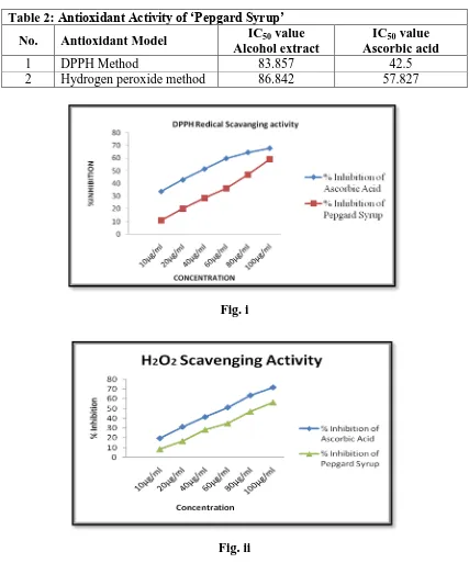
model comparing with ascorbic acid as standard antioxidant. Results are given in Table. 2
[image:6.595.84.511.181.694.2]And graphical presentation is given in Fig. i & ii.
Table 2: Antioxidant Activity of ‘Pepgard Syrup’
No. Antioxidant Model IC50 value
Alcohol extract
IC50 value
Ascorbic acid
1 DPPH Method 83.857 42.5
2 Hydrogen peroxide method 86.842 57.827
Fig. i
Fig. ii
There was a significant reduction in the concentration of DPPH and H2O2 radicals due to the
scavenging ability by increasing the dose of alcohol extract of Pepgard syrup and Ascorbic
ability with 100µg/ml of alcoholic extract of Pepgard syrup and Ascorbic acid was exhibited
59.259% and 67.836% in DPPH assay and 56.39% and 71.78% in H2O2 assay respectively.
The IC50 values were 42.5µg/ml and 83.857µg/ml in DPPH radical scavenging assay and
57.827 µg/ml and 86.842µg/ml H2O2 radical scavenging assay for Ascorbic acid and alcohol
extract of Pepgard syrup respectively.
Anti-ulcer activity of Pepgard syrup by Aspirin induced ulcers model.
Normal animal had shown 0.333 ulcer index, aspirin induced; disease control had elevated
ulcer index 4.0, standard (ranitidine) reduced ulcer index to 0.416±0.250. Pepgard syrup also
inhibited ulcer in a dose of 1ml showing ulcer index 2.75±0.822, 2 ml ulcer index
21.58±0.343 and 4 ml ulcer index 2 0.833±0.519. Result indicated that Pepgard syrup
exhibited antiulcer activity in a dose dependent manner. The photographs of open stomach of
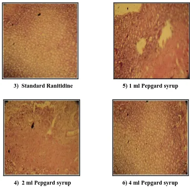
each group are given in Fig. No.iii, Histopathological microphotographs are given in Fig. No.
iv and the results are summarized in Table No.3 with graphically presentation in Fig. No.v
Fig.iii. Macroscopical view of Aspirin induced gastric ulceration in Rats.
3) Standard Ranitidine 5) 1 ml Pepgard syrup
[image:8.595.101.490.67.455.2]
4) 2 ml Pepgard syrup 6) 4 ml Pepgard syrup
[image:8.595.33.565.497.756.2]Fig.iv Histopathological microphotograph of Rat Stomach in different group.
Table 3. Mean ulcer index
Groups Body Wt.
(gm)
ULCER INDEX Total
score
Mean ulcer
IndexSEM
% Protection
0 0.5 1 1.5 2
Normal 170 180 200 190 185 175 0.5 0.5 - 0.5 - 0.5 - - - - - - - - - - - - - - - - - - - - - - - - 0.5 0.5 - 0.5 - 0.5
0.333 ±0.110 -
Disease Control (100mg/kg Aspirin) 155 180 175 155 160 175 0.5 0.5 0.5 0.5 0.5 0.5 1.0 1.0 1.0 1.0 1.0 1.0 1.5 1.5 1.5 1.5 1.5 1.5 2.0 - 2.0 - - 2.0 - - - - - - 5.0 3.0 5.0 3.0 3.0 5.0
4.0±0.4701 -
Std. Ranitidine (100mg/kg) 170 165 175 155 - 0.5 - 0.5 - - - 1.0 - - - - - - - - - - - - 0 0.5 0 1.5
180 170 - 0.5 - - - - - - - - 0 0.5 1 ml pepgard syrup 150 160 155 170 165 175 0.5 0.5 0.5 0.5 0.5 0.5 1.0 1.0 1.0 1.0 1.0 - 1.5 - 1.5 - 1.5 - - - 2.0 - 2.0 - - - - - - - 3.0 1.5 5.0 1.5 5.0 0.5
2.75±0.822 31.5
2ml pepgard syrup 175 150 185 165 160 160 0.5 0.5 0.5 0.5 0.5 0.5 1.0 1.0 - 1.0 1.0 1.0 1.5 - - - - - - - - - - - - - - - - - 3.0 1.5 0.5 1.5 1.5 1.5
1.58±0.343** 60.5
4 ml pepgard syrup 160 175 165 180 155 160 0.5 - 0.5 - 0.5 - 1.0 - - - 1.0 - - - - - 1.5 - - - - - - - - - - - - - 1.5 0 0.5 0 3.0 0
0.833±0.519*** 79.18
Values are the Mean±SEM of six rats/treatment.
Significance *P <0.05, **P<0.001 and *** P<0.001 vs. Disease Control. Statistically
Analysed by one-way analysis of variance (ANOVA) followed by Dennett’s multiple
comparison test.
Effect of Pepgard syrup on Aspirin Induced gastric ulcer in Rats
Groups M e a n Ul c e r In d e x ± S E M Gr-1 Gr-2 Gr-3 Gr-4 Gr-5 Gr-6 0 1 2 3 4 5 Gr-1 Normal
Gr-2 Disease control
Gr-3 Std.Ranitidine 100 mg/kg Gr-4 1 ml Pepgard syrup Gr-5 2 ml Pepgard syrup Gr-6 4 ml pepgard syrup
***
**
***
HISTOPATHOLOGICAL STUDIES
Histopathological microphotograph of rat stomach in normal, Disease control group,
ranitidine treated, 1 ml pepgard syrup, 2 ml pepgard syrup and 4 ml Pepgard syrup are given
in Fig.v. respectively.
1) Normal control: Histopathological microphotograph revealed that No congestion, no
necrosis only gastric lesion .supercritical mucosal layer and muscularis mucosa unaffected.
2) Disease control (100 mg/kg Aspirin): that necrosis and a granulocytosic area were
observed and gastric lesions were predominant over vast surface area, perforations with
complete mucosal destruction.
3) Standard Ranitidine (100mg/kg Ranitidine): that no congestion, no necrosis and gastric
lesions in Ranitidine treated Group. Superficial mucosal layer and muscularis mucosa were
remain unaffected
4) 1 ml Pepgard syrup: moderate congestion and muscularis mucosa.
5) 2 ml Pepgard syrup: no congestion, no necrosis, very less ulcer site.
6) 4 ml Pepgard syrup: there were histological changes limited to the superficial layers with
no congestion and no necrosis in 4 ml Pepgard syrup treated group.
DISCUSSION
Although in most of the cases the etiology of ulcer is unknown, it is generally accepted that
ulcer results from an imbalance between aggressive factors and the maintenance of the
mucosal integrity through the endogenous defence mechanism. To regain the balance,
different therapeutic agents including herbal preparations are used to inhibit the gastric acid
secretion or to boost the mucosal defence mechanism by increasing mucus production.
Different studies give highlights to various causes for the development of peptic ulcer. Ulcers
caused by aspirin could be due to their direct effect or release of noxious substances
including free radicals.[11] Antioxidants play a major role in repairing the gastric damage.
Total antioxidant activity, metal chelation, radical scavenging (DPPH) effects and reducing
power as well as activities destructive to active oxygen species such as the superoxide anion
radical, hydroxyl radical, and hydrogen peroxide are widely used for this purpose.[10]
Hydrogen peroxide (H2O2) is a strong oxidizer. To prevent that, our cells also contain
antioxidant enzymes known as peroxiredoxins that degrade H2O2 molecules. In the present
Aspirin can cause ulcers by interfering with the stomach's ability to protect itself from gastric
acids. While stomach acids are vital to the digestive process, they can cause damage if the
protective barriers of the stomach are compromised. Normally, the stomach has three
protections against gastric acid 1) Mucus produced by foveolar cells that line the stomach. 2)
Bicarbonate produced by foveolar cells which helps neutralize stomach acid. 3) Blood
circulation that aids in the repair and renewal of cells in the stomach’s mucosal layer.
Disruption of prostanoid synthesis is another contributing factor for aspirin-induced ulcers. It
appears that the development of gastric mucosal damage by aspirin, and possibly other
ulcerogenic NSAIDs, involves hypersecretion of tissue-destructive free radicals which may
come from (a) enhanced conversion of hydroperoxy to hydroxy fatty acids in the
lipooxygenase pathway, (b) accelerated xanthine-oxidase activity in the mucosa, and (c)
possibly from the drugs themselves. The inhibition of gastric mucosal prostaglandin
production occurs rapidly following oral administration of ulcerogenic drugs. This is
correlated with the rapid absorption of these drugs through the mucosa. Inhibition of
prostglandin coincides with the earlier stages of injury to the cell membranes of the mucosa
with the concomitant loss of the permeability characteristics of the mucus and electron
microscopic evidence of damage to mucosal, parietal, and endothelial cells. The later changes
also reflect rapid ischemia which appears to develop in the mucosa during aspirin injury.[12]
NSAID’s such as aspirin and indomethacin are known to induce gastric ulceration. The
reason being attributed principally to inhibition of biosynthesis of cytoprotective
prostaglandins such as prostaglandin E and prostacyclin by inhibition of cyclooxygenase
pathway of arachidonic acid metabolism, resulting in overproduction of leukotrienes and
other products of 5-lipoxygenase pathway.[13] Role of cytoprotection in preventing ulcer
generation is clearly shown.[14] Different studies show role of ROS, for the genesis of gastric
ulcer.[15] A small part of oxygen used is converted to ROS, i.e., the superoxide anion radical
(O2−), hydrogen peroxide (H2O2), and the hydroxyl radical (•OH). They are highly toxic to
cells and can cause oxidative damage. Ischemia-reoxygenation-induced gastric mucosal
injury shows role of ROS in pathogenesis of gastric ulcer.[16] H. pylori is an important causative agent leading to chronic Type B gastritis, peptic ulcer, adenocarcinoma, and
mucosa associated lymphoid tissue lymphoma affecting stomach.[17] Various studies show the
role of ROS in NSAID’s and H. pylori [18] induced gastric mucosal injury. These ROS decreases the level of endogenous antioxidants such as α tocopherol, glutathione and
their direct effect or release of noxious substances including free radicals.[19] A specific •OH
scavenger, dimethyl sulfoxide found to inhibit gastric ulceration produced by ischemia,
stress,[20] showing the role of •OH in causing mucosal ulceration. Antioxidant potential of
melatonin was found to be the reason for its anti-ulcer property by scavenging ROS.
The present study revealed that Mean ulcer Index and histopathology study revealed that
Pepgard syrup had shown Anti ulcer activity in a dose dependent manner. The Pepgard Syrup
shows a significant inhibitor of gastric mucosal lesions caused by aspirin in rats thereby
confirming its anti-ulcerogenic activity. The cytoprotective effect could be partially due to
the presence of phyto-constituents like saponin, flavonoid, tannin alkaloid, glycoside,
Carbohydrates and triterpenoid content in Pepgard syrup and its reactive oxygen species
scavenging property. The antioxidant mechanism of Pepgard syrup against gastric mucosal
lesions was supported by its in vitro antioxidant potency as evidenced by its high DPPH and H2O2 value.
CONCLUSION
The present study with Pepgard syrup revealed that it has significant oxidant and
anti-ulcer activity. Pepgard syrup was found to possess anti-oxidant and anti-anti-ulcer activity
properties in in-vivo and in-vitro models, and these findings suggest that the significant
gastroprotective activity could be mediated by its antioxidant activity. Therefore, Pepgard
syrup is relatively safe for peptic ulcers and related disorders in human for long period.
REFERENCES
1. Haubrich WS, Schaffner F, Berk JE. Bockus Gastroenterology. 5th ed., Vol. 1.
Philadelphia, PA: WB Saunders Company, 1995; 714-32.
2. Rao TS, Basu N, Siddiqui HH. Anti-inflammatory activity of curcumin analogues. Indian
journal of medical research, 2013; 137(4): 574-8.
3. Kasper D, Fauci A, Hauser S, Longo D, Jameson J, Loscalzo J. Harrison's principles of
internal medicine. McGraw-Hill Professional Publishing, 2018.
4. Paget GE, Barnes JM. Evaluation of drug activities. In: Laurence DR, Bacharach AL,
editors. Pharmacometrics. Vol. 1. New York: Academic Press, 1964; 189.
5. Oktay M, Gülçin İ, Küfrevioğlu Öİ. Determination of in vitro antioxidant activity of
fennel (Foeniculum vulgare) seed extracts. LWT-Food Science and Technology, 1; 36(2):
6. Valko M, Leibfritz D, Moncol J, Cronin MT, Mazur M, Telser J. Free radicals and
antioxidants in normal physiological functions and human disease. The international
journal of biochemistry & cell biology, 2007; 39(1): 44-84.
7. Konturek SJ, Piastucki I, Brzozowski T, Radecki T, Dembińska-Kieć A, Źmuda A,
Gryglewski R. Role of prostaglandins in the formation of aspirin-induced gastric ulcers.
Gastroenterology, 1981; 80(1): 4-9.
8. Parmar N, Desai JK. A review of the current methodology for the evaluation of gastric
and duodenal anti-ulcer agents. Indian journal of pharmacology, 1993; 25(3): 120.
9. Shimada K, Fujikawa K, Yahara K, Nakamura T. Antioxidative properties of xanthan on
the autoxidation of soybean oil in cyclodextrin emulsion. Journal of agricultural and food
chemistry, 1992; 40(6): 945-8.
10.Chainani-Wu N. Safety and anti-inflammatory activity of curcumin: a component of
tumeric (Curcuma longa). The Journal of Alternative & Complementary Medicine, 2003;
9(1): 161-8.
11.Rainsford KD. Mechanisms of gastro-intestinal ulceration by non-steroidal
anti-inflammatory/analgesic drugs. Adv Inflam Res, 1984; 6: 51-64.
12.Rainsford KD. The effects of 5-lipoxygenase inhibitors and leukotriene antagonists on the
development of gastric lesions induced by nonsteroidal antiinflammatory drugs in mice.
Agents and Actions, 1987; 21(3-4): 316-9.
13.Dedieu-Chaufour, C.A.T.H.E.R.I.N.E., Hertz, F., Caussade, F. and Cloarec, A., 1991.
Pharmacological profile of UP 5145-52, an original antiulcer and antisecretory
agent. Journal of Pharmacology and Experimental Therapeutics, 259(1): 190-197.
14.Halliwell B, Gutteridge JM. Free radicals, ageing and disease. Free Radicals in Biology
and a Medicine. 2nd ed. Oxford: Clarendon Press, 1989; 416.
15.Perry MA, Wadhwa S, Parks DA, Pickard WE, Granger DN. Role of oxygen radicals in
ischemia-induced lesions in the cat stomach. Gastroenterology, 1986; 90(2): 362-7.
16.Ananthanarayanan, Jayarampaniker RC. Textbook of Microbiology. 10thed. Oxford:
Orient Longman Pvt. Ltd, 2017; 407-8.
17.Davis GR, Simmonds NJ, Stevens TR, Sheaff MT, Banatwala N, Laurenson IF, et al. Helicobacter pylori stimulates antral mucosal reactive oxygen metabolite production in vivo. Gut, 1993; 35: 179.
18.Mizui T, Doteuchi M. Lipid peroxidation: A possible role in gastric damage induced by
19.Davies GR, Simmonds NJ, Stevens TR, Sheaff MT, Banatvala N, Laurenson IF, Blake
DR, Rampton DS. Helicobacter pylori stimulates antral mucosal reactive oxygen
metabolite production in vivo. Gut, 1994; 35(2): 179-85.
20.Rajakrishnan V, Viswanathan P, Rajasekharan KN, Menon VP. Neuroprotective role of
curcumin from Curcuma longa on ethanol‐induced brain damage. Phytotherapy Research:
An International Journal Devoted to Pharmacological and Toxicological Evaluation of