www.wjpr.net Vol 4, Issue 2, 2015. 1491
COMPARISON BETWEEN RECOMMENDED DAILY DOSE AND
MODIFIED REDUCED DOSES OF IRON PROPHYLAXIS IN
NON-ANAEMIC PREGNANT WOMEN
Dr.Rajarshi Rahut1, Dr.Subinay Datta1, Dr.Mrinal Pal*1, Dr.Amit Kumar Pradhan1,
Dr.Sudipta Sil2
1
Department of Biochemistry, Burdwan Medical College, Burdwan, West Bengal, India. 2
Department of Pharmacology, Burdwan Medical College, Burdwan, West Bengal, India.
ABSTRACT
Background: Recommended prophylactic daily dose iron supplementation in non-anaemic pregnant mothers increases the body iron store that leads to unfavourable pregnancy outcome. So the present study was conducted on pregnant mothers with reduced dose of elementary iron and to investigate whether the reduce dose of iron supplementation can reduce the adverse effects. Material and methods: A prospective observational randomized hospital based clinical study was performed on 299 pregnant women with haemoglobin ≥ 10gm/dl and divided into three groups. One group received 100mg elementary iron daily, second group same dose but in alternate day and other received 200 mg elementary iron twice weekly. Blood ferritin concentration was measured at 28-32 weeks and was compared across the three study groups and also compared with the birth weight of their babies within a group.
Result: There was significant increased concentration of blood ferritin level and this increased ferritin level were significantly associated with higher rates of intrauterine growth restricted baby in pregnant mothers receiving daily iron supplementation. Among the reduced and modified dose of iron supplementation alternate day 100 mg elementary iron prophylaxis strategy is much more subject friendly. Conclusion: The rationale of routine iron supplementation in non-anemic pregnant women needs to be re-examined.
KEYWORDS: Iron prophylaxis, intrauterine growth restriction, Ferritin, Non-anaemic pregnant mother.
Volume 4, Issue 2, 1491-1500. Research Article ISSN 2277– 7105
Article Received on 10 Dec 2014,
Revised on 04 Jan 2015, Accepted on 29 Jan 2015
*Correspondence for
Author
Dr. Mrinal Pal
Department of
Biochemistry, Burdwan
Medical College,
Burdwan, West Bengal,
www.wjpr.net Vol 4, Issue 2, 2015. 1492 1. INTRODUCTION
Iron is an essential element for both pregnant women and the growing foetus and so pregnant women are routinely recommended for iron supplementation ,[1-3] despite the fact that such prophylactic iron supplementation is still a matter of controversy. Indeed, animal studies suggest that the administration of iron daily at the current recommended dosages may be neither desirable nor innocuous[4] as elevated iron stores during pregnancy have been associated with increased risk of maternal and neonatal morbidity such as intrauterine growth restriction (IUGR).[5] So, the present study was conducted to evaluate the level of serum ferritin in pregnant mother between 28-32 weeks of gestation, which could point out whether the intermittent iron supplementation can reduce the incidence of this type of detrimental effect in pregnancy than daily dose and can stands for possible alternative strategy for giving supplementation in non-anemic subjects. As serum concentration of ferritin is in correlation with total iron reserve in the human and the decrease levels is in correlation with the decrease of iron reserves in the mother resulting from increased uptake (by the mother, placenta and foetus) as well as from hemodilution[6] therefore it was used as a reliable parameter for the estimation of iron status in body of study population.[7-9]
2. MATERIAL AND METHODS
2.1 Study area
The present study was conducted in the department of Biochemistry with the collaboration of department of Gynaecology and Obstetrics of Burdwan Medical College, Burdwan, West Bengal, India.
2.2 Selection of subjects
www.wjpr.net Vol 4, Issue 2, 2015. 1493
ten subjects were selected by a simple random method for this study after informed consent had been received. The proposal of this study was approved by the Ethics Committee of Burdwan Medical College, West Bengal.
2.3 Iron Supplementation
The mothers were divided into three groups. One group (n = 107) was received traditional prophylactic daily dose (100mg) of elementary iron, in second group (n = 119) given same dose of iron in alternate day and third group (n = 84) given 200mg elementary iron in twice weekly. All the groups were getting their respective way of supplementation with same compound of iron throughout the pregnancy. Complains of missing iron tables and gastrointestinal upsets such as nausea, vomiting, constipation, diarrhoea, epigastric pain was noted carefully.
2.4 Collection of samples
Peripheral venous blood from the entire pregnant mother under study were drawn at 28-32 weeks of gestation and allowed to coagulate at room temperature for 30–45 min, followed by centrifugation at 2500 rpm for 5 min. All serum samples were stored at -700C and kept under these conditions until chemical analysis was performed. The gestation of 28-32 weeks was chosen for the blood sampling because iron storage decreases with advancing gestation and the lowest of ferritin are recorded at this period after which the concentration stay on constant levels.[6] Furthermore, the demand for the power of the absorption of iron is greatest in the third trimester.[11]
2.5 Laboratory investigation
Serum ferritin estimation was done using micro-particle enzyme immunoassay, IMX system of Abbott Park, IL, USA. Intra-assay CV% and inter-assay CV% was 3.4 and 3.5 respectively.
2.6 Physical features at birth
After delivery, the data on infant characteristics for the presence of intrauterine growth restriction were retrieved from the records in our hospital for analysis.[12]
2.7 Statistical Analysis
www.wjpr.net Vol 4, Issue 2, 2015. 1494 3. RESULT
Of the 310 pregnant women with a gestational age of 30–32 weeks included in the investigation, 11 were excluded during the investigation period for the following reasons: 3 pregnant women developed hypertension at 32 gestational weeks, 2 presented with gestational diabetes, 4 women delivered before the 37th completed week of gestation, whereas 2 blood sample was contaminated by the presence of fibrin and estimations of ferritin could not have been done.
3.1 Personal profile and clinical details of the pregnant mothers and their babies of
Group I, II and III
Selected characteristics of the women at the beginning of the study as shown in the Table 1
Table 1: Personal profiles and clinical parameters of the pregnant mothers and their
babies under the study.
Population
characteristics Group I Group II Group III
Group I vs Group II (p value)
Group I vs Group III
(p value)
Group II vs Group III
(p value)
n 105 111 83
Mother’s age
(years) 26.34 ± 5.82 27.68 ± 6.36 28.12 ± 7.53 > 0.05 > 0.05 > 0.05 Gestational age
(days) 272 ± 6.73 270 ± 6.17 271 ± 5.96 > 0.05 > 0.05 > 0.05 Singletons 47 (44.76) 53 (47.75) 39 (47) >0.05 > 0.05 > 0.05 IUGR baby ⃰ 51 (48.57) 13 (11.71) 10 (12.5) < 0.001 < 0.001 >0.05 Birth weight (gm) 2263 ± 159 2987 ± 336 2911 ± 225 < 0.001 < 0.001 >0.05 Hemoglobin
(gm/dl) 11.4 ± 2.67 12.6 ± 1.59 12.1 ± 2.16 > 0.05 > 0.05 > 0.05 Complain of
gastrointestinal upsets ⃰
61 (58.09) 13 (11.71) 69 (83.13) < 0.001 0.011 < 0.001 Missing iron
tables ⃰ 2(1.9) 72 (64.86) 59 (71.08) < 0.001 < 0.001 0.034 n = number of subject; p > 0.05 is considered statistically not significant; p < 0.05 is significant; Values are mean ± SD
⃰ Data are expressed as numbers (group percentages in parentheses)
3.2 Comparison of the serum ferritin level between different groups
[image:4.595.26.574.348.602.2]www.wjpr.net Vol 4, Issue 2, 2015. 1495
and Group III (12.95 µg/L) but there was no significant difference between Group II and III as shown in the Table 2.
Table 2: Comparison between concentrations of serum ferritin in Group I, Group II and III study population
Concentration of serum ferritin (µg/L)
Group I vs Group II
(p value)
Group I vs Group III
(p value)
Group II vs Group III
(p value) Group I
Group II Group III
29.12 ± 19.44 13.27 ± 2.65 12.95 ± 2.32
< 0.001 < 0.001 >0.05 Values are mean ± SD; p value p < 0.05 considered statistically significant
3.3Correlation of blood ferritin with birth weight of baby in the study groups
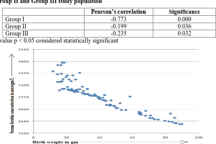
[image:5.595.78.520.158.250.2]In case of traditional iron supplementation there was a more significant negative correlation between serum ferritin and birth weight (r = -0.773, p = 0.000) than in case of two modified types of iron supplementation (r = -0.199, p = 0.036; r = -0.235, p 0.032) as depicted in the Table 3 and Figure 1, 2 and 3. So it is clear that only in higher concentration of serum ferritin seriously affects in reduction of birth weight.
Table 3: Bivarient correlation between birth weight and serum ferritin in Group I,
Group II and Group III study population
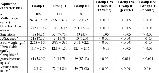
Pearson’s correlation Significance
Group I -0.773 0.000
Group II -0.199 0.036
Group III -0.235 0.032
p value p < 0.05 considered statistically significant
Figure 1: Correlation of birth weight (g) and blood ferritin levels (µg/L) in pregnant
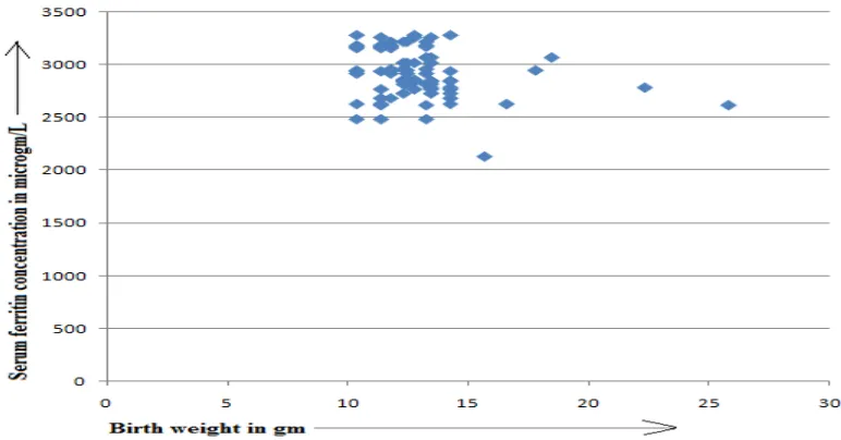
[image:5.595.82.509.437.721.2]www.wjpr.net Vol 4, Issue 2, 2015. 1496 Figure 2: Correlation of birth weight (g) and blood ferritin levels (µg/L) in pregnant
[image:6.595.105.489.77.282.2]women between 30 and 32 weeks of gestation of Group II study population.
Figure 3: Correlation of birth weight (g) and blood ferritin levels (µg/L) in pregnant
women between 30 and 32 weeks of gestation of Group III study population.
4. DISCUSSION
[image:6.595.105.491.330.532.2]www.wjpr.net Vol 4, Issue 2, 2015. 1497
versus daily iron dosing in an attempt to observed effect of pregnancy outcome and also identify the optimum supplementary protocol.
First of all, it was found that traditional iron supplementation causes elevated maternal serum ferritin level than other reduced dose of iron supplementation. Numerous recent studies have shown that iron supplementation in reduced dose in non-anemic pregnant women is an effective option for prophylaxis.[14]
Another important finding in the study was that elevated level of serum ferritin was associated with an increased risk of IUGR. Various previous studies have been shown that increased maternal iron store appear to have a negative impact on birth weight,[10,15] as raised serum ferritin level can point to impaired transfer of micronutrients from the mother to foetus.[6] In addition, there is the potential concern that some women who are not truly anemic may taking large doses of daily supplemental iron during pregnancy. Such a strategy has been suggested as a way to build up the mother’s iron store and to increase blood viscosity to impair uteroplacental flow.[16-18] It is also possible that increased levels of plasma free iron as a result of daily iron supplementation at recommended dose also increase oxidative damage to DNA in the maternal-foetal unit.[1] But reduced dose iron supplementation has recently been associated with a reduced risk of low birth weight infants in pregnant women with no anemia.[19] Moreover Iron absorption is suppressed for at least 24 h after consumption of a high iron meal or iron supplement, principally by controlled suppression of intestinal mucosal cell uptake.[20-23] So, it has been suggested that less frequent supplementation may reduce the transient iron overload and may decrease the cost of routine daily supplementation[24] but there is a chance of missing table because mothers have to remember the particular date in a week when has to take.
Another important observation in this study was that though both the reduced modes of iron prophylaxis can effectively reduced the chance of IUGR baby but in respect to patient compliance alternate day 100mg elementary iron supplementation is far better than 200mg twice weekly supplementation.
5. CONCLUSION
www.wjpr.net Vol 4, Issue 2, 2015. 1498
applied they can effectively reduce the chance of development iron overload and consequently IUGR. But person compliance limits the use of 200 mg twice weekly supplementation. So, alternate day 100 mg supplementation as a possible alternative strategy that may enhance the effectiveness of operational programs.
6. ACKNOWLEDGEMENT
Authors are thankful to Mr. Karthik Basu, Naba Kumar Das and Chandi Charan Das of Burdwan Medical College and Hospital for technical support.
7. CONFLICT OF INTEREST
We do not have any conflict of interest.
REFERENCES
1. Scholl TO. Iron status during pregnancy: setting the stage for mother and infant. Am.J. Clin. Nutr, 2005; 81(suppl.): 1218-1222.
2. Siddappa AM, Rao R, Long JD, Widness A, Georgieff MK. The assessment of newborn iron stores at birth: a review of literature and standard for ferritin concentrations. Neonatology, 2007; 92: 73-82.
3. Rao R, Georgieff M. Iron in foetal and neonatal nutrition. Sem. Fetal Neonatal Med, 2007; 12: 54-63.
4. Viteri F, Berger J. Importance of pre-pregnancy and pregnancy iron status: can long-term weekly preventive iron and folic acid supplementation achieve desirable and safe status? Nutrf. Rev, 2005; 63: 65-76.
5. Rayman M, Barlis J, Evans R, Redman C, King L. Abnormal iron parameters in the pregnancy syndrome Preeclampsia. Am. J. Obstet. Gynecol, 2002; 1872: 412-418.
6. Višnjevac N, Segedi ML, Ćurčić A, Višnjevac J, Stajić D. Blood ferritin levels in pregnant women and prediction of the development of fetal intrauterine growth restriction. J Med Biochem, 2011; 30: 317–322.
7. Uberos J, Molina A, Munoz A. Blood ferritin levels in pregnant women as an estimator of low birth weight? Prenat Neonat Med, 2000; 5: 177–181.
8. Lipschitz DA. Cook JD, Finch CA. A clinical evaluation of serum ferritin as an index of iron store. N. Engl. J. Med, 1974; 290: 1213-1216.
www.wjpr.net Vol 4, Issue 2, 2015. 1499
10.Lao TT, Tam KF, Chan LY. Third trimester iron status and pregnancy outcome non-anaemic women; pregnancy unfavourably affected by maternal iron excess. Human reproduction, 2000; 15(8): 1843-1848.
11.Whittaker PG, Lind T, Williams JG. Iron absorption during normal pregnancy: a study using stable isotopes. Br. J. Nutr, 1991; 65: 457-463.
12.Dutta DC. Low birth weight baby. In: Text book of Obstetrics including perinatology and contraception, 5th Edition, Kolkata, New Central Book Agency Private Limited, 2001; 496-501.
13.Soubasi V, Petridau S, Sarafidis K, Tsantali Ch, Diamanti E, Buonocore G, Drossou-Agakidou V. Association of increased maternal ferritin levels with gestational diabetes and intrauterine growth retardation. Diabetes and Metabolism, 2010; 36: 58-63.
14.Mukhopadhyay A, Bhatla N, Kriplani A, Pandey RM, Saxena R. Daily versus intermittent iron supplementation in pregnant women: hematological and pregnancy outcome. J. Obstet. Gynaecol. Res, 2004; 30(6): 409-17.
15.Lao TT, Tam KF, Chan LY, Ho LF. Maternal haemoglobin and risk of gestational diabetes mellitus in Chinese women. Obstet. Gynecol, 2002; 99: 807-812.
16.Scanlon KS, Yip R, Schieve LA, Cogswell ME. High and low haemoglobin levels during pregnancy: differential risk for preterm birth and small for gestational age. Obstet. Gynecol, 2000; 96: 741-748.
17.Zhou LM, Yang WW, Hua JZ, Deng CQ, Tao X, Stolzfus RJ. Relation of haemoglobin measured at different times in pregnancy to preterm birth and low birth weight in Shanghai, China. Am. J. Epidemiol, 1998; 148: 998-1006.
18.Rush D. Nutrition and maternal mortality in the developing world. Am. J. Clin. Nutr, 2000; 72(suppl.): 212S-240S.
19.Palma S, Perez-Iglesias R, Prieto D, Pardo R, Llorca J, Delgado-Rodriguez M. Iron but not folic acid supplementation reduces the risk of low birth weight in pregnant women without anaemia: a case control study. J. Epidemiol. Community Health, 2008; 62: 120-124.
20.Fairweather-Tait SJ, Swindell TE, Wright AJA. Further studies on rats on the influence of previous intake on the estimation of bio-availability of Fe. Br. J. Nutr, 1985; 51: 185–191. 21.Fairweather-Tait SJ, Minski MJ. Studies on iron-availability in man using stable isotope
techniques. Br. J. Nutr, 1986; 55: 279–285.
www.wjpr.net Vol 4, Issue 2, 2015. 1500
23.Solomons NW, Pineda O, Viteri F, Sandstead HH. Studies on the bio-availability of zinc in human: mechanism of the intestinal interaction on non-heme Fe and Zinc. J. Nutr, 1983; 113: 337–349.