A STUDY OF THE CURING POTENTIAL OF ASCORBIC ACID ON A
SELECTED MULTIDRUG RESISTANT
Escherichia coli
UROPATHOGEN STRAIN
1*
F. D. Otajevwo and 2H. A. Z. Obiazi
1
Department of Microbiology, Western Delta University, Oghara, Nigeria.
2
Department of Microbiology, Ambrose Alli University, Ekpoma, Nigeria.
INTRODUCTION
Background to the Study
World Health Organization (WHO) has identified multi drug resistant
(MDR) bacteria as one of the top three threats to human health
(Bassetti et al., 2011). Antibiotics are naturally occurring compounds
produced by microorganisms. They have the capacity to kill
susceptible microorganisms or to inhibit their growth (Todar, 2012).
The use of antibiotics has been one of the reasons for the increase in
life expectancy in the 20th century (Todar, 2012). Despite this impressive feat, there have been records of disease causing microbes
that have become resistant to antibiotics (Livermore, 2004; Pereira et
al., 2013).
Resistance to antibiotics is of two types and they are intrinsic (natural) and acquired
resistance. Intrinsic resistance occurs when microbes naturally do not possess target sites for
antibiotics and therefore, the drugs do not affect them or they naturally have low permeability
to those agents due to differences in the chemical nature of such drugs and the microbial
membrane structures especially for antibiotics that selectively attack cell membrane (Fluit et
al., 2001). Others are due to overproduction of the target sites of the antimicrobial agent,
expression or suppression of a gene invivo in contrast to the situation invitro, production of
antibiotic inactivating enzymes and exotoxin production.
Multidrug resistance develops when a pathogen is resistant or acquires resistance to two or
more antibiotics which do not have to be related (WHO, 2012). The increasing number of
multiple antibiotic resistant pathogens has become a serious threat to human health (CDC,
Article Received on 21 March 2018,
Revised on 10 April 2018, Accepted on 01 May 2018,
DOI: 10.20959/wjpr20189-12152
*Corresponding Author
F. D. Otajevwo
Department of
Microbiology, Western
Delta University, Oghara,
Nigeria.
2013; WHO, 2014). Moreover, there has been an upsurge in antibiotic resistant strains of
clinically important pathogens in recent times and this has led to the emergence of new
bacterial strains that are multi resistant (WHO, 2001; Albinu et al., 2003; Albinu et al.,
2004). The primary causes of antibiotic resistance in bacteria are mobile elements called
plasmids, conjugative transposons and integrons (Su et al., 2012; Amaral et al., 2013).
Curing is the process of removing plasmids from a bacterial cell (Trevors, 1986). The
resulting bacterial organism then becomes sensitive to the selective agent. It was initially
thought that this phenomenon would proffer solution in controlling the development of
antibiotic resistance in formerly antibiotic susceptible bacteria. Novobiocin, ethidium
bromide, acriflavine, acridine orange, ascorbic acid and elevated temperatures have been used
as curing agents (Ramesh et al., 2000). There are a number of reports demonstrating the
ability of various chemical and physical agents to increase the rate of loss of plasmid DNA
from bacteria (Sonstein and Baldwin, 1972; Stanier, 1984; Kumar and Surendran, 2006).
Ingram et al. (1972) found that drug resistance of P. aeruginosa could be eliminated by
treatment with SDS and Pattnakik et al. (1995) reported that acridine orange could not affect
P.aeruginosa due to impermeability of its cell wall while ethidium bromide and SDS cured
antibiotic resistance plasmid at a concentration of 1-2% and 700-3000ug/ml for SDS and
ethidium bromide respectively. Al-Amar et al. (1999) treated an isolate of P. aeruginosa with
1000ug/ml dilution of acridine orange and reported that there were no cured cells as the
plasmid profile of cured cells was the same as that of untreated samples with a conclusion
that acridine orange had no effect on Pseudomonas aeruginosa as a curing agent. It was also
reported that ethidium bromide was effective as a curing agent at 600, 700 and 1000ug/ml
dilutions for the same isolate of Pseudomonas aeruginosa.
It has been reported that phenothiazines have the ability to control overexpression of efflux
pump systems and thus are able to remove or reduce antibiotic resistance (Viveiros et al.,
2010; Amaral et al., 2013). Mukherjee et al. (2012) reported the use of 1000-3000ug/ml
dilutions of a type of phenothiazine and an anti-psychotic drug called thioridaxine to cure a
multidrug resistant strain of Pseudomonas aeruginosa.
Justification of the Study
The non-availability and high cost of new generation antibiotics with limited effective
proliferation of multidrug resistant pathogens continues within and around us. It is important
that their resistance trend be put under check through intensive research and antibiotic
surveillance (Akortha and Filgona, 2009).
Besides, it is common these days for physicians to report increasing number of treatable
infections that fail to respond to antibiotic treatment. Antibiotic resistant bacteria may keep
people sick longer and sometimes unable to recover. The development of microbial resistance
to antibiotics has resulted in the search for new antibiotics in order to maintain a pool of
effective drugs at all times and unless antibiotic resistance problems are detected and tackled
as they emerge and action taken to enforce the necessary and judicious use of antibiotics, the
society could be faced with previously treatable diseases such as tuberculosis, pneumonia,
gonorrhea, typhoid etc that have become untreatable as in the era before antibiotics were
discovered (Todar, 2012).
Multidrug resistance is now common among familiar pathogens such as Escherichia coli,
Staphylococcus aureus, Klebsiella pneumoniae to mention but a few. Multidrug resistant
(MDR) E.coli and other Enterobacteriaceae isolates are characterized by non-susceptibility to
at least one agent in three or more antibiotic categories (Magiorakos et al., 2012).
Escherichia coli has high resistance for older generation of human and veterinary antibiotics
including ampicillin, streptomycin, tetracycline as well as increasing resistance to newer
antibiotics such as fluoroquinolones and cephalosporins (Tadesse et al., 2012). Moreso, as a
gram negative organism, E.coli is resistant to many antibiotics that are effective against gram
positive organisms (Johnson et al., 2006). E.coli bacteria often carry multidrug resistance
plasmids and under stress, readily transfer those plasmids to other species (Salyers et al
2004). Extended spectrum beta lactamase producing E.coli is highly resistant to an array of
antibiotics and infections by these strains of E.coli are highly difficult to treat (Paterson and
Bonomo, 2005). Virulent strains of E.coli can cause infantile gastro-enteritis, pelvic
inflammatory disease, neonatal meningitis, 85% community acquired UTIs, 50% hospital
acquired UTIs and more than 80% cases of uncomplicated pyelonephritis (Bergeron, 1995;
Todar, 2007).
Treatments that increase frequency of elimination of plasmids will certainly enhance
sensitivity (effectiveness) of antibiotics in situ. Ramesh et al. (2000) and Potrus (2009) have
used dilutions of ascorbic acid to induce antibiotic resistance loss in Pediococcus acidilactivi
to cure antibiotic resistance plasmid genes in Azotobacter chroococcum (Garg et al., 2003)
and Staphylococcus aureus (Amabile-Ceuvas, 1988, 1991). Hence, there is not much
documented work on use of dilutions of ascorbic acid (an anti-oxidant and a non-antibiotic
agent) to reduce resistance loss in MDR bacterial pathogens and particularly Escherichia coli
isolated from an infected urinary tract in terms of removal of antibiotic resistance genes (if
plasmid mediated).
Overall Aim of the Study
The overall aim of the study is to investigate the curing potency of ascorbic acid dilutions on
a selected multidrug resistant Escherichia coli uropathogen strain.
The Specific Objectives of the study are:
i. To determine the antibiotic susceptibility patterns or antibiograms of fifteen (15)
Escherichia coli strains (coded EC1 – EC15) before exposure to dilutions of ascorbic acid
after incubation at 37oC for 24hr.
ii. To determine the occurrence and distribution of multidrug resistant E.coli uropathogen
strains among the isolates.
iii. To determine the dilutions of ascorbic acid that induced ≤50% loss in resistance
after treatment on a selected uropathogen strain and incubation at 37oC for 6hr,12hr,18hr and 24hr.
iv. To determine the cumulative means of ≤50% loss of resistance as induced by ascorbic
acid dilutions on the selected MDR E.coli uropathogen after 6hr, 12hr, 18hr and 24hr
incubation at 37oC.
v. To determine effect of ascorbic acid dilutions treatment on the selected MDR E.coli
uropathogen strain on the minimum inhibitory concentration (MIC) of ampicillin invitro.
MATERIALS AND METHODS
Study Area
This study will be carried out in the Medical Microbiology Department of the University of
Benin, Benin City, Nigeria. Benin City is the capital of Edo State in Midwestern part of
Southern Nigeria. It has a population of 1, 147, 188 (Census, 2006) with a density of 950/km2 (2, 500/sq miles), occupies an area of 1,204 km2 (465sq miles) and is approximately 40 kilometres (25miles) north of Benin River. Benin City is also situated 320kilometres
Study Design/Sampling
Pure cultures of Escherichia coli are to be randomly obtained from 24hr Cystine Lactose
Electrolyte Deficient (CLED) agar and MacConkey agar plates which had been inoculated
with freshly voided midstream urine samples obtained from in-patients and out-patients of the
University of Benin Teaching Hospital, Edo State, Nigeria diagnosed with symptoms of
urinary tract infections (UTIs).
Power and sample size estimation
To ensure that power will be high to detect reasonable departures from the statement of
research question, a power analysis will be carried out to determine that effect, using the
following formula described by Naing et al. (2006).
N = Z P (1-P)/d²
Where N = sample size
Z = statistics for a level of 95% confidence interval = 1.96
P = urinary tract infection prevalence rate = 59.2% (2015)
d = precision (allowable error) =5% = 0.05
A power of 0.60 will give an estimated sample size of 250 samples and a mean difference of
0.25 at p=0.05.
Selection Criteria
Inclusion criterion
Only Escherichia coli pure cultures grown on 18-24hr sterile CLED and MacConkey agar
plates from midstream urine samples will be used for the study.
Exclusion criterion
All pure cultures of Escherichia coli grown on other culture media and on CLED or
MacConkey agar but not from urine samples will not be recruited for this study.
Ethical Approval and Informed Consent
Ethical approval for this study will be sought from the ethical committee of University of
Benin Teaching Hospital (UBTH), Benin City, Edo State as well as the informed consent of
Sampling and Processing
The status of the E.coli isolates (strains) will be re-confirmed by gram reaction, biochemical
and sugar fermentation tests by standard methods (Cowan and Steel, 1993; Cheesbrough,
2003; Oyeleke and Manga, 2008). After confirmation, all gram negative, raised, entire,
circular, motile, indole positive, methyl red positive, voges praskauer negative, citrate
negative, urease negative, lactose and glucose fermenting colonies will be stocked aseptically
on sterile Nutrient agar slants and incubated at 37oC for 24hr. The resulting axenic cultures will be kept at 4oC in the refrigerator for further use after appropriate labeling. All stocked E.coli strains will be subjected to antibiotic sensitivity testing to determine their antibiograms
as well their multidrug resistance statuses after which they will be treated with dilutions of
ascorbic acid.
Antibiotic Sensitivity Testing
Each of the fifteen (15) stocked E.coli strains will be subcultured on sterile CLED agar
medium and incubated at 37oC for 24hr. Antibiotic sensitivity testing will then be carried out on the resulting pure culture colonies using the agar disc diffusion method on sterile Mueller
Hinton agar (MHA) plates initially described by Bauer et al. (1966) and recommended by the
Clinical Laboratory Standard Institute (2006). A loopful of each colony of the uropathogen
strains will be picked aseptically using a flamed and cooled wire loop and placed in the centre
of the sterile MHA plates. This will then be spread all over the plates applying the caution of
not touching the edges of the plates. The seeded plates will be allowed to stand for about
2mins to allow the agar surface to dry. A pair of forceps will be flamed and cooled and used
to pick an antibiotic multidiscs (Abitek, Liverpool) containing ciprofloxacin (5ug), ampicillin
(10ug), gentamycin (10ug), ceftazidime (30ug), cefuroxime (30ug), ofloxacin (5ug),
amoxicillin/clauvulanic acid (30ug) and nitrofurantoin (300ug). The discs will be placed at
least 22.0mm from each other and 14.0mm from the edge of the plates (Ochei and Kolhatkar,
2008). Antibiotic discs will be selected on the basis of their clinical importance and efficacy
on various pathogenic strains of Escherichia coli. The seeded plates will be allowed to stand
for 10mins before incubation (Mbata, 2007).
At the end of incubation, the diameters of the zones of inhibition from one edge to the
opposite will be measured to the nearest millimeter using a transparent ruler (Byron et al.,
2003). Isolates will be grouped as resistant or sensitive based on the scheme provided by
antibiotics will be termed multiple drug resistant strains (Jan et al., 2004; Santo et al., 2007)
and will be noted.
Preparation of Ascorbic acid Dilutions
The dilutions of ascorbic acid (100 – 1000ug/ml) will be prepared according to Ramesh et al.
(2000) using RV/O. Ten Spartan vitamin C tablets (Kunimed Pharmachem Ltd, Lagos) each
having a dosage of 100mg will first be dissolved in 100ml of sterile water to obtain a stock
concentration of 10,000ug/ml. To obtain 100ug/ml dilution, 0.5ml of stock will be mixed
with 49.5ml sterile water (diluent). The next dilution of 200ug/ml will be prepared by mixing
2ml of stock solution with 98ml of diluent. Other dilutions will be prepared by mixing
various volumes of stock with their corresponding volumes of sterile water thus:
1.5ml+48.5ml (300ug/ml), 3ml+72ml (400ug/ml), 4ml+76ml (500ug/ml), 3ml+47ml
(600ug/ml), 7ml+93ml (700ug/ml), 6ml+69ml (800ug/ml), 4.5ml+45.5ml (900ug/ml) and
6ml+54ml (1000ug/ml).
Growing Broth Culture of a Selected MDR Escherichia coli uropathogen
A pure stock culture strain will be selected from among the eleven MDR strains based on
their antibiograms from the sensitivity testing earlier done. An inoculum of the selected MDR
organism will be aseptically picked from its slant stock culture using flamed and cooled wire
loop and inoculated into 10ml sterile Nutrient broth (LabM, UK). The inoculated broth will
be incubated at 37oC for 18hr. The resulting turbid broth culture will then be diluted according to a modified method of Shirtliff et al. (2006). Using a sterile pipette, 0.1ml of the
overnight broth culture will be mixed with 19.9ml (1:200 dilution) of sterile nutrient broth
(LabM, UK). This will be properly mixed and used as working inoculum and should contain
105 to 106 organisms and will be used within 30mins (Ochei and Kolhatkar, 2008).
Treatment of Selected MDR E.coli strain with prepared Ascorbic acid dilution
The treatment of the selected MDR Escherichia coli isolate with the prepared ascorbic acid
dilutions will be done according to a modified method previously described by Byron et al.
(2003). Eleven (11) sterile medium sized test tubes (capped with cotton wool) will be
arranged on a test tube rack and labeled with the ascorbic acid dilutions of 100-1000ug/ml
and the selected E.coli strain. The 11th tube will be labeled as organism control. Using a sterile 5ml needle and syringe, 4.5ml of Mueller Hinton broth (Liofilchem Diagnostics, Italy)
each tube. With another sterile 2ml needle and syringe, 0.5ml of each of the prepared
ascorbic acid dilutions will be dispensed rinsing the syringe intermittently with sterile water.
No ascorbic acid agent will be introduced into the control tube. The content of each tube will
be mixed and capped properly. All tubes will then be incubated in a water bath at 37oC for 6hr, 12hr, 18hr and 24hr (the same set of tubes will be used).
Antibiotic Sensitivity Testing of Ascorbic acid treated selected E.coli strain
At the end of 6hr, 12hr, 18hr and 24hr incubation periods, all tubes will be removed from the
water bath and arranged on test tube racks. Sterile Mueller Hinton agar (Biotech Lab. USA)
plates labeled corresponding to the number of test tubes will be prepared and also arranged.
Using sterile 2ml needle and syringe, an aliquot (about 0.04ml) of the mixture content of each
will be taken and inoculated on the surface of each plate as labeled with the ascorbic acid
dilutions. Inoculation on sterile MH agar plates will be done after 6hr, 12hr, 18hr and 24hr
incubation and plates will be labeled as such. A sterile wire loop will be used to spread the
inoculum on the surface of the plate aseptically and the plates will be allowed to stand for
10mins. Using a pair of sterile forceps, the same gram negative multidiscs used before
treatment will be picked and impregnated on the seeded MH agar plates. Seeded plates will
be allowed to stand for 10mins before incubation (Mbata, 2007). Plates will then be incubated
at 37oC for 24hr.
Zones of inhibition will be measured as previously described for before treatment with
ascorbic acid dilutions. Results will be recorded as after ascorbic acid treatment. Differences
in zones of inhibition before and after treatment will be calculated in terms of ≤50%
resistance reduction as benchmark (Akortha and Filgona, 2008).
Ascorbic acid Dilutions’ Treatment of selected E.coli strain and the Effect on MIC of
One of the resisted antibiotic drugs (based on antibiogram before treatment)
Ascorbic acid effect on the minimum inhibitory concentration (MIC) of one of the antibiotic
drugs resisted by the selected multidrug resistant E.coli uropathogen will be determined.
Serial doubling dilutions of the randomly selected antibiotic (using its standard MIC as a
basis) will be carried out. The idea is that any ascorbic acid dilution that can reduce the MIC
of a particular antibiotic below its known invitro MIC may consequently assist in reducing
the resistance (hence, increase the sensitivity) of the selected MDR E.coli strain to that
particular antibiotic. At the molecular level, this has implication in partial removal of
known MIC being in the middle, higher and lower dilutions of the selected antibiotic will be
aseptically prepared. For instance, if the selected antibiotic has an MIC of 10ug and the
capsule or tablet has a dose of 250mg, hence, 80, 40, 20, 10, 5, 2.5, 1.25, 0.63 and 0.32ug/ml
dilutions of the antibiotic will be prepared from 250mg capsule or tablet of the antibiotic.
Nine sterile 100ml transparent screw capped bottles will be labeled with the above dilutions.
The 250mg capsule will initially be dissolved in 100ml sterile water in a separate sterile
100ml transparent bottle and mixed properly and this will give 2,500ug/ml stock solution
concentration. This sterile antibiotic stock solution will then be diluted in turn, into each of
the dilutions. Therefore, to prepare 80ug/ml from 2,500ug/ml stock, 0.4ml of stock will be
added to 12.1ml of diluent (sterile water). To obtain 40ug/ml ampicillin, 0.2ml stock and
12.3ml diluent. To reduce contamination to the barest minimum, the diluents will first be
measured and dispensed appropriately into their labeled bottles and sterilized at 121oC for 15mins in an autoclave after which they will be allowed to cool and the various calculated
mini-volumes of the stock drug solutions will aseptically be added. The dilutions of ascorbic
acid as already prepared earlier will be used.
Statistical Analysis
Simple percentages and averages will be used in data analyses. Significant resistance loss will
be calculated as equal to or greater than 50% obtained from the percentage differential of
antibiotic disc zones of inhibition after and before treatment with the inducing agent.
RESULTS
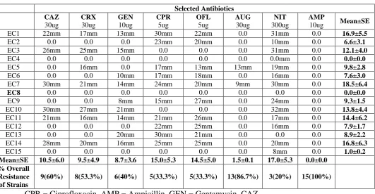
The antibiotic sensitivity patterns of Escherichia coli (EC1 – EC15) uropathogens strains
(isolated from midstream samples) to ciprofloxacin, ofloxacin (fluoroquinolones),
ceftazidime, cefuroxime (cephalosporins), gentamycin (aminoglycoside), ampicillin,
amoxicillin/clauvulanic acid (penicillins) and nitrofurantoin (urinary antiseptic) are shown in
Table 1. In terms of overall resistance of the strains to each antibiotic, 15(100.0%),
13(86.7%), 9(60.0%), 8(53.3%), 6(40.0%), 5(33.3%), 5(33.3%) and 3(20.0%) of the E.coli
uropathogen strains were resistant to ampicillin, amoxicillin/clauvulanic acid, ceftazidime,
cefuroxime, gentamycin, ciprofloxacin, ofloxacin and nitrofurantoin in that decreasing order.
This means the most and least resisted drugs were ampicillin and nitrofurantoin respectively.
Vertically, in decreasing order, zones of inhibition recorded were 17.0±5.3mm, 15.0±5.3mm,
14.5±5.0mm, 10.5±6.0mm, 9.5±4.9mm, 8.7±3.6mm, 1.5±0.1mm and 0.0±0.0mm for
amoxicillin/clauvulanic acid and ampicillin respectively. Hence, nitrofurantoin and ampicillin
recorded the highest and lowest mean± standard error zones of inhibition.
Horizontally, Table 1 also showed the mean±standard error zones of inhibition of each E.coli
strain to all the selected antibiotics. Uropathogen strains EC4, EC8, EC15, EC2, EC6, EC12,
EC13, EC9, EC5, EC3 and EC10 recorded mean± standard error zones of inhibition of
0.0±0.0mm, 0.0±0.0mm, 1.0±0.2mm, 6.6±3.1mm, 7.6±3.0mm, 7.9±1.7mm, 8.9±2.2mm,
9.3±1.5mm, 9.8±2.8mm, 12.1±4.0mm and 13.8±4.4mm respectively in that increasing order.
Other strains such as EC11, EC14, EC1 and EC7 recorded much higher mean± standard error
zones of inhibition of 14.4±6.2mm, 16.8±6.3mm, 16.9±5.5mm and 18.5±6.4mm respectively
in that increasing order. Out of the fifteen strains, EC7 strain recorded the highest mean±
standard error zones of inhibition.
Table 1: Antibiotic Susceptibility Patterns of Fifteen (15) Escherichia coli isolates bef
before exposure to dilutions of ascorbic acid after incubation at 37oC for 24hr.
Selected Antibiotics CAZ
30ug
CRX
30ug
GEN
10ug
CPR
5ug
OFL
5ug
AUG
30ug
NIT
300ug
AMP
10ug Mean±SE
EC1 22mm 17mm 13mm 30mm 22mm 0.0 31mm 0.0 16.9±5.5
EC2 0.0 0.0 0.0 23mm 20mm 0.0 10mm 0.0 6.6±3.1
EC3 26mm 25mm 15mm 0.0 0.0 0.0 31mm 0.0 12.1±4.0
EC4 0.0 0.0 0.0 0.0 0.0 0.0 0.0mm 0.0 0.0±0.0
EC5 0.0 16mm 0.0 17mm 13mm 13mm 19mm 0.0 9.8±2.8
EC6 0.0 0.0 10mm 17mm 18mm 0.0 16mm 0.0 7.6±3.0
EC7 30mm 21mm 14mm 24mm 20mm 9mm 30mm 0.0 18.5±6.4
EC8 0.0 0.0 0.0 0.0 0.0 0.0 0.0 0.0 0.0±0.0
EC9 0.0 0.0 8mm 15mm 27mm 0.0 24mm 0.0 9.3±1.5
EC10 30mm 27mm 21mm 0.0 0.0 0.0 32mm 0.0 13.8±4.4
EC11 21mm 16mm 14mm 21mm 26mm 0.0 17mm 0.0 14.4±6.2
EC12 0.0 0.0 0.0 22mm 25mm 0.0 16mm 0.0 7.9±1.7
EC13 0.0 0.0 20mm 30mm 21mm 0.0 0.0 0.0 8.9±2.2
EC14 28mm 20mm 16mm 25mm 25mm 0.0 20mm 0.0 16.8±6.3
EC15 0.0 0.0 0.0 0.0 0.0 0.0 8mm 0.0 1.0±0.2
Mean±SE 10.5±6.0 9.5±4.9 8.7±3.6 15.0±5.3 14.5±5.0 1.5±0.1 17.0±5.3 0.0±0.0
% Overall Resistance of Strains
9(60%) 8(53.3%) 6(40%) 5(33.3%) 5(33.3%) 13(86.7%) 3(20%) 15(100%)
CPR = Ciprofloxacin, AMP = Ampicillin, GEN = Gentamycin, CAZ
= Ceftazidime, CRX = cefuroxime, OFL = Ofloxacin, AUG =
Amoxicillin/Clauvulanic acid, NIT = Nitrofurantoin
Table 2 shows the occurrence and distribution of multidrug resistant E.coli uropathogen
isolates after antibiotic sensitivity was carried out. Whereas EC5 strain was resistant to three
[image:10.595.37.569.363.639.2]penicillin), EC3, EC6, EC9 and EC10 strains each resisted 4(50.0%) drugs as shown.
Uropathogen strains EC2, EC12 and EC13 resisted 5(62.5%) drugs each as shown also.
Strain EC15 resisted 7(87.5%) drugs while strains EC4 and EC8 each resisted all 8(100%)
drugs tested. In increasing order 1(9.0%) strain, 4(36.4%) strains, 3(27.3%) and 3(27.3%)
strains resisted 3drugs, 4drugs, 5drugs and more than 6drugs respectively. On the whole,
11(73.3%) out of the 15(100%) strains were multidrug resistant.
Table 2: Distribution of Multidrug Resistant E.coli Uropathogen Strains Among Total
Isolates.
Isolates’ Strain Codes
No of Antibiotics Resisted
3drugs 4drugs 5drugs ≤ 6drugs Drugs Resisted
EC2 - - + - CAZ, CRX, GEN, AUG, AMP
EC3 - + - - CPR, OFL, AUG, AMP
EC4 - - - + CAZ, CRX, CPR, OFL, AUG,
AMP, GEN, NIT
EC5 + - - - CAZ, GEN, AMP
EC6 - + - - CAZ, CRX, AUG, AMP
EC8 - - - + ALL EIGHT DRUGS
EC9 - + - - CAZ, CRX, AUG, AMP
EC10 - + - - CPR, OFL, AUG, AMP
EC12 - - + - CAZ, CRX, GEN, AUG, AMP
EC13 - - + - CAZ, CRX, AUG, AMP, NIT
EC15 - - - + ALL EIGHT EXCEPT NIT.
TOTAL
n=11 1(9.0%) 4(36.4%) 3(27.3%) 3(27.3%)
CAZ, CRX (cephalosporins), CPR, OFL (fluoroquinolones), AUG, AMP (penicillins), GEN
(aminoglycoside), NIT (urinary antiseptic).
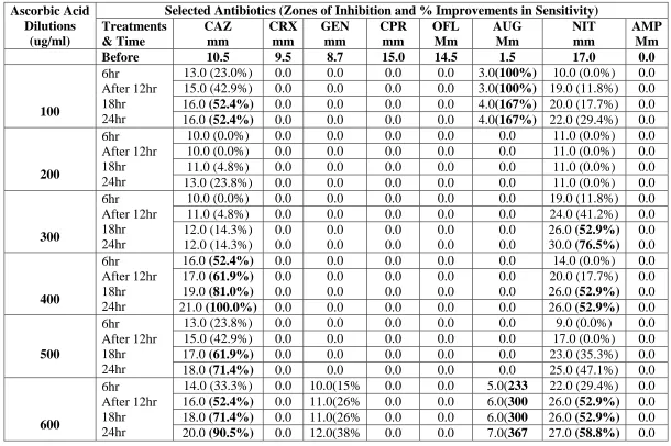
Data of zones inhibition of EC8 strain before and after exposure to the prepared dilutions of
ascorbic acid are shown in Table 3. Zones of inhibition before and after treatment were
mathematically computed to obtain ≤50% loss of resistance according to a scheme provided
by Akortha et al. (2011). Before treatment with the doses of ascorbic acid, EC8 uropathogen
strain was resistant to 8(100%) of antibiotics tested. After treatment and incubation for 6hr,
12hr, 18hr and 24hr, EC8 strain became sensitive to only 6 (75.0%) of the eight tested
antibiotics and these were ceftazidime (a cephalosporin), gentamycin (an aminoglycoside),
ciprofloxacin, ofloxacin (fluoroquinolones), amoxicillin/clauvulanic acid (branded
augmentin) and nitrofurantoin (a urinary antiseptic). Multidrug resistant EC8 strain remained
Treatment with 100ug/ml ascorbic acid dilution recorded more than 50% reduction in EC8
resistance to ceftazidime after 18hr and 24hr incubation each. No significant effect was seen
after 6hr incubation and more than 40% resistance reduction was recorded after 12hr
incubation. With the same ascorbic acid dilution treatment, EC8 strain recorded less than
20% resistance reduction to nitrofurantoin after 18hr incubation and there was no effect
recorded after 6hr incubation. More than 100% resistance reduction to augmentin was
recorded by 100ug/ml AA treatment after 18hr and 24hr incubation while 100% resistance
reduction was recorded after 6hr and 12hr incubation each (Table 3).
Ascorbic acid treatment dose of 200ug/ml on EC8 strain recorded no significant resistance
reduction to both ceftazidime and nitrofurantoin after 6hr to 24hr incubation. The same
organism recorded more than 50% and 70% resistance reduction to nitrofurantoin after 18hr
and 24hr incubation respectively with 300ug/ml ascorbic acid. The same dilution induced no
significant reduction to ceftazidime. The test organism (EC8) after 6hr, 12hr, 18hr and 24hr
incubation with 400ug/ml ascorbic acid concentration, recorded 52.4%, 61.9%%, 81% and
100% resistance reduction respectively to ceftazidime. Incubation of EC8 strain with
400ug/ml dose yielded 52.9% and 52.9% resistance reduction after 18hr and 24hr
respectively to nitrofurantoin.
Similarly, after 18hr and 24hr incubation of EC8 strain treated with 500ug/ml dilution, more
than 60% and 70% resistance reduction was recorded to ceftazidime and less than 40% and
50% resistance reduction to nitrofurantoin was noted after 18hr and 24hr incubation periods.
In the case of 600ug/ml dilution, 12hr, 18hr and 24hr incubation period with EC8 strain
recorded 52.4%, 71.4% and 90.5% resistance reduction to ceftazidime respectively and
52.9%, 52.9% and 58.8% RR to nitrofurantoin after the same incubation periods respectively.
Noteworthy is that the same dilution induced 233%, 300%, 300% and 367% RR to augmentin
after 6hr, 12hr, 18hr and 24hr incubation periods respectively and also less than 20%, 30%,
30% and 40 RR to gentamycin after the same incubation periods respectively (Table 3).
After 18hr and 24hr incubation of EC8 strain with 700ug/ml dilution, significant resistance
reduction (RR) of 71.4% and 81.0% respectively to ceftazidime was recorded. Lower
incubation periods yielded less than 5% and 40% reduction in resistance. The same curing
agent dilution treatment induced no significant RR to nitrofurantoin after all four incubation
periods (Table 3). Similar to the effect of 700ug/ml AA dilution, 71.4% and 71.4% RR to
respectively. Ascorbic acid dose of 900u/ml after 18hr and 24hr incubation effected 71.4%
and 109.5% respectively to ceftazidime with no significant RR to nitrofurantoin after all four
incubation periods with the same dilution.
Ascorbic acid dilution of 1000ug/ml treatment of EC8 strain after 6hr, 12hr, 18hr and 24hr
incubation periods yielded resistance reduction to six of the antibiotics tested and they
included ceftazidme, gentamycin, ciprofloxacin, ofloxacin, augmentin and nitrofurantoin
respectively. Eighty one percent, 100%, 100% and 128% RR to ceftazidime were recorded
respectively after the four incubation periods. The same dilution yielded 52%, 52%, 79% and
86% RR respectively to ofloxacin after the four incubation periods. The same dilution after
the four incubation periods also induced 150%, 150%, 167% and 233% RR respectively to
amoxicillin/clauvulanic acid. One thousand microgram per milliliter dilution of AA after 18hr
and 24hr incubation periods recorded 64.7%% and 70.6% RR respectively to nitrofurantoin.
The same dilution recorded less than 50%, less than 50%, less than 50% and 53% RR to
ciprofloxacin after the four incubation periods respectively. Although resistance reduction to
gentamycin was induced by 1000ug/ml, none was significant after the same incubation
Table 3: Dilutions of ascorbic acid that induced ≤50% loss in resistance after treatment on EC8 uropathogen strain and incubation at
37oC for 6hr, 12hr, 18hr and 24hr.
Ascorbic Acid Dilutions
(ug/ml)
Selected Antibiotics (Zones of Inhibition and % Improvements in Sensitivity) Treatments & Time CAZ mm CRX mm GEN mm CPR mm OFL Mm AUG Mm NIT mm AMP Mm
Before 10.5 9.5 8.7 15.0 14.5 1.5 17.0 0.0
100
6hr
After 12hr 18hr 24hr
13.0 (23.0%) 0.0 0.0 0.0 0.0 3.0(100%) 10.0 (0.0%) 0.0
15.0 (42.9%) 0.0 0.0 0.0 0.0 3.0(100%) 19.0 (11.8%) 0.0
16.0 (52.4%) 0.0 0.0 0.0 0.0 4.0(167%) 20.0 (17.7%) 0.0
16.0 (52.4%) 0.0 0.0 0.0 0.0 4.0(167%) 22.0 (29.4%) 0.0
200
6hr
After 12hr 18hr 24hr
10.0 (0.0%) 0.0 0.0 0.0 0.0 0.0 11.0 (0.0%) 0.0
10.0 (0.0%) 0.0 0.0 0.0 0.0 0.0 11.0 (0.0%) 0.0
11.0 (4.8%) 0.0 0.0 0.0 0.0 0.0 11.0 (0.0%) 0.0
13.0 (23.8%) 0.0 0.0 0.0 0.0 0.0 11.0 (0.0%) 0.0
300
6hr
After 12hr 18hr 24hr
10.0 (0.0%) 0.0 0.0 0.0 0.0 0.0 19.0 (11.8%) 0.0
11.0 (4.8%) 0.0 0.0 0.0 0.0 0.0 24.0 (41.2%) 0.0
12.0 (14.3%) 12.0 (14.3%) 0.0 0.0 0.0 0.0 0.0 0.0 0.0 0.0 0.0 0.0 26.0 (52.9%) 30.0 (76.5%) 0.0 0.0 400 6hr After 12hr 18hr 24hr
16.0 (52.4%) 0.0 0.0 0.0 0.0 0.0 14.0 (0.0%) 0.0
17.0 (61.9%) 19.0 (81.0%) 0.0 0.0 0.0 0.0 0.0 0.0 0.0 0.0 0.0 0.0 20.0 (17.7%) 26.0 (52.9%) 0.0 0.0
21.0 (100.0%) 0.0 0.0 0.0 0.0 0.0 26.0 (52.9%) 0.0
500
6hr
After 12hr 18hr 24hr
13.0 (23.8%) 0.0 0.0 0.0 0.0 0.0 9.0 (0.0%) 0.0
15.0 (42.9%) 0.0 0.0 0.0 0.0 0.0 17.0 (0.0%) 0.0
17.0 (61.9%) 0.0 0.0 0.0 0.0 0.0 23.0 (35.3%) 0.0
18.0 (71.4%) 0.0 0.0 0.0 0.0 0.0 25.0 (47.1%) 0.0
600
6hr
After 12hr 18hr 24hr
14.0 (33.3%) 0.0 10.0(15% 0.0 0.0 5.0(233 22.0 (29.4%) 0.0
16.0 (52.4%) 0.0 11.0(26% 0.0 0.0 6.0(300 26.0 (52.9%) 0.0
18.0 (71.4%) 0.0 11.0(26% 0.0 0.0 6.0(300 26.0 (52.9%) 0.0
700
6hr
After 12hr 18hr 24hr
12.0 (14.8%) 0.0 0.0 0.0 0.0 0.0 17.0 (0.0%) 0.0
14.0 (43.3%) 0.0 0.0 0.0 0.0 0.0 18.0 (5.9%) 0.0
18.0 (71.4%) 0.0 0.0 0.0 0.0 0.0 19.0 (11.8%) 0.0
19.0 (81.0%) 0.0 0.0 0.0 0.0 0.0 22.0 (29.4%) 0.0
800
6hr
After 12hr 18hr 24hr
11.0 (8.0%) 0.0 0.0 0.0 0.0 0.0 12.0 (0.0%) 0.0
15.0 (48.9%) 0.0 0.0 0.0 0.0 0.0 14.0 (0.0%) 0.0
18.0 (71.4%) 0.0 0.0 0.0 0.0 0.0 14.0 (0.0%) 0.0
18.0 (71.4%) 0.0 0.0 0.0 0.0 0.0 18.0 (5.9%) 0.0
900
6hr
After 12hr 18hr 24hr
11.0 (10.0%) 0.0 0.0 0.0 0.0 0.0 14.0 (0.0%) 0.0
12.0 (14.8%) 0.0 0.0 0.0 0.0 0.0 16.0 (0.0%) 0.0
18.0 (71.4%) 0.0 0.0 0.0 0.0 0.0 17.0 (0.0%) 0.0
22.0 (109.5%) 0.0 0.0 0.0 0.0 0.0 20.0 (17.7%) 0.0
1000
6hr
After 12hr 18hr 24hr
19.0 (81.0%) 0.0 9.0(3.5% 21.0(40 22.0(52 3.0(150 24.0 (41.2%) 0.0
21.0 (100.0%) 0.0 10.0(15% 21.0(40 22.0(52 3.0(150 24.0 (41.2%) 0.0
21.0 (100.0%) 0.0 11.0(26% 22.0(47 26.0(79 4.0(167 28.0 (64.7%) 0.0
24.0 (128.6%) 0.0 11.0(26% 23.0(53 27.0(86 5.0(233 29.0 (70.6%) 0.0
A summary of the means of percentage losses (reduction) of antibiotic resistance after 6hr, 12hr, 18hr and 24hr incubation and ≤50% loss of
resistance are shown in Table 4. Mean percentage reduction of resistance after 6hr, 12hr, 18hr and 24hr incubation of EC8 strain with ascorbic
acid dilutions of 400ug/ml, 500ug/ml, 600ug/ml, 700ug/ml, 800ug/ml, 900ug/ml and 1000ug/ml recorded 73.8%, 50.0%, 61.9%, 52.6%, 50.0%,
51.3% and 102.4% RR to ceftazidime respectively. Only 1000ug/ml dilution recorded ≤50% loss of resistance of 67.6%, 175.3% and 54.5% to
ofloxacin, amoxicillin/clauvulanic acid and nitrofurantoin respectively.
Ascorbic acid dilutions of 100ug/ml and 600ug/ml also induced ≤50% loss of resistance of 133.4% and 300.4% respectively to
amoxicillin/clauvulanic acid. More than 45% resistance reduction to nitrofurantoin was recorded each for 300ug/ml and 600ug/ml ascorbic acid
Table 4: Cumulative means of ≤50% loss of resistance as induced by exposure of EC8
uropathogen strain to ascorbic acid dilutions after 6hr, 12hr, 18hr and 24hr incubation
at 37oC.
Ascorbic Acid Dilutions
ug/ml
Antibiotics That Recorded ≤50% loss of resistance Treatments & Time CAZ Mm OFL mm AUG mm NIT mm
Before 10.5 14.5 1.5 17.0
100 6hrs After 12hrs 18hrs 24hrs 23.0% 42.9% (42.7%) 52.4% 52.4%
0.0% 100.0%
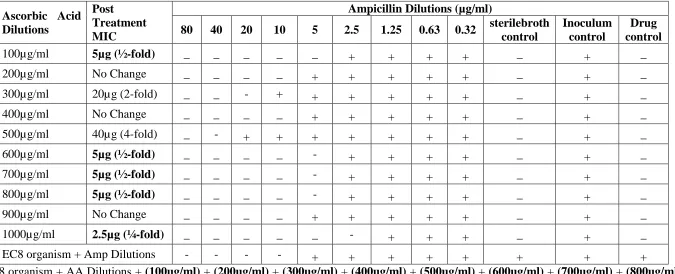
[image:16.595.33.566.137.754.2]The data on the effect of ascorbic acid dilutions on the minimum inhibitory concentration
(MIC) of ampicillin on the MDR EC8 strain are shown in Table 5. After 18hr incubation at
37oC, inoculum control tubes showed turbidity (cloudiness) as expected. Sterile broth control and drug control tubes separately remained clear at the end of incubation as expected.
Ascorbic acid dilutions of 200, 400 and 900ug/ml did not affect the minimum inhibitory
concentration of ampicillin (a penicillin and one of the two antibiotics that EC8 strain
remained resistant to after treatment) as the MIC remained 10ug. Dilutions of 100, 600, 700
and 800ug/ml reduced ampicillin MIC to 5ug each which is a two-fold reduction. Ampicillin
MIC was reduced to 2.5ug (a four-fold reduction) by the effect of 1000 ug/ml dilution only
and this was the highest MIC reduction. Ascorbic acid dilutions of 300ug/ml and 500ug/ml
Table 5: Effect of Ascorbic acid dilutions treatment of EC8 strain on the minimum inhibitory concentration (MIC) of Ampicillin
Invitro.
Ascorbic Acid Dilutions
Post Treatment MIC
Ampicillin Dilutions (µg/ml)
80 40 20 10 5 2.5 1.25 0.63 0.32 sterilebroth
control
Inoculum control
Drug control
100µg/ml 5µg (½-fold) _ _ _ _ _ + + + + _ + _
200µg/ml No Change _ _ _ _ + + + + + _ + _
300µg/ml 20µg (2-fold) _ _ - + + + + + + _ + _
400µg/ml No Change _ _ _ _ + + + + + _ + _
500µg/ml 40µg (4-fold) _ - + + + + + + + _ + _
600µg/ml 5µg (½-fold) _ _ _ _ - + + + + _ + _
700µg/ml 5µg (½-fold) _ _ _ _ - + + + + _ + _
800µg/ml 5µg (½-fold) _ _ _ _ - + + + + _ + _
900µg/ml No Change _ _ _ _ + + + + + _ + _
1000µg/ml 2.5µg (¼-fold) _ _ _ _ _ - + + + _ + _
EC8 organism + Amp Dilutions - - - - + + + + + + + +
EC8 organism + AA Dilutions + (100ug/ml) + (200ug/ml) + (300ug/ml) + (400ug/ml) + (500ug/ml) + (600ug/ml) + (700ug/ml) + (800ug/ml)
[image:18.842.89.764.113.387.2]DISCUSSION
The increasing number of multiple –antibiotic resistant pathogens has become a serious threat
to human health (CDC, 2013; RAR, 2014; WHO, 2014). Hence, the emergence and
dispersion of antibiotic resistance caused by these multiple antibiotic resistant or MDR
pathogens) has reduced the susceptibility of such pathogens to antibiotics in medical
treatment (Allen et al., 2010). The overall outcome of this is long hospital stay (by patients
suffering from diseases caused by these MDR pathogens), increased hospital bills, spread of
such pathogens internationally, increased mortality rate among others (Bassetti et al., 2011).
Non effective antibiotics due to MDR bacteria will affect areas of medicine such as surgery,
cancer chemotherapy, transplantation etc. (Spellberg et al., 2007).
The prevalence of resistance to specific antibiotics within a microbial population may be
explained by the presence of mobile genetic elements such as integrons, plasmids and
transposons found within the population encoding such antibiotic resistance (Tamma et al.,
2012). Moreso, the existence of plasmid-bearing extended spectrum beta-lactamases (ESBL)
and fosfomycin resistance determinants that can spread effectively in Enterobacteriaceae (of
which Escherichia coli is a member) have been discovered and are of great clinical concern
(Zhao et al., 2015).
This study was focused on seeking a scientific pathway to reclaim some commonly used
antibiotics which have lost therapeutic value or usefulness owing to development of
resistance genes against them by gram negative pathogens such as Escherichia coli.
Antibiotic sensitivity patterns of fifteen (15) strains (uropathogens) of Escherichia coli before
ascorbic acid treatment in this study showed that the fifteen uropathogen strains were
sensitive to nitrofurantoin except strains EC4, EC8 and EC13. Therefore, 15(100%),
13(86.7%), 9(60.0%), 8(53.3%), 5(33.3%), 5(33.3%) and 3(20.0%) Escherichia coli strains
were resistant to ampicillin, amoxicillin/clauvulanic acid, ceftazidime, cefuroxime,
gentamycin, ciprofloxacin, ofloxacin and nitrofurantoin respectively. This implied that all
strains resisted ampicillin (a penicillin antibiotic) while 12(80.0%) strains were sensitive to
nitrofurantoin (a urinary antiseptic). Strain EC8 selected and used in this work was resistant
to all eight antibiotics used.
In terms of sensitivity, all fifteen strains recorded (in decreasing order) mean±standard zone
of inhibition sensitivity of 17.0±5.3mm, 15.0±5.3mm, 14.5±5.0mm, 10.5±6.0mm,
ofloxacin, ceftazidime, cefuroxime, gentamycin, amoxicillin/clauvulanic acid and ampicillin
respectively. This sensitivity pattern suggests that nitrofurantoin followed by ciprofloxacin,
by ofloxacin, ceftazidime and cefuroxime (singly or in synergistic combination of any two
different classes) may be drug of choice for treatment of urinary tract infections caused by
E.coli. While the choice of nitrofurantoin may be cheering to low income patients, the
alternative of choosing any of ciprofloxacin, ofloxacin (both fluoroquinolones), ceftazidime
or cefuroxime (both cephalosporins) may be a sad one owing to their high cost and
sometimes, non-availability. The total resistance recorded by ampicillin was expected owing
to its abuse arising from its availability and low cost. Worrisome is the almost total resistance
recorded by amoxicillin/clauvulanic acid (branded augmentin) and the reason is because it is
popularly or commonly prescribed and used by physicians to treat a good number of human
diseases. Some authors have also expressed similar worry over augmentin efficacy (Oluremi
et al., 2011). The sensitivity patterns obtained for all fifteen E.coli strains used in this study
are however, subject to verification and confirmation by other authors.
The multidrug resistant (MDR) statuses of 11(73.3%) out of the fifteen E.coli strains showed
that 1(9.0%), 4(36.4%), 3(27.3%) and 3(27.3%) strains were resistant to 3drugs, 4drugs,
5drugs and more than 6drugs respectively. Escherichia coli strain 8 used in this study was
resistant to all the eight antibiotics sampled. Uropathogen EC5 strain resisted 3drugs which
belonged to cephalosporin, aminoglycoside and penicillin antibiotic groups. Strain EC3
resisted four drugs which were of fluoroquinolone and penicillin groups. Strain EC13 resisted
five drugs belonging to cephalosporin, penicillin and urinary antiseptic groups. The other
strains resisted antibiotics of various groups. These strains therefore, qualified as multiple
resistant or MDR strains. Isolates are considered MDR if they are resistant to at least three of
the antibiotics tested (Santo et al., 2007; Jan et al., 2004). According to Chethana et al.
(2013), one of the methods used by various authors to characterize organisms as MDR is
based on invitro antimicrobial susceptibility test results when they test resistant to multiple
antimicrobial agents, classes or subclasses of antimicrobial agents.
Multidrug resistant organisms are microorganisms that are resistant to one or more
therapeutic classes of antimicrobial agents (Adam et al., 2008). The MDR statuses of
11(73.3%) of the E.coli strains used in this study is further established by the report of
previous authors which stated that multidrug resistant E.coli and other enterobacteriaceae
categories (Magiorakos et al., 2012). The incidence of 11(73.3%) MDR E.coli strains in this
study is a high one and this is in line with a report which stated that E.coli had been
recognized as a contributor to the dissemination of antibiotic resistance genes in natural
environments (Henriques et al., 2006; Zhao and Dang, 2012; Alm et al., 2014; Alves et al.,
2014).
Antibiotic resistant E.coli and other enteric bacteria that survived the extensive antibiotic
treatments in the gut of humans or animals can enter aquatic systems through discharge from
poultry and livestock production and through hospital municipal wastewaters (Pruden et al.,
2006; Pereira et al., 2013) and therefore, the rivers that are used for recreational activities,
irrigation etc can be potent sources through which antibiotic resistant bacteria are
disseminated (Su et al., 2012; Pereira et al., 2013).
In this study, ascorbic acid dilutions of 100, 200, 300, 400, 500, 600, 700, 800, 900 and
1000ug/ml were used to treat and possibly cure an MDR EC8 strain which was resistant to
8(100.0%) of all antibiotics tested with the aim of reducing its resistance significantly or
eliminating it completely (via complete or partial plasmid removal). The use of 100 –
1000ug/ml was according to the scheme provided originally by Ramesh et al. (2000) and
adopted by Potrus (2009). Ascorbic acid or vitamin C is a non-hazardous, non-antibiotic
anti-oxidant agent which is cheap, safe to handle and readily available. The loss of 50-100% or
≤50% of resistance after treatment with the above stated dilutions of ascorbic acid was used
as the basis of establishing the curing effects of these dilutions. The use of 50% and above
loss in resistance as a criterion to determine the extent of plasmid curing was according to the
scheme provided by Akortha et al. (2011). Stainer et al. (1984) reported that the elimination
of plasmids by dyes and other natural agents reflects the ability of such an agent to inhibit
plasmid replication at a concentration that does not affect the chromosome.
After treatment with the above stated ten (10) invitro ascorbic acid dilutions, Escherichia coli
strain 8 still remained completely resistant to cefuroxime and ampicillin. Strain 8 E.coli
however, became sensitive to ceftazidime, gentamycin, ciprofloxacin, ofloxacin, augmentin
and nitrofurantoin as a result of the invitro treatment. Hence, there was reduction from
8(100.0%) resistant drugs to 2(25.0%) resistant drugs or from 8(100.0%) resistant drugs to
6(75.0%) sensitive drugs after treatment. Continued resistance to the 2(25.0%) antibiotics
listed above (even after treatment) may be due to the fact that resistance genes responsible for
(Akortha and Filgona, 2009). After treatment with the doses of ascorbic acid, incubation was
carried out for 6hr, 12hr, 18hr and 24hr. This was to avail the dividing cells in EC8 strain
pure broth culture the opportunity to respond effectively to the curing effect (if any), of
ascorbic acid at the various liquid growth phases of the organism during incubation in
Mueller Hinton broth medium.
Growth in liquid medium is synonymous with growth of bacterial pathogens invivo (in the
blood circulatory system, tissues, lymphatic system etc) of man and related primates.
Incubation in water bath rather than conventional incubator was to ensure direct heat
(temperature) contact of diluted broth culture cells and the ascorbic acid dilutions to ensure
optimal biochemical and physiological activities of the EC8 strain in the set up. Based on the
results therefore, 100ug/ml, 400ug/ml, 600ug/ml and 1000ug/ml dilutions recorded
significant resistance loss (and hence, significant sensitivity improvement) of EC8 strain to
amoxicillin/clauvulanic acid, ceftazidime, amoxicillin/clauvulanic acid and nitrofurantoin
respectively after 6hr incubation (Table 3). It is possible that the lag phase (i.e. phase of
physiological and biochemical inactivity) of this strain of E.coli is short. This is not in
agreement with the report of Thiel (2015) which stated that culture harvest is ideal after
12-16hr of incubation which is the period of transition from logarithmic phase to stationary
phase and that it is during the 12-16hr period that bacterial DNA or plasmid is not degraded.
The presence of resistance loss after 6hr (or 360mins) incubation however, is in agreement
with the report of a previous author which stated that generation time of E.coli in the
laboratory is between 15-20mins (Todar, 2012).
After 12hr incubation, ≤50% loss of resistance was also recorded by 100ug/ml, 400ug/ml,
600ug/ml and 1000ug/ml ascorbic acid dilutions in that they induced100%, 61.9%, 52.4%
and 100% resistance reduction respectively to amoxicillin/clauvulanic acid and ceftazidime.
After 18hrs incubation, 100, 400, 500, 600, 700, 800, 900 and 1000ug/ml ascorbic acid
dilutions induced resistance reduction of 52.4%, 81%, 61.9%, 71.4%, 71.4%, 71.4%, 71.4%
and 100% respectively to ceftazidime. During the same incubation period, 100ug/ml,
600ug/ml and 1000ug/ml dilutions recorded 167%, 300% and 167% RR respectively to
amoxicillin/clauvulanic acid. Other significant RR during the 18hr incubation period was
recorded by 300ug/ml and 400ug/ml dilutions which induced 52.9% and 52.9% resistance
losses respectively to nitrofurantoin and lastly, 1000ug/ml dilution induced 79%, 167% and
the same incubation period. It is clear therefore that 8(75.0%) of the dilutions tested induced
significant RR after 18hr incubation and this may suggest that 18hr incubation is optimal for
invivo expression of the physiological and biochemical activities of Escherichia coli.
Similarly, after 24hr incubation, 100, 400, 500, 600, 700, 800, 900 and 1000ug/ml dilutions
yielded RR of 52.4%, 100%, 71.4%, 90.5%, 81%, 71.4%, 109.5% and 128.6% respectively to
ceftazidime. Also, after 24hrs incubation, only 100, 600 and 1000ug/ml recorded 167%,
367% and 233% RR respectively to amoxicillin/clauvulanic acid. During the same incubation
period, 300, 400, 600 and 1000ug/ml dilutions induced 76.5%, 52.9%, 58.8% and 70.6% RR
to nitrofurantoin. The treatment outcomes under the same incubation period as recorded by
600ug/ml and 1000ug/ml with respect to amoxicillin/clauvulanic acid were 367% and 233%
resistance reduction respectively. A resistance reduction of 53% and 86% was recorded by
1000ug/ml dilution with respect to ofloxacin and ciprofloxacin (both fluroquinolones).
Clearly, optimal Mueller Hinton broth culture activity of EC8 uropathogen was attained after
18hr and 24hr incubation. This may be because there were more actively dividing cells in the
logarithmic phase of the Mueller Hinton broth culture. Hence, this finding tends to lend
credence to the choice of 18-24hr incubation period for both agar and broth cultures after
appropriate inoculation. Besides, this finding is in line with the report of a previous author
(Thiel, 2015).
This longer incubation period may be suggestive of the fact that the cells of EC8 strain are
still dividing even up to this period and the fact that dilutions that induced ≤50% loss of
resistance losses were achieved more and most after 18hr and 24hr incubation respectively
may indicate that the highest biochemical and physiological activities of EC8 uropathogen
strain will be attained after 18-24hr incubation at 37oC - the conventionally prescribed temperature for human pathogens such as E.coli urinary tract pathogens. After 24hr
incubation, 100, 400, 500, 600, 700, 800, 900 and 1000ug/ml dilutions recorded ≤50% loss of
resistance to ceftazidime and 100, 300, 600 and 1000ug/ml recorded ≤50% loss of resistance
to nitrofurantoin (Table 3). This may be because these dilutions have the capacity to remove
resistance genes carried on the inherent plasmids present in the organism.
The means of all resistance losses after 6hr, 12hr, 18hr and 24hr periods of incubation for all
dilutions showed that only ascorbic acid dilutions of 400, 500, 600, 700, 800, 900 and
of resistance in EC8 strain to both ceftazidime and nitrofurantoin. Resistance losses of ≤50%
of 67.6% and 175.3% to ofloxacin and amoxicillin/clauvulanic acid respectively were also
induced by 1000ug/ml AA dilution (Table 4). This particular finding in this study singles out
1000ug/ml and to a lesser extent, 600ug/ml for industrial and pharmaceutical trial/application
in terms of drug modification with regard to ceftazidime, ofloxacin, amoxicillin/clauvulanic
acid and nitrofurantoin. The implication of this is that 1000ug/ml dilution and to a lesser
extent, 600ug/ml ascorbic acid dilutions may possess the capacity to eliminate resistance
genes (if plasmid mediated) in MDR EC8 and indeed other MDR E.coli strains and therefore,
could be administered in the therapeutic control of various infections caused by such bacteria.
According to Ramesh et al. (2000), the toxic effect of ascorbate (derivative of ascorbic acid)
on cells and bacterial cells was dose dependent. Ascorbic acid causes conformational damage
to unprotected cells (Ramesh et al., 2000). L- ascorbic acid inhibits a wide range of biological
functions and modifies the properties of DNA, generation of hydrogen peroxide and hydroxyl
radicals by auto-oxidation and lipid peroxidation of membrane components (Morgan et al.,
1976; Shamberger, 1984; Sugiyama et al., 1991; Halliwell and Aruoma, 1991). The reactive
oxygen species such as OH- and H2O2 are involved in DNA damage by Fenton – type
reaction which has been shown to occur in bacterial cells (Imlay and Linn, 1989).
In this study, higher doses (dilutions) of ascorbic acid (i.e. 400, 500, 600, 700, 800, 900 and
1000ug/ml especially 1000ug/ml) recorded significant resistance losses and perhaps,
increased curing or plasmid removal. Lower doses did not induce or yielded insignificant
losses. This finding is supported by the report of some authors which stated that the effect of
ascorbate in terms of antibiotic resistance genes curing is dose dependent. Since L-ascorbic
acid has the capacity to modify the properties of DNA (Morgan et al., 1976), it can be
reasoned that it will also have effect on extra-chromosomal DNA (which is plasmid).
That EC8 uropathogen significantly became sensitive to ceftazidime, ofloxacin,
amoxicillin/clauvulanic acid and nitrofurantoin after 1000ug/ml ascorbic acid treatment
(Table 4) may open up a new era of therapy. The simultaneous application of this dose of
ascorbic acid dilution and the implicated drugs (antibiotics) to patients however, may not
only act as an additional antibacterial agent but may help to eliminate the drug resistance
genes (if plasmid mediated) from the infectious bacterial cells (Spendler et al., 2006). Hence,
patients suffering from urinary tract infections caused by MDR E.coli may be administered
along with ceftazidime, ofloxacin, amoxicillin/clauvulanic acid and nitrofurantoin or any of
their combinations (combination therapy). Besides, Escherichia coli is catalase negative and
catalase is a protective enzyme that which reduces the effects of reactive oxygen species (i.e
OH- and H2O2) mediated damage to the bacterial DNA (Hardy, 1987) and this is
advantageous for curing by ascorbic acid in EC8 uropathogen.
Resistance loss effect of the ten ascorbic acid dilutions was also tested on the minimum
inhibitory concentration (MICs) of ampicillin (a penicillin drug). The idea is that a fast and
accurate determination of MIC can indicate optimal effective treatment of patients while at
the same time, avoiding over prescription. This will save money for healthcare providers as
well as reduce development of resistance (NCCLS, 2000; McGowan and Wise, 2001).
Findings in this study showed that MIC of ampicillin (which is 10ug based on long standing
research), was reduced to 5ug (a fold reduction), 5ug (a fold reduction), 5ug (a
two-fold reduction), 5ug (a two-two-fold reduction) and 2.5ug (a four-two-fold reduction) by the
synergistic action of ascorbic acid dilutions of 100ug/ml, 600ug/ml, 700ug/ml, 800ug/ml and
1000ug/ml respectively.
According to Dimitru et al. (2006), there is a significant correlation between MIC values and
the inhibition zone diameters obtained by an antibiotic disc containing a drug in microgram.
Dilutions of ascorbic acid reduced the MIC of ampicillin by two, two, two, two and four
folds. The lower the MIC and the larger the zone of inhibition, the more susceptible the
microorganism is to the antimicrobial agent (in this regard, ampicillin) and conversely, the
higher the MIC and smaller the zone of inhibition, the more resistant the microorganism
(Dimitru et al., 2006). Again, it is noteworthy the four-fold reduction of the MIC of
ampicillin induced by 1000ug/ml ascorbic acid dilution which was the highest reduction. This
was followed by two-fold reduction recorded each by 100ug/ml, 600ug/ml, 700ug/ml,
800ug/ml and 1000ug/ml. It should be noted that whereas the first control which contained
only EC8 organism and ampicillin dilutions did not show any MIC change, the second
control which contained only EC8 organism and ascorbic acid dilutions showed turbidity for
all the dilutions. This suggested to some extent, that ampicillin MIC reductions as stated in
this study could be solely due to the synergistic action of ascorbic acid and the drug dilutions.
Cursino et al. (2005) has reported the synergistic interaction of ascorbic acid with antibiotics
Tunicamycin at a concentration of 0.08ug/ml and in synergy with beta-lactam antibiotics has
been reported to reduce the MIC of oxacillin against methicillin resistant Staphylococcus
aureus from 50ug/ml to 0.4ug/ml, a 125-fold reduction (Campbell et al., 2011). Farha et al.
(2012) reported the use of the anti-platelet drug-ticlopidine in synergy with cefuroxime to
lower the MIC of MRSA by up to 64-fold. The therapeutic importance (in terms of
application) therefore of findings in this regard is that when doses of one of these ascorbic
acid dilutions or a combination of any two are incorporated into the manufacture of
ampicillin or any other related antibiotic and administered to a patient diagnosed to be
suffering from a disease caused by an MDR E.coli organism, a better result in terms of
therapeutic outcome (cure of the disease) may be achieved. These results are supported by
reports of previous authors who carried out related studies (Kohler, 2010; Crowle et al.,
1992; Shiram et al., 2008).
CONCLUSION
The results of antibiotic sensitivity testing showed that to treat any urinary tract infection
caused by an MDR E.coli strain, any of nitrofurantoin, ciprofloxacin, ofloxacin, ceftazidime
or cefuroxime or a synergistic combination of any two may be effective. Resistance losses (or
reduction) of ≤50% were achieved most between 18-24hr incubation suggesting that optimal
biochemical and physiological activities may be attained by EC8 strain and indeed other
MDR E.coli uropathogens during this incubation period at 37oC for E.coli uropathogens. Ascorbic acid dilutions of 100ug/ml, 400ug/ml, 500ug/ml, 600ug/ml, 700ug/ml, 800ug/ml,
900ug/ml and 1000ug/ml recorded ≤50% mean loss of resistance in EC8 strain to ceftazidime. Noteworthy was 1000ug/ml dilution which induced ≤50% mean loss of
resistance to ceftazidime, ofloxacin, amoxicillin/clauvulanic acid and nitrofurantoin.
Ascorbic acid dilutions of 100ug/ml, 600ug/ml, 700ug/ml, 800ug/ml and 1000ug/ml as
treated with EC8 strain reduced ampicillin MIC to 5ug (two-fold reduction), 5ug (two-fold
reduction), 5ug (two-fold reduction), 5ug (two-fold reduction) and 2.5ug (four-fold
reduction) respectively. Uropathogen EC8 strain treated with 1000ug/ml reduced ampicillin
MIC to 2.5ug (four-fold reduction) which was the highest reduction. The administration
therefore of ascorbic acid at standard human doses using 1000ug/ml in particular or even
higher dose as a basis along with ceftazidime, ofloxacin, amoxicillin/clauvulanic acid and
nitrofurantoin or any of their appropriate combinations may assist in eliminating inherent
will bring about faster treatment outcomes and perhaps, help reclaim some hitherto first line
antibiotics which have long lost their therapeutic usefulness.
REFERENCES
1. Adam, L., Cohen, B., David, C., Scott, K., Susan, S., John, A and Robert A. (2008).
Recommendations for metrics for multidrug resistant organisms in healthcare settings:
SHEA/HICPAC Position Paper on infection control and hospital epidemiology, 29(10):
901-913.
2. Allen, H.K., Donato, J., Wang, H.H., Cloud-Hansen, K.A., Davies, J and Handelsman J.
(2010). Call of the wild: antibiotic resistance genes in natural environments.
Nat.Rev.Microbiol, 8: 251–259.
3. Akortha, E.E and Filgona, J. (2009). Transfer of gentamicin resistance genes among
enterobacteriaceae isolated from the outpatients with urinary tract infections attending 3
hospitals in Mubi, Adamawa State. Scientific Research and Essay, 4(8): 745-752.
4. Akortha, E.E., Aluyi, H.S.A and Enerijiofi, K.E. (2011). Transfer of amoxicillin
resistance gene among bacterial isolates from sputum of pneumonia patients attending the
University of Benin Teaching Hospital, Benin City, Nigeria. Journ. Med. Med. Sci, 2(7):
1003-1009.
5. Alm, E., Zimbler, D., Callahan, E and Plomaritis, E. (2014). Patterns and persistence of
antibiotic resistance in faecal indicator bacteria from freshwater recreational beaches.
Journ. Appl. Microbiol, 117: 273–285.
6. Alves, M.S., Pereira, A., Araújo, S.M., Castro, B.B., Correia, A.C., and Henriques, I.
(2014). Sea water is a reservoir of multiresistant Escherichia coli including strains
hosting plasmid-mediated quinolones resistance and Antibiotic resistance in urban
waterways extended-spectrum beta-lactamases genes. Front. Microbiol, 5: 426-434.
7. Al-mar, L.A.K. (1999). Molecular study of virulence factor in P. aeruginosa. Ph.D thesis.
College of Science, University of Baghdad, Iraq.
8. Aminov, R.I. (2011). Horizontal gene exchange in environmental microbiota. Front.
Microbiol, 2: 158-162.
9. Amara, L., Spendler, G., Martins, A and Molnar, J. (2013). Efflux pumps that bestow
multidrug resistance of pathogenic gram negative bacteria. Biochem. Pharmacol. Journ,
2(3): 119-121.
10.Barth, V.N., Charnet, E., Martin, L.J and Need, A. (2006). Comparison of rat dopamine
78(26): 3007-3019.
11.Bassetti, M., Ginocchio, F and Mikulska, M. (2011). New treatment options against gram
negative organisms. Crit. Care, 15(2): 215-224.
12.Bauer, A.W, Kirby, W.M.M, Sherris, J.C, Turk, M. (1966). Antibiotic susceptibility testing
by a standardized single disc method. Am. Journ. Clin. Pathol, 45: 493-496.
13.Baral, P., Neupane, S., Marasini, B.P and Shrestha, B. (2012). High prevalence of
multidrug resistance in bacterial uropathogens from Nepal. BMC Research, 30(1):
411-417.
14.Bergeron, M.G. (1995). Treatment of pyelonephritis in adults. Med. Clin. Nor. Amer, 75:
619 - 649.
15.Byron, F, Brehm, S, Eric, A.J. (2003). Sensitization of Staphylococcus aureus and
Escherichia coli to antibiotics by these sesquiterpenoids. Antimicrob. Agents Chemother,
47(10): 3357-3360.
16.Campbell, J., Singh, A.K., Santa-Maria, J.P., Kim, Y., Brown, S., Swoboda, J.G and
Walker, S. (2011). Synthetic lethal compound combinations reveal a fundamental
connection between wall teichoic acid and peptidoglycan biosynthesis in Staphylococcus
aureus. ACS Chem Biol, 6(1): 106-116.
17.Centers for Disease Control and Prevention [CDC]. (2013). Antibiotic Resistance Threats
in the United States, 2013. Washington, DC: US Department of Health and Human
Services.
18.Chakrabarthy, P.K., Mishra, A.K and Chakrabarti, S.K. (1984). Loss of plasmid linked
Linked drug resistance after treatment with iodo-deoxyuridine. Indian Journ Experim.
Biol, 22: 333-334.
19.Crowle, A. J., Douvas, S.G and May, M.H. (1992). Chlorpromapine: a drug potentially
useful for treating mycobacterial infections. Chemotherapy, 38: 410-419.
20.Chakrabarthy, P.K., Mishra, A.K and Chakrabarti, S.K. (1984). Loss of plasmid-like drug
resistance after treatment with iodo-deoxyuridine. Indian Journ. Expt. Biol, 22: 333-334.
21.Cheesbrough, M. (2003). Medical Laboratory Manual. Tropical Health Technology. Low
priced edition. Doddington, Cambridgeshire, England, 20-35.
22.Chethana, G.S., Hari, V., Farhad, M and Gopinath, S.M. (2013). Review on multidrug
resistant bacteria and its implication in medical sciences. Journal Biological & Scientific
Opinion, 1: 32-37.