Original Article
Cerulenin induces apoptosis in hepatic cancer
HepG2 cells in vitro and in vivo
Fang Fang1, Jiaqing Shen2
1Health Management Center, The First Affiliated Hospital of USTC, Division of Life Sciences and Medicine, University of Sciences and Technology of China, Hefei 230001, Anhui, China;2Department of Gastroenterology, The First Affiliated Hospital of Soochow University, Suzhou 215006, Jiangsu, China
Received December 22, 2018; Accepted April 9, 2019; Epub July 15, 2019; Published July 30, 2019
Abstract: Cerulenin is believed to play an anti-tumor role in several types of cancers, but its impact on hepatic cancer is not fully clarified. In this study, the growth inhibitory effect of cerulenin on hepatic cancer cell line HepG2 was detected in vitro and in vivo. Hoechst 33342/PI and Annexin V/PI assay were applied to detect apoptosis and necrosis in HepG2 cells. Expression changes of fatty acid synthase (FASN) and apoptosis related proteins were also investigated. An obvious growth inhibition was found in HepG2 cells after cerulenin treatment. Hoechst 33342/PI and Annexin V/PI assay showed that cerulenin could induce apoptosis in vitro. Expression of Bax and Caspase-3, 8, 9 was up-regulated after cerulenin treatment, while Bcl-2 expression was down-regulated. The in vivo anti-tumor effect of cerulenin performed as inhibiting the growth of transplanted tumors in nude mice. In conclusion, cerulenin is a pro-apoptotic agent for hepatic cancer cell line HepG2 cells and may be an anti-tumor agent in hepatic cancer treatment.
Keywords:Cerulenin, hepatic cancer cell line, HepG2, apoptosis, transplanted tumor
Introduction
The incidence of liver cancer is increasing year-ly. The current clinical treatment of liver cancer bases on surgery, chemotherapy and
radiother-apy, while the five year survival rate is still low. Therefore, finding an effective treatment is
urgent [1]. Endogenous fatty acids are consid-ered to be important sources of fatty acids which are required for tumor cell growth. Different from normal tissues, the fatty acid metabolism of tumor cells depends on fatty acid synthase (FASN) to synthesize fatty acids to meet the need of cancer cell division and proliferation [2]. Regarded as a cancer antigen gene at present, FASN is highly expressed in a variety of tumors [3-8]. The expression level of FASN is associated with the occurrence and development of tumors, pathological grade, degree of malignancy and prognosis. According to the characteristics of FASN distribution, selective inhibition of FASN has become a new way of tumor treatment.
As a metabolite of fungus Cephalosporium ceruleans, cerulenin is reported to inhibit the
activity of FASN [9]. Research has shown that cerulenin can suppress tumor cell growth and induce apoptosis of cancer cells, but there have been few studies on its role in hepatocellular carcinoma. In the present study, the effect of cerulenin on the growth of hepatocellular
carci-noma cell line HepG2 was analyzed and to a
possible mechanism was uncovered.
Methods
Cell lines and culture
Hepatic carcinoma cell line HepG2 was kindly
gifted by the Laboratory of Cellular and Mole- cular Tumor Immunology of Soochow University,
and was cultured in RPMI-1640 (Gibco, USA)
supplemented with 10% fetal bovine serum (FBS), 100 U/ml penicillin and 100 mg/ml
streptomycin in a humidified incubator at 37°C
in 5% CO2.
Cell viability assay
HepG2 cells were plated in 100 ml medium per
the primary antibodies (FASN, Bax, Bcl-2, Ca- spase-3, 8 and 9) (Santa Cruz, CA, USA) at
4°C overnight. The membranes were then
washed with PBST three times and then incu-bated with the peroxidase conjugated second-ary antibody at a dilution of 1:1000 for 45 min-utes. The membranes were washed with PBST three times again, and was developed using
the ECL-detection system, quickly dried, and exposed to ECL film. β-actin was used as an
internal standard.
Animals
Specific pathogen free female nude mice (athy
-mic, Balb/c nu/nu) aged 5 weeks (15-16 g;
Slac, Shanghai, China) were housed in indivi-
dual ventilated caging system at 23±5°C with 12-hour cycled light and dark environment,
being allowed free access to sterilized water and food. The experimental protocol was approved by the guidelines of Animal Care and Use Committee of Soochow University.
In vivo experiment
HepG2 cells were injected subcutaneously into
the right anterior armpit of nude mice to estab-lish an animal model of transplanted tumors.
HepG2 cells were resuspended in serum-free
RPMI-1640. The cell suspension was then injected subcutaneously (5 × 10⁷ cells; total volume 0.5 mL) into the nude mice. Then mice were divided into two groups: normal saline-treated control group, cerulenin-saline-treated group. Five days after the transplantation, cerulenin
(80 mg/kg) was administered intraperitoneally daily until the end of the study. Four weeks after the transplantation, mice were
sacrific-ed and the tumor tissues were collectsacrific-ed and
stored at -80°C for RNA and protein
extra-ction.
Immunohistochemistry
After dewaxing and hydration, paraffin sections
were boiled in citrate for antigen retrieval.
Endogenous peroxidase activity was blocked by 3% H2O2 for 15 minutes at room temperature.
The slides were blocked by incubating in 5% bovine serum albumin (BSA) at 37°C for 30
minutes. Sections were then incubated with primary antibodies against FASN (Santa Cruz, CA, USA) and PCNA (Novusbio, CO, USA)
over-night at 4°C. The slides subsequently proceed
-ed to the protocol of GTVision™ III Detection cerulenin (0 μg/ml, 2.5 μg/ml, 5 μg/ml, 10 μg/
ml, 20 μg/ml and 40 μg/ml; Sigma-Aldrich,
USA) was added in six replicates per concentra-tion. Cell viability was measured after 12, 24, and 48 hour incubation with Cell Counting Kit-
8 (Peptide Institute Inc., Osaka, Japan), and the inhibition rate was calculated. Enzyme linked
immunosorbent assay (450 nm) was used to measure the absorbance (A) value, and the cell culture medium without cells was adjusted to zero. The cell relative inhibition rate (%) = (1- drug group mean A value/control group mean A value) × 100%.
Hoechst 33342/PI staining
HepG2 cells were inoculated in 6-well plates
and cultured overnight. After cell adhesion, cells were cultured by medium containing
dif-ferent concentrations of cerulenin (5 μg/ml, 10 μg/ml and 20 μg/ml). After 48 hour incuba
-tion, cells were fixed for 10 minutes, and
th-en washed 2 times by PBS. 5 min after add-
ing Hoechst 33342/PI staining (Sigma-Aldrich, USA), cells were observed by fluorescence
microscopy and photographed. Apoptosis rate (%) = (apoptotic cell number/total cell number) × 100%.
Annexin V/PI assay
HepG2 cells were plated in six-well plate. Cells of blank group, normal saline-treated control group and cerulenin-treated (5 μg/ml, 10 μg/ml and 20 μg/ml) group after 48 hour incubation
were collected, washed in cold PBS twice and then the cells were mixed in 100 ml of 1 ×
bind-ing buffer and incubated with an Annexin V/PI
double-staining solution (Sigma-Aldrich, USA) at room temperature for 15 minutes. The st-
ained cells were analyzed by flow cytometry
and the percentage of apoptotic and necrotic cells were calculated with ModFitLT software
(Verity Software House, Topsham, ME, USA).
The percentage of apoptotic and necrotic cells were calculated.
Western blot assay
Sample proteins were separated on a 10% po- lyacrylamide gel electrophoretically and then
transferred on to a PVDF membrane. The mem
Figure 1. Cerulenin reduces viability of HepG2 cells
in a dose- and time-dependent manner, assessed by
CCK-8 kit assay. Growth inhibition rates of SW-1990
cells were counted as mentioned in Methods sec-tion. The data represent mean ± SE.
System/Mo&Rb (Genetech, China), followed by
counter staining with hematoxylin. Cell death was analyzed using the in situ cell death de-
tection kit for TUNEL assay (Beyotime, China).
Positive immunohistochemistry signals were shown in brown. For nucleus positive cells, the percentage of positive cells was calculated in each image. For cytoplasm or cytomembrane positive cells, area of positive reactions was analyzed by Image J software. A negative buffer replaced the primary antibody.
Statistical analysis
Data are expressed as mean ± standard error (SE) and were analyzed using SPSS PC version 18.0 (SPSS Inc, Chicago, Ill, USA). Statistical analysis was performed using one-analysis of
variance (ANOVA) followed by SNK tests as post hoc test. Kruskal-Wallis test was used to evalu -ate the differences of c-ategorical values
fol-lowed by Mann-Whitney U tests as post hoc
test. The criterion of significance was a p value of less than 0.05.
Results
Survival and growth inhibition of HepG2 cells
was measured with or without cerulenin
treat-ment, using the CCK-8 kit assay. The results
show that the cell growth was inhibited in a dose- and time-dependent manner (Figure 1).
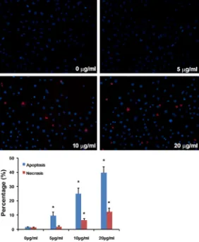
Results of Hoechst 33342/PI assay showed that both apoptosis and necrosis of HepG2
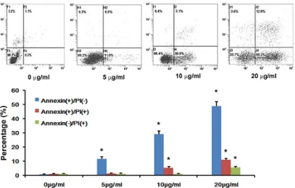
cells were observed and increased in a dose-dependent manner, 48 hours after the co-cul-ture of cerulenin, compared with the normal saline treated control (Figure 2). A similar
phe-nomenon was found in Annexin V/PI analysis
(Figure 3). Western blot analysis showed that
the expression of FASN protein was deceased in a dose-dependent manner, 48 hours after the co-culture of cerulenin. The change of Bcl- 2 protein expression increased after cerulenin
treatment in HepG2 cells, while the changes of
Bax, caspase-3, -8 and -9 protein expression had an opposite pattern (Figure 4). These re- sults suggest that cerulenin had a cytotoxic
effect and promote apoptosis in HepG2 cells.
According to the results of the in vivo
experi-ment, treatment of cerulenin exerted a benefi -cial effect to the hepatic cancer in terms of the decreased tumor volume and weight, dur-ing the whole observation period (Figure 5). Immunochemical staining showed that the ex- pression of FASN protein was decreased by cerulenin treatment in vivo. PCNA was stained to evaluate cell proliferation, and the result showed that the expression of PCNA was
sig-nificantly decreased in the cerulenin group
compared to the control group. TUNEL assay was applied to evaluate the cell apoptosis in transplanted tumor. Apoptosis was increased after cerulenin, compared with the normal saline treatment (Figure 6).
Discussion
Even in a nutrient starvation state, cancer cells have a high potential for proliferation and sur-vival, which may be based on a high potential for cellular fatty acid synthesis [10]. Synthesiz- ed from acetyl-CoA and malonyl-CoA, fatty ac- ids are aliphatic acids fundamental to energy production and storage, cellular structure and as intermediates in the biosynthesis of hor-mones and other biologically important mole-cules [11]. Endogenous fatty acids are the main source of fatty acids which are required for the growth of many cancer cells. Fatty acid metab-olism of tumor cells is different from that in the normal tissue cells, which depends on the fatty acid synthase (FASN)-based synthesis of fatty acids in order to meet the needs of the cancer cells’ division and proliferation [12]. Fatty acid
synthase (FASN) is a key enzyme in the biosyn -thesis of fatty acids during the polymerization of long chain fatty acids with small molecular carbon units. The gene that codes for FASN has been investigated as a possible oncogene. In normal tissues, no expression or low expres-sion of FASN was found, except in liver, uterus,
colorectal cancer, ovarian cancer, melanoma
and multiple myeloma [5-8, 13, 14]. High
expression of FASN was reported to be related with tumor development, pathological grade, clinical degree of malignancy and poor
progno-sis. High expression of FASN found in the
tumors will contribute to the synthesis of high amounts of fatty acids and high cell viability under nutrient starvation [15]. Recent studies suggest that the inhibition of FASN could be a therapeutic target of some tumors [16]. As an antifungal antibiotics, cerulenin inhibits fatty
acid and steroid synthesis. It can bind to β-keto-acyl-ACP synthase in fatty acid synthesis, block -ing the interaction of malonyl-CoA, and is also able to stimulate fatty acid oxidation through the activation of CPT1, another enzyme normal-ly inhibited by malonyl-CoA. These two behav-iors may increase the availability of energy in the form of ATP. Therefore, cerulenin is often
otal mechanism for a large number of chemo-therapeutic agents, in which process the func-tions of Bcl-2 (an anti-apoptotic factor) and Bax (a pro-apoptotic factor and natural antagonist of Bcl-2) are well investigated. As a major inhibi- tor of apoptosis, Bcl-2 can promote the forma-tion of tumor by resisting various forms of cell death, prolonging cell life and increasing the number of cells in tumor [19, 20]. In this study, down-regulation of Bcl-2 and the up-regulation of Bax may be a plausible reason for the pro-apoptotic effect of cerulenin treatment. The- refore, cerulenin may have the anti-proliferative and pro-apoptotic effects on hepatic cancer cells, through its inhibition of FASN. In other words, FASN is essential for the proliferation and survival of hepatic cancer, suggesting that depletion of fatty acids is fatal for hepatic can-cer cells. It has been reported that FASN inhi-
[image:4.612.91.373.72.415.2]bition reduces cell proliferation by blocking
Figure 2. Apoptosis of HepG2 cells were measured by Hoechst33342/PI staining. Apoptosis of HepG2 cells was observed and increased in a
dose-dependent manner 48 hours after cerulenin treatment. The data represent mean ± SE. *p<0.05 versus control.
applied as an inhibitor of FASN in experiments recently [17, 18]. In the present study, the expression of FASN was ob-
served in HepG2 cells, and
cerulenin, a FASN inhibitor, co- uld reduce the FASN expres-sion. Then, the effect of
ceru-lenin on HepG2 cells was stud -ied in vitro and in vivo in this study.
In in vitro experiments, ceru-lenin displayed an obvious inhibitory effect on the
prolif-eration of HepG2 cells in a dose-dependent manner.
Hoe-chst 33342/PI staining reve- aled the characteristic featur- es of apoptosis in
cerulenin-treated HepG2 cells, repre -sented as chromatin conden-sation and the formation of
apoptosis body. Annexin V/PI
analysis also showed that ce- rulenin could induce the
apop-tosis of HepG2 cells. These
piv-Figure 3. Apoptosis of HepG2 cells were measured by Annexin V/PI assessment. Apoptosis of HepG2 cells was in -creased in a dose-dependent manner 48 hours after cerulenin treatment. The data represent mean ± SE. *p<0.05 versus control.
Figure 4. The changes of FASN and apoptosis related proteins expression in HepG2 cells following cerulenin treat
-ment. A, B: Expression of FASN, Bax, Bcl-2, caspase-3, caspase-8, and Caspase-9 proteins in HepG2 cells 48 hours
after ceruleinin treatment were detected by Western blot analysis. The data represent mean ± SE. *p<0.05 versus
control.
DNA replication during S-phase [21]. However,
the biological mechanisms responsible for the FASN inhibition-induced apoptosis are still not clear. The extrinsic pathway of apoptosis trig-gered by death domains was described in br- east cancer cells because of the accumulation of malonyl-CoA and ceramide after FASN si- lencing with siRNA [22]. Mitochondrial involve-ment, evidenced by increased levels of the
[image:5.612.95.523.409.542.2]Figure 5. Inhibitory effect of the combined treatment with cerulenin on the growth of xenografts of hepatic cancer cells in nude mice. A. Tumor volume changes in nude mice after cerulenin treatment. B. Tumor weights were
mea-sured 4 weeks after cerulenin treatment. Data are shown as mean ± SE. *P<0.05 versus NS-treated mice.
Figure 6. Expression of FASN, apoptosis and acinar regeneration in the transplanted tumors were assessed as
described in the Method section 4 weeks after cerulenin treatment. A, D. Representative histochemical-stained
sections of FASN. B, E. Representative immunohistochemical staining of PCNA. C, F. Representative TUNEL-stained
sections of apoptotic cells. G-I. Alterations of FASN-positive cells, PCNA-positive cells and TUNEL-positive cells in the transplanted tumors after cerulenin treatment. Original magnification × 400; data are presented as mean ± SE;
[image:6.612.89.524.288.646.2]hepatic cancer cells was also studied in the present study. The in vivo experiment revealed that expression of FASN in tumor tissue was reduced after cerulenin treatment. Further- more, cerulenin could inhibit growth of the transplanted tumor and could also regulate proliferation and apoptosis, as evidenced by the results of the histochemical staining of PCNA and TUNEL.
In conclusion, cerulenin may be an anti-prolifer-ative and pro-apoptosis agent for hepatic can-cer cells in vitro and in vivo by down-regulating FASN expression and inhibiting intracellular FASN activity. Our results suggest that ceru- lenin could be applied in treatment of liver can-cer. There are a lot of reports of the application of cerulenin in cancer treatment experiments, which may supply useful ideas and new clues in developing target-directed anti-cancer drugs for further studies.
Disclosure of conflict of interest
None.
Acknowledgements
This study was supported by Jiangsu Provincial Youth Medical Talent program (QNRC2016722) and Suzhou Municipal Science and Technology Development Program (SYS2018041).
Address correspondence to: Dr. Jiaqing Shen, De- partment of Gastroenterology, The First Affiliated Hosptial of Soochow University, Suzhou 215000, Jiangsu, China. E-mail: sjqsz@126.com
References
[1] Augustine MM, Fong Y. Epidemiology and risk factors of biliary tract and primary liver tumors. Surg Oncol Clin N Am 2014; 23: 171-188. [2] Swierczynski J, Hebanowska A, Sledzinski T.
Role of abnormal lipid metabolism in develop-ment, progression, diagnosis and therapy of pancreatic cancer. World J Gastroenterol 2014; 20: 2279-2303.
[3] Ookhtens M, Kannan R, Lyon I, Baker N. Liver and adipose tissue contributions to newly formed fatty acids in an ascites tumor. Am J Physiol 1984; 247: 146-53.
[4] Kuhajda FP, Katumuluwa AL, Pasternack GR. Expression of haptoglobin-related protein and its potential role as a tumor antigen. Proc Natl Acad Sci U S A 1989; 86: 1188-1192.
[5] Alò PL, Visca P, Trombetta G, Mangoni A, Lenti L, Monaco S, Botti C, Serpieri DE, Di Tondo U. Fatty acid synthase (FAS) predictive strength in poorly differentiated early breast carcinomas. Tumori 1999; 85: 35-40.
[6] Welsh JB, Sapinoso LM, Su AI, Kern SG, Wang-Rodriguez J, Moskaluk CA, Frierson HF Jr, Hampton GM. Analysis of gene expression identifies candidate markers and pharmaco -logical targets in prostate cancer. Cancer Res 2001; 61: 5974-5978.
[7] Zecchin KG, Alberici LC, Riccio MF, Eberlin MN, Vercesi AE, Graner E, Catharino RR. Visualizing inhibition of fatty acid synthase through mass spectrometric analysis of mitochondria from melanoma cells. Rapid Commun Mass Spec- trom 2011; 25: 449-452.
[8] Uddin S, Jehan Z, Ahmed M, Alyan A, Al-Dayel F, Hussain A, Bavi P, Al-Kuraya KS. Overexpression of fatty acid synthase in Middle Eastern epithe-lial ovarian carcinoma activates AKT and its inhibition potentiates cisplatin-induced apop-tosis. Mol Med 2011; 17: 635-645.
[9] Lawrence DS, Zilfou JT, Smith CD. Structure-activity studies of cerulenin analogues as pro-tein palmitoylation inhibitors. J Med Chem 1999; 42: 4932-4941.
[10] Huang, Li T, Wang L, Zhang L, Yan R, Li K, Xing S, Wu G, Hu L, Jia W, Lin SC, Dang CV, Song L, Gao P, Zhang H. Hepatocellular carcinoma re -directs to ketolysis for progression under nutri -tion depriva-tion stress. Cell Res 2016; 26: 1112-1130.
[11] Bastos DC, Paupert J, Maillard C, Seguin F, Carvalho MA, Agostini M, Coletta RD, Noël A, Graner E. Effects of fatty acid synthase inhibi-tors on lymphatic vessels: an in vitro and in vivo study in a melanoma model. Lab Invest 2017; 97: 194-206.
[12] Vazquez-Martin A, Colomer R, Brunet J, Lupu R, Menendez JA. Overexpression of fatty acid synthase gene activates HER1/HER2 tyrosine kinase receptors in human breast epithelial cells. Cell Prolif 2008; 41: 59-85.
[13] Gu Z, Wang X, Qi R, Wei L, Huo Y, Ma Y, Shi L, Chang Y, Li G, Zhou L. Oridonin induces apop-tosis in uveal melanoma cells by upregulation of Bim and downregulation of Fatty Acid Synthase. Biochem Biophys Res Commun 2015; 457: 187-193.
[14] Wang WQ, Zhao XY, Wang HY, Liang Y. Increased fatty acid synthase as a potential therapeutic target in multiple myeloma. J Zhejiang Univ Sci B 2008; 9: 441-447.
[22] Migita T, Ruiz S, Fornari A, Fiorentino M, Priolo C, Zadra G, Inazuka F, Grisanzio C, Palescandolo E, Shin E, Fiore C, Xie W, Kung AL, Febbo PG, Subramanian A, Mucci L, Ma J, Signoretti S, Stampfer M, Hahn WC, Finn S, Loda M. Fatty acid synthase: a metabolic enzyme and can- didate oncogene in prostate cancer. J Natl Cancer Inst 2009; 101: 519-32.
[23] Zhang Y, Fu R, Liu Y, Li J, Zhang H, Hu X, Chen Y, Liu X, Li Y, Li P, Liu E, Gao N. Dephosphoryla- tion and mitochondrial translocation of cofilin sensitizes human leukemia cells to cerulenin-induced apoptosis via the ROCK1/Akt/JNK sig -naling pathway. Oncotarget 2016; 7: 20655-20668.
[24] Li JN, Gorospe M, Chrest FJ, Kumaravel TS, Evans MK, Han WF, Pizer ES. Pharmacological inhibition of fatty acid synthase activity pro-duces both cytostatic and cytotoxic effects modulated by p53. Cancer Res 2001; 61: 1493-1499.
Proc Natl Acad Sci U S A 2000; 97: 10619-10624.
[16] Lupu R, Menendez JA. Pharmacological inhibi-tors of Fatty Acid Synthase (FASN)--catalyzed endogenous fatty acid biogenesis: a new fami-ly of anti-cancer agents? Curr Pharm Biotechnol 2006; 7: 483-493.
[17] Nishi K, Suzuki K, Sawamoto J, Tokizawa Y, Iwase Y, Yumita N, Ikeda T. Inhibition of fatty acid synthesis induces apoptosis of human pancreatic cancer cells. Anticancer Res 2016; 36: 4655-4660.
[18] Yasumoto Y, Miyazaki H, Vaidyan LK, Kagawa Y, Ebrahimi M, Yamamoto Y, Ogata M, Katsuyama Y, Sadahiro H. Inhibition of fatty acid synthase decreases expression of stemness markers in glioma stem cells. PLoS One 2016; 11: e0147717.
[19] Adams JM, Cory S. The Bcl-2 protein family: ar-biters of cell survival. Seienee 1998; 281: 1332-1326.
[20] Jo M, Kim TH, Seol DW. Apoptosis induced in normal human hepatocytes by tumor necrosis factor-related apoptosis-inducing ligand. Nat Med 2000; 6: 564-567.