Original Article
Mechanisms of renal sympathetic denervation on
improving ventricular arrhythmias after acute
myocardial infarction in rats
Yong-Quan Lu1,2, Li-Yuan Zhu3, Yin-Fen Zhang2, Zi-Guan Zhang2,3, Hong-Lang Huang1, Lin Lin4, Jun Li3,
Wu-Yang Zheng1, Xin Jin3, Qiang Xie1
1Department of Cardiology and Xiamen Institute of Cardiovascular Diseases, The First Affiliated Hospital of
Xiamen University, Xiamen 361003, China; 2Fujian University of Traditional Chinese Medicine, Fuzhou 350122,
China; 3Medical College, Xiamen University, Xiamen 361102, China; 4The First Clinical Medical College, Fujian
Medical University, Fuzhou 350108, China
Received April 1, 2019; Accepted April 10, 2019; Epub July 15, 2019; Published July 30, 2019
Abstract: Background: More than 50% of acute myocardial infarction (MI) survivors died from malignant ventricular arrhythmias (VA). Renal sympathetic denervation (RSD) has been demonstrated to exert remarkable effects on VA, but the mechanism remains unclear. Methods: Thirty Sprague Dawley rats were divided into three groups ran-domly, that is Sham, MI (ligation of left anterior descending artery) and MI+RSD (ethanol ablation). Six hours after modeling, electrocardiogram was recorded. Four weeks later, the left ventricular function indexes were obtained through echocardiography, and cardiac tissues were stained by Masson trichrome for fibrotic analysis. Whole-cell patch-clamp recordings were performed to record the transient outward K+ current (I
to) and the protein expression of
Kv4.2 and Kv4.3 in the left ventricle were detected using Western blot. Results: Compared to that in MI group, RSD group showed reduced incidence of premature ventricular contractions and ventricular tachycardia, increased left ventricular ejection fraction and fractional shortening, and decreased left ventricular end diastolic diameter and left ventricular end systolic diameter. RSD attenuated collagen deposition in the cardiac tissue. RSD group alleviated prolonged action potential duration (p < 0.05) especially APD20. The Ito current density was significantly decreased in the MI group compared to the sham group, and was reversed by RSD. MI-induced a decreased cardiac protein expression of Kv4.2, but not that of Kv4.3, and it was restored by RSD. Conclusions: RSD reduced the incidence of VA after MI in rats. This may be due to the improvement of left ventricular function, the recovery of cardiac Ito density and Kv4.2 protein expression.
Keywords: Renal sympathetic denervation, ventricular arrhythmias, myocardial infarction, left ventricular function, Ito current, Kv4.2
Introduction
Ventricular arrhythmia (VA) is the most com-mon complication of acute myocardial infarc-tion (AMI), and it is also an important factor affecting prognosis [1]. Malignant ventricular arrhythmias, such as persistent ventricular tachycardia or ventricular fibrillation, are the main causes of sudden death in AMI patients [2]. The mechanism of ventricular arrhythmias mainly involves reentry and triggering [3, 4], but the VA treatment including antiarrhythmic dr- ugs and catheter ablation are both unsatisfac-tory at present [5]. More than 50% of AMI
trans-mits activation signals to the kidney, meanwhile taking signals to the heart or other highly sym-pathetic dominating organs [9-11]. Tsai provid-ed the direct evidence that RSD could rprovid-educe the cardiac sympathetic nerve activity, which prompts that RSD has a potential role in reduc-ing the incidence rate of ventricular arrhyth-mias. Krum [12] first proposed RSD as a new therapeutic measure for refractory hyperten-sion, and follow-up studies found that RSD also had a therapeutic effect on ventricular arrhyth-mia. Ukena [13] first reported that two patients with chronic heart failure complicated with refractory ventricular arrhythmias were suc-cessfully treated by RSD. Boris [14] also report-ed that a patient with ventricular arrhythmias after acute ST-segment elevation myocardial infarction was effectively controlled by RSD. Armaganijan [15] took 10 patients with refrac-tory arrhythmia and implanted them with ICD, and the results showed that the average times of the VT/VF, antitachycardia and electrical car-dioversion before RSD were 28.5 (1~106), 20.5 (0~52), 8 (0~88), but they were all reduced after RSD operation 6 months later, respec- tively times were 0 (0~9), 0 (0~7), 0 (0~3). Evranos [16] made a similar clinical trial to Armaganijan and the results also showed that RSD could reduce the average times of the VT/ VF, antitachycardia and electrical cardiover-sion, which suggested that RSD was an effec-tive adjuvant therapy in the treatment of refrac-tory arrhythmia. Linz [17] found that RSD was capable of inhibiting the occurrence of ven- tricular arrhythmias after acute myocardial ischemia in pigs. Conclusively, a growing body of evidence [18, 19] has confirmed that RSD could reduce the incidence of ventricular arrhythmias, but the mechanism is still uncl- ear. In the research work of this paper, we work to reveal the mechanism of RSD on improv- ing VA after acute myocardial infarction, by studying the relationship between RSD and multiple physiological features of the rat heart, based on the rat model of AMI-induced arrhythmias.
Materials and methods
Animals and model preparation
The Male Sprague-Dawley rats (200-250 g) were purchased from Animal Center (Wushi, Fuzhou, China) and were housed in a 12 h
dark/light cycle. The temperature was 22~ 25°C, and the relative humidity was 55%~ 60%. Thirty experimental rats were divided into three groups randomly, that is sham group (n=10), MI group (n=10) and MI+RSD group (n=10).
In the MI and MI+RSD groups, the rats were anesthetized by intraperitoneal injection of 10% chloral hydrate (3.5 mL/kg), and were ven-tilated at 75 breaths/min, 5.5 mL tidal volume (Rodent Ventilator, ALC-V8S, Shanghai, China). The heart was exposed and pericardium was incised. The model was produced by ligation of the left anterior descending coronary artery with 6-0 polypropylene suture approximately 3-4 mm distal from its origin. The chest was closed and lungs were reinflated using posi-tive end-expiratory pressure. In the MI+RSD group, the bilateral renal artery and vein was exposed immediately after MI operation and the sympathetic innervation was surgically denervated by cutting all visible nerves, and then the vascular wall was daubed with abso-lute ethanol for 10 minutes. The rats in the sham group, the surgical procedure was per-formed without coronary artery ligation and RSD was performed.
TTC staining
Infarct size was assessed using TTC staining as described previously [20]. Briefly, the rat hearts were harvested and frozen at -80°C for 5 min, and then rapidly cut into 6 slices approxi-mately 1 mm thick each. The slices were incu-bated with 1% triphenyl-tetrazolium chloride (TTC, Solarbio, Beijing, China) for 20 min at 37°C and then fixed in 4% formaldehyde for another 30 min. The viable perfused myoca- rdium was colored in red and the infarcted area in white. The ratio of infarcted area to to- tal area was calculated by computerized pla-nimetry with ImageJ software (NIH, USA). The results were expressed in average of percent-age of infarcted area on total area in the 6 slices.
H&E staining
idly excised and the infarct border zones were harvested. Myocardial tissue was first decom -posed by Ca2+-free Tyrode’s solution containing
II type collagenase (0.5 g/L), bovine serum albumin (1 g/L) and XIV type protease (0.1 g/L) for 50 min, followed by decomposition with the similar solution containing II type collagenase (0.5 g/L) and bovine serum albumin (1 g/L) for 40 min. The whole process was carried out in constant oxygen perfusion at 37°C. The freshly isolated myocytes were centrifuged at 1000 rpm for 3 min and resuspended in KB solution, followed by resting for 3 hours and then pre-served at 4°C before electrophysiological re- cording. The rod-shaped cells with clear cross-striation and without spontaneous contraction were selected for patch recording.
The Whole-cell patch-clamp techniques were performed by an Axopatch 200B amplifier (Ax-on instrument, USA). Recording pipettes with a tip resistance of 3-5 MΩ when filled with the internal pipette solution were used. For Ito recordings, the pipette solution contained in mM: 20 KCl, 110 K-Asparate, 1 MgCl2·6H2O, 5 Na2-Phosphocreatine, 10 HEPES, 5 K2-EGTA, 0.1 GTP and 5 Mg2-ATP (pH adjusted to 7.2 with
KOH). 0.2 µM BaCl2 was used to block ICa and IK1, respectively. Ito was elicited by a 300 ms depolarizing current stepped from -30 mV to 60 mV with 10 mV increments from a holding potential of -80 mV. The data were acquired with pCLAMP system and analyzed by Origin 7.5 software.
Western blot
The ventricle samples of infarct border zone were lysed, and total protein was extracted using RIPA Lysis Buffer. Before the experiment, the BCA method was used to determine the protein concentration. The 2 mg/mL BSA stan-dard sample was diluted into 8 groups of 0-2000 ug/mL gradient concentrations using deionized water, and the absorbance value cor-responding to 562 nm was used as the ordi-nate to draw the standard concentration curve of the protein sample, and then the protein con-centration of the sample to be tested was cal-culated according to the standard curve. The protein samples were separated on 8% sodium dodecyl sulfate-polyacrylamide gel electropho-resis (SDS-PAGE) and transferred to polyvinyli-ECG assessment
A standard II lead of electrocardiogram (ECG) was recorded after modeling for 6 hours by using Multichannel physiological signal acquisi-tion system (RM6240CD type, Chengdu, China). Ventricular arrhythmias observed in the ECG were classified as premature ventricular con -tractions (PVCs), ventricular tachycardia (VT) and ventricular fibrillation (VF) according to the Lambeth standard.
Echocardiographic assessment of left ventricu -lar function
The echocardiogram was performed with high-resolution ultrasound (20 MHz) imaging system (Vevo 2100, Visual Sonics, Ontario, Canada). The left ventricular ejection fraction (LVEF), fractional shortening (FS), left ventricular end diastolic dimension (LVEDD) and left ventricular end systolic dimension (LVESD) were measur- ed before the surgery and after 28 days of left anterior descending coronary artery ligation. The LVEF were defined by the formula LVEF = (LVEDD-LVESD)/LVEDD * 100%. All echocardio-grams were measured in triplicate by the same sonographer, and the averages were taken for analysis.
Masson’s staining
The rats were sacrificed by an overdose of chlo -ral hydrate, and then the left ventricles were rapidly harvested and fixed in 4% neutral buff -ered formalin for 18 hours and embedded in optimal cutting temperature compound (OCTc). Longitudinal sections (6 um) of hearts were prepared using a microtome. The sections were stained with Masson’s trichrome (Maixing, Fuzhou, China) for measurement of fibrosis. The collagen volume fraction in the peri-infarct-ed areas of left ventricular was calculatperi-infarct-ed by measuring the optical density of fibrotic area using ImageJ software (NIH, USA).
Cardiomyocyte isolation and whole-cell patch-clamp recording
rap-tion system (Image Starap-tion 4000R, Kodak, USA). Auto- radiography images were analyzed by Carestream Molecular Imaging analysis software.
Statistical analysis
Experimental data were obtained from the statisti-cal average of multiple samples in the same group of subjects and all values were presented as the mean ± standard deviation. Statistical analysis was per-formed with SPSS 20.0 software. First, the experi-mental values were statisti-cally analyzed, and the experimental values col-lected from each sample of sham, MI and MI+RSD group were consistent with normal distribution and odd variance. And then one-way analysis of variance was applied to compare mean differences among the sh- am, MI and MI+RSD groups. Furthermore, LSD method was used for multiple com-parisons, and sham, MI, MI+RSD groups were com-pared in pairs to analyze whether there were signifi -cant differences between them. The statistical meth-od of t test was used for sig-nificance test and the p -val-dene fluoride (PVDF) membrane. The mem
-branes were blocked with 5% nonfat milk for 1 hour at room temperature, and were incubated overnight at 4°C with the primary antibodi- es (rat anti-Kv4.2 and anti-Kv4.3, monoclonal antibody, Abcam, Cambridge, UK) and the pri-mary anti-Kv4.2 and Kv4.3 protein antibodies were diluted with TBST solution at a ratio of 1:1000 according to the instructions, followed by incubation for 1 hour at room temperature with secondary antibody (Goat anti-Mouse, Multi Sciences, Zhejiang, China), this concen-tration is also diluted according to the product instructions. The membranes were visualiz- ed by an enhanced chemiluminescence
detec-ue of less than 0.05 was considered to be sta-tistically significant.
Results
MI induced by ligating left anterior descending coronary artery
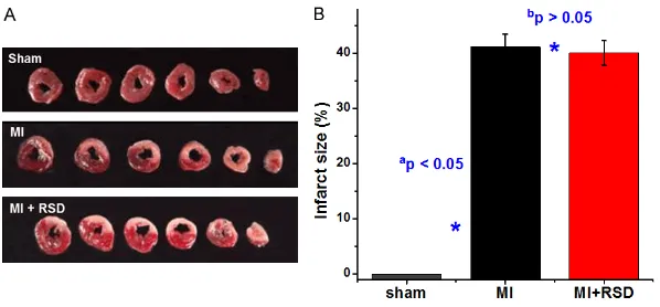
[image:4.612.89.388.70.209.2]TTC staining was performed to confirm whether the rat model of MI was successfully estab-lished. The result showed that the myocardium infarction region was stained white, while the non-infarct area remained red (Figure 1A). There was no significant difference in infarct size between MI group and MI+RSD group Figure 1. TTC staining was performed to measure the infarct size in rat hearts.
A. Photos showed the myocardium infarction region was stained white, whereas the non-infarct area remained red. B. Infarct sizes were presented as a percent-age of the infarct area to the total left ventricular area. Values are presented as the mean ± SD. ap < 0.05 versus the sham group; bp > 0.05 versus the MI
group.
[image:4.612.90.385.297.457.2](41.22±2.23 vs 40.06±2.22, p > 0.05) (Figure 1B).
Histology of the renal artery and nerves
The renal artery and surrounding nerves were stained with H&E (Figure 2A). The results sh- owed that sympathetic nerves in RSD group were significantly decreased in comparison with sham group (0.25±0.50 vs 2.75±0.50,
p < 0.05) (Figure 2B). Some nerves lost histo-logical details after RSD. In contrast, these alterations were not observed in rats without ablation.
Effect of RSD on occurrence of AMI-induced
arrhythmias
[image:5.612.91.524.71.543.2]The ECG manifestations in rats were showed in Figure 3A and ST-segment elevation was Figure 3. A. The standard II lead of electrocardiogram (ECG) manifestations of normal, post ligation of coronary artery, PVCs and VT in rats. B. Ventricular arrhythmias could be induced by myocardial infarction. RSD (renal sym-pathetic denervation) treatment reduced the incidence of PVCs and VT. ap < 0.05 versus the sham group; bp < 0.05
Figure 4. Echocardiographic images of rat hearts in three groups.
clearly observed after coronary artery ligation. Ventricular arrhythmias could be significantly induced by MI. Compared with MI group, MI+RSD group has a lower incidence of PVCs (25.00±2.16 vs 42.50±10.88, p < 0.05) and VT (4.80±1.64 vs 10.00±4.08, p < 0.05) (Figure 3B).
Effect of RSD on cardiac function after MI
The echocardiographic images were showed in Figure 4. There were no significant differences in pre-operative echocardiogram parameters among the three groups. The MI group had sig-nificantly decreased LVEF (32.39±10.64 vs 75.28±8.72%, p < 0.05) and FS (16.13±5.68 vs 45.49±7.53%, p < 0.05), and increased LVEDD (8.26±0.79 vs 5.70±0.33 mm, p < 0.05) and LVESD (7.01±0.91 vs 3.37±0.71 mm, p < 0.05) at 4 weeks after coronary artery ligation compared to that of the sham group. The decline of LVEF (32.39±10.64 vs 51.49±8.91%,
p < 0.05) and FS (16.13±5.68 vs 27.07±5.42%,
p < 0.05) was significantly attenuated by the
MI+RSD group, and the elevation of LVEDD (8.26±0.79 vs 6.63±0.28 mm, p < 0.05) and LVESD (7.01±0.91 vs 4.85±0.73 mm, p < 0.05) was also attenuated by the MI+RSD group (Table 1).
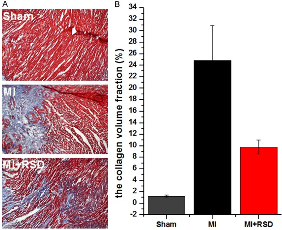
Effect of RSD on myocardial fibrosis
Masson staining showed that the cardiac myo-cytes were dyed red and the collagen fibers were dyed blue under light microscope (Figure 5A). The area of the collagen fraction was sig-nificantly increased in MI group compared with sham group (24.84±6.07 vs 1.23±0.15, p < 0.05). The extent of collagen deposition was attenuated in MI+RSD group compared to MI group (9.75±1.22 vs 24.84±6.07, p < 0.05) (Figure 5B).
Effect of RSD on APD in rat hearts
Action potentials were recorded in current clamp mode (Figure 6A). The result showed that MI group had prolonged action potential duration (APD, 137.5±6.81 vs 115.9±6.36, p < 0.05) and APD of 50% repolarization (APD50, 58.45±1.44 vs 43.32±2.39, p < 0.05). Compared with MI group, prolonged APD and APD50 have been shortened in the MI+RSD group (Figure 6B).
Effect of RSD on Ito in rat hearts
To detect the effect of RSD on Ito in MI rats, we examined Ito under whole-cell voltage- clamp mode. At the test potential of +60 mV, the current density of Ito was significantly decreased in MI group than that in sham gr- oup (11.30±5.81 vs 22.10±10.55, p < 0.05), while RSD treatment could obviously increa- se the suppressed Ito current density (15.82± 7.80 vs 11.30±5.81, p < 0.05). The current-voltage (I-V) curves demonstrating the changes of Ito in different groups were plotted in Figure 7B.
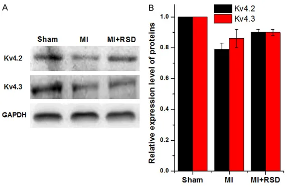
Effect of RSD on protein expression of Kv4.2 and Kv4.3
tion I phase of the action potential, so we detected Ito and its relevant ion channel proteins.
In the rat AMI model, we showed that the Ito density and protein expression level of Kv4.2 and Kv4.3 were decreased in the marginal zone of myocardial infarction. Furthermore, we first found that RSD could alleviate the decline of Ito density and up regulate Kv4.2 protein expres-sion level. Ito is the main cur-rent of repolarization I phase of action potentials in cardiac myocytes, and it can affect the action potential morpholo-gy and duration. Kv4.2 and Kv4.3 are the major subuni- ts of Ito in cardiomyocytes. Previous studies have shown that the protein expression levels of Kv4.2 and Kv4.3 in cardiomyocytes were decr- eased after MI, and it could decrease Ito density and then prolonged action potential duration (APD) [22, 23], whi- ch can cause the occurrence of early after depolarization and contribute to ventricular arrhythmias. Moreover, the decrease of Ito current density can increase the sensitivity of the myocardium to hypokale-mia, ischemia and acidosis, which can increase the sus-vs 1.00±0.00, p < 0.05) (Figure 8A). Inter-
estingly, the RSD treatment could significantly increase the Kv4.2 protein expression level (0.90±0.02 vs 0.79±0.04, p < 0.05) while mak-ing no difference on Kv4.3 protein level (Figure 8B).
Discussion
In our experiment, we first observed the effect of RSD on the occurrence of VA. Second, we recorded the APD and found the prolonged APD in MI group could be shortened by RSD espe-cially APD50. APD50 mainly reflected repolariza
-ceptibility to arrhythmias. Some studies have showed that cardiac nerve sprouting or isopro-terenol could suppress Ito and increased the susceptibility to ventricular fibrillation. Ven-tricular electrical remodeling is one of the important factors of the occurrence of ventricu-lar arrhythmia after myocardial infarction, and myocardial electrophysiological heterogenei- ty in different positions of the myocardium increases the risk of ventricular arrhythmias. Here we show that RSD can reduce the inci-dence of ventricular arrhythmias, and its mech-anism is possibly related to attenuation of the decline of Ito current density through the up-Table 1. Changes of echocardiographic parameters in the three
groups
Group n LVEF (%) FS (%) LVEDD (mm) LVESD (mm)
Sham 10 75.28±8.72 45.49±7.53 5.70±0.33 3.37±0.71
MI 8 32.39±10.64a 16.13±5.68a 8.26±0.79a 7.01±0.91a
MI+RSD 6 51.49±8.91a,b 27.07±5.42a,b 6.63±0.28a,b 4.85±0.73a,b
[image:7.612.92.375.95.149.2]Note: LVEF (left ventricular ejection fraction), FS (fractional shortening), LVEDD (left ventricular end diastolic dimension), LVESD (left ventricular end systolic dimen-sion), MI (myocardial infarction), RSD (renal sympathetic denervation). Values are presented as the mean ± SD. ap < 0.05 versus the sham group; bp < 0.05 versus the MI group.
Figure 5. Masson’s trichrome staining for assessment of myocardial fibrosis. A. Photos showed that the cardiac myocytes were dyed red and the collagen fibers were dyed blue under light microscope (×100). B. The area of the col-lagen fraction was significantly increased in MI groups compared with sham groups. RSD treatment attenuated myocardial fibrosis in MI rats. ap < 0.05,
[image:7.612.88.375.224.458.2]antiarrhythmic role. In addi-tion, our results demonstrat- ed RSD’s effect of improving cardiac function after MI by increasing EF and FS and reducing LVEDD and LVESD. Collagen fiber hyperplasia is an important factor in myo- cardial fibrosis and cardiac remodeling after MI, the Masson staining showed that RSD can reduce the degree of myocardial fibrosis, which may be an important pathological mechanism of its role im- proving cardiac function after MI.
Conclusions
Electrical remodeling and st- ructure remodeling play a cri- tical role in the occurrence and maintenance of ventricu-lar arrhythmias after myocar-dial infarction. Our results indicated that the mechanism of RSD on inhibition of VA after MI was linked to the reverse of Ito density through the up-regulation of Kv4.2 protein expression and the improve-ment of cardiac function.
Acknowledgements
This study was supported by funds from Young and Middle-Aged High-Level Back- bone Talent Training Project of Fujian Health System (2013-ZQN-ZD-32), Key Pro- ject of Fujian Science and Technology Program (2014- D023), Natural Science Fou- ndation of Fujian Province (2016J01637) and Science and Technology Project of Xiamen (3502Z20154007). regulation of Kv4.2 protein expression level.
The recovery of Ito current can mitigate electro-physiological heterogeneity and thus play an
Disclosure of conflict of interest
[image:8.612.89.370.71.291.2]None. Figure 7. The effect of RSD on Ito in MI rat hearts. A. The transient outward K+
current (Ito) was elicited by a 300 ms depolarizing current stepped from -30
to +60 mV with 10 mV increments from a holding potential of -80 mV. 0.2 µM BaCl2 was used to block ICa and IK1. B. The current voltage (I-V)
relation-ships in three groups rat hearts. ap < 0.05, versus the sham group; bp > 0.05
[image:8.612.89.374.368.582.2]versus the MI group.
Figure 6. The effect of RSD on APD in MI rat hearts. A. The representative action potential traces in three groups rat hearts. B. ap < 0.05, versus the
sham group; bp < 0.05 versus the MI group; cp > 0.05 versus the sham
[5] Tanner H, Hindricks G and Kottkamp H. [Frequent ven-tricular tachycardias: antiar-rhythmic drug treatment or catheter ablation?]. Herz 2005; 30: 613-618.
[6] Wyman MG, Wyman RM, Cannom DS and Criley JM. Prevention of primary ven-tricular fibrillation in acute myocardial infarction with prophylactic lidocaine. Am J Cardiol 2004; 94: 545-551.
[7] De Ferrari GM and Sch- wartz PJ. Autonomic ner-vous system and arrhyth-mias. Annals of The New York Academy of Sciences 2010; 601: 247-262. [8] Katra RP and Laurita KR.
Cellular mechanism of calci-um-mediated triggered ac-tivity in the heart. Circ Res 2005; 96: 535-542. Address correspondence to: Dr. Qiang Xie, Depart-
ment of Cardiology and Xiamen Institute of Car- diovascular Diseases, The First Affiliated Hospital of Xiamen University, 55 Zhenhai Road, Xiamen 361003, China. Tel: 592-2139716; Fax: +86-592-2139550; E-mail: arthur2014@sina.com; Dr. Xin Jin, Medical College, Xiamen University, Xiang’an South Road, Xiamen 361102, China. Tel: +86-592-2188676; Fax: +86-592-+86-592-2188676; E-mail: xinjin@ xmu.edu.cn
References
[1] Bharamato J, Davies W and Agarwal S. Ven- tricular arrhythmia after acute myocardial in-farction: ‘the perfect storm’. Arrhythm Elec- trophysiol Rev 2017; 6: 134-139.
[2] Mehta RH, Yu J, Piccini JP, Tcheng JE, Farkouh ME, Reiffel J, Fahy M, Mehran R and Stone GW. Prognostic significance of postprocedural sus-tained ventricular tachycardia or fibrillation in patients undergoing primary percutaneous coronary intervention (from the HORIZONS-AMI trial). Am J Cardiol 2012; 109: 805-812. [3] Nattel S, Maguy A, Le Bouter S and Yeh YH.
Arrhythmogenic ion-channel remodeling in the heart: heart failure, myocardial infarction, and atrial fibrillation. Physiol Rev 2007; 87: 425-456.
[4] Anumonwo JM and Pandit SV. Ionic mecha-nisms of arrhythmogenesis. Trends Cardiovasc Med 2015; 25: 487-496.
[9] Wu B, Hong M, Wu H and Lin R. Renal sympa-thetic denervation might be an adjunctive treatment approach for managing ventricular arrhythmia. Int J Cardiol 2015; 184: 257-258. [10] DiBona GF. Physiology in perspective: the wis-dom of the body. Neural control of the kidney. Am J Physiol Regul Integr Comp Physiol 2005; 289: R633-641.
[11] Esler M. The 2009 Carl Ludwig lecture: patho-physiology of the human sympathetic nervous system in cardiovascular diseases: the transi-tion from mechanisms to medical manage-ment. J Appl Physiol (1985) 2010; 108: 227-37.
[12] Krum H, Schlaich M, Whitbourn R, Sobotka PA, Sadowski J, Bartus K, Kapelak B, Walton A, Sievert H, Thambar S, Abraham WT, Esler M. Catheter-based renal sympathetic denervation for resistant hypertension: a multicentre safety and proof-of-principle cohort study. Lancet 2009; 373: 1275-81.
[13] Ukena C, Bauer A, Mahfoud F, Schreieck J, Neuberger HR, Eick C, Sobotka PA, Gawaz M and Bohm M. Renal sympathetic denervation for treatment of electrical storm: first-in-man experience. Clin Res Cardiol 2012; 101: 63-67. [14] Hoffmann BA, Daniel S, Stephan W and Kar-
sten S. Renal sympathetic denervation as an adjunct to catheter ablation for the treatment of ventricular electrical storm in the setting of acute myocardial infarction. J Cardiovasc Ele- ctrophysiol 2013; 24: E21-E21.
[image:9.612.89.377.70.256.2][15] Armaganijan LV, Staico R, Moreira DA, Lopes RD, Medeiros PT, Habib R, Melo Neto J, Katz M, Figure 8. Western blot analysis of Kv4.2 and Kv4.3 protein expression levels
in three groups. GAPDH was used as a loading control. A. The protein expres-sion levels of myocardial Kv4.2 and Kv4.3 were significantly decreased in the MI group than that in sham group. B. RSD treatment increased the Kv4.2 level, but not the Kv4.3 level. ap < 0.05, versus the sham group; bp > 0.05
Armaganijan D, Sousa AG, Mahfoud F and Abizaid A. 6-month outcomes in patients with implantable cardioverter-defibrillators under-going renal sympathetic denervation for the treatment of refractory ventricular arrhyth-mias. JACC Cardiovasc Interv 2015; 8: 984-990.
[16] Evranos B, Canpolat U, Kocyigit D, Coteli C, Yor- gun H and Aytemir K. Role of adjuvant renal sympathetic denervation in the treatment of ventricular arrhythmias. Am J Cardiol 2016; 118: 1207-1210.
[17] Linz D, Wirth K, Ukena C, Mahfoud F, Pöss J, Linz B, Böhm M, Neuberger HR. Renal dener-vation suppresses ventricular arrhythmias dur-ing acute ventricular ischemia in pigs. Heart Rhythm 2013; 10: 1525-30.
[18] Bazoukis G, Korantzopoulos P and Tsioufis C. The impact of renal sympathetic denervation on cardiac electrophysiology and arrhythmias: a systematic review of the literature. Int J Cardiol 2016; 220: 87-101.
[19] Alizadeh A, Dyck SM, Kataria H, Shahriary GM, Nguyen DH, Santhosh KT and Karimi-Abdolre- zaee S. Neuregulin-1 positively modulates glial response and improves neurological recovery following traumatic spinal cord injury. Glia 2017; 65: 1152-1175.
[20] Liu C, Liu Y and Yang Z. Myocardial infarction induces cognitive impairment by increasing the production of hydrogen peroxide in adult rat hippocampus. Neurosci Lett 2014; 560: 112-116.
[21] Li X, Chu W, Liu J, Xue X, Lu Y, Shan H, Yang B. Antiarrhythmic properties of long-term treat-ment with matrine in arrhythmic rat induced by coronary ligation. Biol Pharm Bull 2009; 32: 1521-6.
[22] Zhang L, Xu CQ, Hong Y, Zhang JL, Liu Y, Zhao M, Cao YX, Lu YJ, Yang BF and Shan HL. Propranolol regulates cardiac transient out-ward potassium channel in rat myocardium via cAMP/PKA after short-term but not after long-term ischemia. Naunyn Schmiedebergs Arch Pharmacol 2010; 382: 63-71.