Persistent right aortic arch (PRAA) along with an aberrant left subclavian artery (SA) is an uncom-mon type of vascular ring anomaly. Whereas, PRAA and retention of left LA is reported to be the most common clinically significant form of vascular ring anomalies and comprises about 95% of develop-mental anomalies of the aortic arches in dog and cats (Ellison, 1990; Yarim et al., 1999; Isako et al., 2000; MacPhail et al., 2001; Ricardo et al., 2001; Koc et al., 2004). In case of PRAA with an aberrant left SA, the oesophagus is dually compressed by a com-plete vascular ring comprised of PRAA with left LA and by a partial ring formed with the aberrant left SA (Ellison, 1980). The PRAA is most frequently diagnosed in young large-breed dogs (Shires and Liu, 1981). The dogs with vascular ring anomaly usually have the history of postprandial regurgita-tion of solid foods soon after weaning (Van Den Ingh and Van Der Linde-Sipman, 1974; Helphrey, 1993). The affected dogs are stunted, thin and un-thrifty (Helphrey, 1993; Muldoon et al., 1997). A presumptive diagnosis can be made based on the clinical history, clinical signs, oesophagraphy and oesophagoscopy whereas; the confirmative diagno-sis is best made after surgical exploration (Muldoon
et al., 1997). Medical treatment (e.g. Liquid diets and supportive care) of PRAA has been shown to be unrewarding (Ellison, 1980; VanGundy, 1989). Thus, the surgical ligation and transection of the ligamentum arteriosum is the recommended meth-od of treatment. There have been numerous reports on PRAA but only a few on PRAA along with an aberrant left subclavian artery (Ketz et al., 2001). The purpose of this report is to present an unusual type of vascular ring anomaly in a Weimaraner dog and its surgical management.
CASE PRESENTATION
A 10-week-old female Weimaraner dog was pre-sented to the Chonbuk National University, Animal Medical Centre for evaluation of regurgitation. The dog had a history of regurgitation soon after wean-ing. It had a ravenous appetite but thin and stunted. The dog was underweight (4.5 kg) and much smaller than a male littermate that weighed 6.5 kg.
The cervical oesophagus was palpable as a flaccid, air-filled cavity. A test meal of dog food was given and the dog regurgitated masticated, non-digested
Persistent right aortic arch and aberrant left
subclavian artery in a dog: a case report
N.S. KIM, M.R. ALAM, I.H. CHOI
Department of Surgery, College of Veterinary Medicine, Chonbuk National University, Jeonju, Republic of Korea
ABSTRACT:A 10-week-old, 4.5 kg female Weimaraner dog was referred to the Chonbuk National University, Animal Medical Centre with signs of regurgitation after weaning. The cervical oesophagus was palpable as a flaccid, air-filled cavity. The thoracic radiographs revealed oesophageal dilatation cranial to the heart and con-striction at the level of third rib. A presumptive diagnosis was made as persistent right aortic arch (PRAA). A left 4th intercostal thoracotomy was performed and the definitive diagnosis was made as PRAA with left ligamentum arteriosum (LA) and an aberrant left subclavian artery (SA). The oesophagus was found dually compressed and severely necrosed. The corrective surgery comprised of transection of the LA as well as resection and anastomosis of the oesophagus, which resulted in a complete alleviation of the clinical signs.
Keywords: persistent right aortic arch; aberrant left subclavian artery; dog
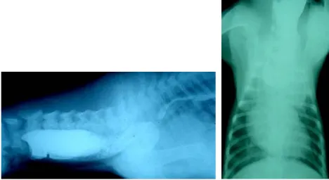
food a few minutes after eating. Thoracic ausculta-tion revealed a harsh inspiratory stridor at the level of the major bronchi, but the lungs were clear. A continuous machine like murmur was heard from all the thoracic fields, with the greatest amplitude being noted over the aorta and lungs. The ECG was normal except for a slightly prolonged P wave. Oesophagraphy revealed oesophageal dilatation cranial to the heart and constriction at the level of third rib (Figure 1). Oesophagoscopy revealed the oesophagus as a terminating blind pouch near the heart base. The blood was submitted to a complete blood count (CBC) and chemistry screening (CS) but no hematological or biochemical abnormalities were found.
A presumptive diagnosis was made as PRAA on the basis of the history, clinical signs, physi-cal examination findings, oesophagraphy and oesophagoscopy. The definitive diagnosis was made after surgical exploration as PRAA with left LA and an aberrant left SA.
The dog was premedicated with atropine sulphate (Atropine Sulfate®, Huons Co. Ltd., Korea) 0.05 mg per kg, SC. Anaesthesia was induced using thiopen-tone sodium (Thionyl®, Daehan Pharmaceutical Co. Ltd., Korea) 25 mg/kg, i.v. and maintained with enflurane and oxygen delivered through a cuffed endotracheal tube. Supportive fluid therapy was
[image:2.595.63.531.462.718.2]maintained throughout the procedure and cepha-lothin sodium (Cephacepha-lothin Sodium®, Kyongbo Pharmaceutical Co. Ltd., Korea) 25 mg/kg, i.v. was administered at the time of induction. The thoracic cavity was entered through a left lateral thoracot-omy incision at the level of the 4th left intercostal space. The heart was exposed with its pleural cover-ing and the vascular rcover-ing anomaly was identified. The oesophagus was found dually compressed, first by a complete ring formed with PRAA and persist-ent left LA, and secondly with a partial ring formed by the aberrant left SA. The left SA originated from the PRAA and compressed the oesophagus on its dorsal aspect during its course from the right to the left. The left vagus nerve, phrenic nerve and recur-rent laryngeal nerve were isolated and retracted ventrally by placement of a temporary ligature. The pleura was incised to expose the LA. Transfixing sutures were placed at both ends of the ligamentum and then it was transected. The oesophagus was then relieved from the first ring and dissected a few centimeters in each direction. There were several necrotic and ulcerative lesions on the oesophagus. The left SA was not manipulated, the necrosed part of the oesophagus was resected and an end-to-end anastomosis was performed. During this the oesophagus was made free from the partial ring of left SA. The temporary sutures were removed
from the nerves. The lungs were repositioned and inflated to eliminate atelectasis. Tension sutures were placed at 1/2 inch intervals behind the 4th rib and in front of the 5th rib. A pleural cavity drain-age tube was inserted through the 6th intercostal space. Then the tension sutures were tied and the wound was closed in a usual manner.
The postoperative treatment was given with ce-phalothin sodium (Cece-phalothin Sodium®, Kyongbo Pharmaceutical Co. Ltd., Korea) 25 mg/kg, i.v., every 8 hrs, and hydromorphone hydrochlo-ride (Hydromorphone Hydrochlohydrochlo-ride®, Mayne Pharmaceutical Co. Ltd., USA) 0.1 mg/kg, i.v., every 6 hrs for 1 week. The chest tube was removed after 3 days. The dog was maintained on Hartmann’s solution with 5% dextrose (Hartmann’s Dex Inj®, Daehan Pharmaceutical Co. Ltd., Korea) 2 ml/kg/h, i.v. for 4 days. Upright feeding was resumed from the 5th postoperative day with some baby foods 5–6 times daily for 2 weeks, after then the dog was fed with small amount of dog foods 4–5 times daily in a normal position.
One day after surgery, the dog looked bright, alert and eager to eat. Postoperatively, as the dog was maintained on intravenous fluids for 4 days, it was not possible to evaluate regurgitation dur-ing this period. However, no episodes of regurgi-tation were noticed during upright feeding stance
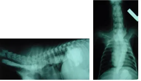
or during feeding at normal position. Fluoroscopic examination 2 days after surgery revealed a normal oesophagus caudal to the anastomosis but the cra-nial part still remained dilated. The motility was normal at the caudal part whereas the amplitude was much less at the cranial part of the oesopha-gus. Before discharge, radiographic (Figure 2) and fluoroscopic examinations were performed and the oesophagus was found to be almost normal except for a little dilatation.
The follow up information was obtained by means of telephone conversations with the dog’s owner up to 10 month after the surgical correction. Fluoroscopic examination after 4 months revealed a normal state of the oesophagus except for a very little dilatation and by this time the dog gained 3 kg body weight. After the surgical correction the dog recovered from regurgitation and regained nearly full function of her oesophagus without any notice-able complications during a 10 months follow-up period.
DISCUSSION
PRAA with retention of left LA along with an aberrant left SA is an unusual type of vascular ring anomaly in dogs (Ketz et al., 2001). During
[image:3.595.66.526.455.720.2]onic development, the aorta, LA and left SA develop from the left aortic arches, and thus, malformations in their development have been related to the right aortic arches (Helphrey, 1993; Ricardo et al., 2001). The PRAA is a developmental anomaly in which the aorta is formed by the right fourth aortic arch instead of the left fourth aortic arch. In this dog, the oesophagus and trachea were encircled by a vascu-lar ring consisting of the aorta on the right, the pul-monary trunk and base of the heart ventrally and the ligamentum arteriosum on the left; just cranial to this the oesophagus was dually compressed by an incomplete ring formed with the aberrant left sub-clavian artery during its course from the right to the left mediastinum. These findings were in consistent with the previous report of similar anomaly in tiger (Ketz et al., 2001). The anomaly was actually more of a gastrointestinal disorder than a heart disease because it often resulted in regurgitation rather than cardiac dysfunction. The dogs with vascular ring anomaly usually have history of postprandial regurgitation of solid foods after weaning (Van Den Ingh and Van Der Linde-Sipman, 1974; Helphrey, 1993). They are stunted, thin, unthrifty and much smaller than the littermates. The age signalment, history and clinical signs in this case were consist-ent with the previous reports of PRAA (Holmberg and Presnell, 1979; Ellison, 1990; Muldoon et al., 1997; Isako et al., 2000; Ketz et al., 2001). The con-servative medical treatment (liquid diet and sup-portive care) has been shown to be unrewarding (Ellison, 1980; VanGundy, 1989). Therefore, it was decided to perform an exploratory thoracotomy via the left 4th intercostal space (Fingeroth and Fossum, 1987; Fingeroth, 1993; Holt et al., 2000; Ketz et al., 2001). Some of the previous reports have suggested that age of the dog at the time of surgical correction of PRAA is an important factor in the long-term prognosis (Ellison, 1980; Berry et al., 1984; Muldoon et al., 1997) and early surgical intervention has been recommended, because it has been thought that oesophageal dilatation and mo-tility disorders would worsen and possibly become irreversible if surgery is delayed (Muldoon et al., 1997). In the present case, the dog was 10 weeks old at the time of surgical correction and the long-term result was satisfactory. The necrosed part of the oesophagus was resected and anastomosed. During this procedure, the oesophagus was made free from the partial ring fomed by the aberrant left SA, so the aberrant left SA was not manipulated. Ketz et al. (2001) also did not manipulate the aberrant left
SA as it did not cause much significant compromise of the esophagus. There have been some differ-ences for the reported outcomes of the long-term results of the surgical correction of PRAA. Shires and Liu (1981) reported 9% dogs did not have any long-term clinical abnormalities or complications; whereas, Muldoon et al. (1997) reported that 92% dogs did not regurgitated at the time of long-term follow up. However, in our case, the dog responded well to the surgical correction and regained nearly full function of the oesophagus. There were no fur-ther episodes of regurgitation and the dog gradually gained weight. The vascular ring anomaly should be considered as a probable cause of postprandial regurgitation with oesophageal dilatation and stric-ture after weaning in any young dog.
REFERENCES
Berry A.P., Brouwer G.L., Tennant B.J. (1984): Persistent right aortic arch in kitten. Veterinary Record, 114, 336–337.
Ellison G.W. (1980): Vascular ring anomalies in the dog and cat. Compendium on Continuing Education for the Veterinary Practitioners, 2, 693–705.
Ellison G.W. (1990): Surgical correction of persistent right aortic arch. In: Bojrab M.J. (ed.): Current Tech-niques in Small Animal Surgery. 3rd ed. Lea & Febiger,
Philadelphia. 508–512 pp.
Fingeroth J.M. (1993): Surgical diseases of the oesophagus. In: Slatter D.H. (ed.): Textbook of Small Animal Surgery. 2nd ed. W.B. Saunders Co, Philadelphia. 538–544 pp.
Fingeroth J.M., Fossum T.W. (1987): Late-onset regur-gitation associated with persistent right aortic arch in two dogs. Journal of American Veterinary Medical As-sociation, 101, 981–983.
Helphrey M. (1993): Vascular ring anomalies. In: Bojrab M.J. (ed.): Disease Mechanisms in Small Animal Sur-gery. 2nd ed. Lea & Febiger, Philadelphia. 350–354 pp.
Holmberg D.L., Presnell K.R. (1979): Vascular ring anomalies: Case report and brief review. Canadian Veterinary Journal, 20, 78–81.
Holt D., Heldmann E., Michel K., Buchanan J.W. (2000): Oesophageal obstruction caused by a left aortic arch and an anomalous right patent ductus arteriosus in two German shepherd littermates. Veterinary Surgery, 29, 264–270.
Corresponding Author:
Dr. Nam-Soo Kim, DVM, MS, PhD., Associate Professor, Chonbuk National University, College of Veterinary Medicine, Department of Surgery, and Director of Animal Medical Centre, Jeonju 561-756, Republic of Korea Tel. +82 63 270 2800, fax +82 63 270 3778, e-mail: namsoo@chonbuk.ac.kr
Ketz C.J., Radlinsky M., Armbrust L., Carpenter J.W., Isaza R. (2001): Persistent right aortic arch and aber-rant left subclavian artery in a white Bengal tiger ( Pan-thera tigris). Journal of Zoo and Wildlife Medicine, 32, 268–271.
Koc Y., Turgut K., Sen I., Alkan F., Birdane F.M. (2004): Persistent right aortic arch and its surgical correction in a dog. Turkish Journal of Veterinary and Animal Science, 28, 441–446.
MacPhail C.M., Monnet E., Twedt D.C. (2001): Thora-coscopic correction of persistent right aortic arch in a dog. Journal of American Animal Hospital Associa-tion, 37, 577–581.
Muldoon M.M., Birchard S.J., Ellison G.W. (1997): Long-term results of surgical correction of persistent right aortic arch in dogs: 25 cases (1980–1995). Jour-nal of American Veterinary Medical Association, 210, 1761–1763.
Ricardo C., Augusto A., Canavese S., Marcose A., Ticona E., Fernandes M., Rita M., Singereti F. (2001): Double aortic arch in a dog (Canis familiaris): A case report. Anatomia, Histologia, Embryologia, 30, 379–381. Shires P.K., Liu W. (1981): Persistent right aortic arch in
dogs: A long-term follow-up after surgical correction. Journal of American Animal Hospital Association, 17, 773–776.
Van Den Ingh T.S., Van Der Linde-Sipman J.S. (1974): Vascular rings in the dog. Journal of American Vet-erinary Medical Association, 164, 939–941.
VanGundy T. (1989): Vascular ring anomalies. Compen-dium on Continuing Education for the Veterinary Practitioners, 11, 36–48.
Yarim M., Gultiken M.E., Ozturk S., Sahal M., Bumin A. (1999): Double aortic arch in a Siamese cat. Veterinary Pathology, 36, 340–341.