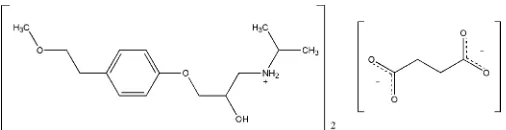
{2-Hydroxy-3-[4-(2-methoxyethyl)-phenoxy]propyl}isopropylammonium
hemisuccinate
Gianluca Bartolucci,aBruno Bruni,aSilvia A. Coranaand Massimo Di Vairab*
aDipartimento di Scienze Farmaceutiche, Universita´ di Firenze, Via U. Schiff 6,
I-50019 Sesto Fiorentino, Firenze, Italy, andbDipartimento di Chimica, Universita´
di Firenze, Via della Lastruccia 3, I-50019 Sesto Fiorentino, Firenze, Italy Correspondence e-mail: massimo.divaira@unifi.it
Received 11 May 2009; accepted 16 May 2009
Key indicators: single-crystal X-ray study;T= 200 K; mean(C–C) = 0.002 A˚; disorder in main residue;Rfactor = 0.045;wRfactor = 0.124; data-to-parameter ratio = 15.1.
Metoprolol, a widely used adrenoreceptor blocking drug, is commonly administered as the succinate or tartrate salt. The structure of metoprolol succinate, C15H26NO3
+
0.5C4H4O4 2
, is characterized by the presence of ribbons in which cations, generated byN-protonation of the metoprolol molecules, are hydrogen bonded to succinate anions. The dicarboxylic acid transfers its H atoms to two metoprolol molecules; the asymmetric unit contains one cation and half an anion, the latter possessing twofold rotational symmetry. There are localized nets of O—H O and N—H O hydrogen bonds along a ribbon, within centrosymmetric arrangements formed by pairs of metoprolol cations and pairs of anions, each of the latter contributing with one of its carboxyl groups to the localized net. This arrangement is repeated along the ribbon by the operation of the twofold axis bisecting the anion, as well as by the lattice translation.
Related literature
For general information on the medical applications of metoprolol, see: Benfieldet al.(1986); Moses & Borer (1981); Brogdenet al.(1977); Hainer & Sugg (2007); Ragnarssonet al. (1987); Sandberget al.(1988).
Experimental
Crystal data
C15H26NO3+0.5C4H4O42
Mr= 326.40 Monoclinic,C2=c a= 26.2630 (4) A˚
b= 7.9396 (2) A˚
c= 17.4629 (4) A˚
= 107.348 (2)
V= 3475.68 (13) A˚3
Z= 8
CuKradiation
= 0.75 mm1
T= 200 K
0.600.200.06 mm
Data collection
Oxford Diffraction Xcalibur PX Ultra CCD diffractometer Absorption correction: multi-scan
(ABSPACKinCrysAlisPro RED; Oxford Diffraction, 2006)
Tmin= 0.732,Tmax= 0.956
22961 measured reflections 3408 independent reflections 3108 reflections withI> 2(I)
Rint= 0.028
Refinement
R[F2> 2(F2)] = 0.045
wR(F2) = 0.124
S= 1.06 3408 reflections 226 parameters
12 restraints
H-atom parameters constrained
max= 0.21 e A˚3
[image:1.610.45.298.638.703.2]min=0.19 e A˚3
Table 1
Hydrogen-bond geometry (A˚ ,).
D—H A D—H H A D A D—H A
O2—H2O O4i
0.84 1.88 2.7231 (15) 179
N—H2N O4ii
0.92 1.89 2.7961 (16) 170
N—H1N O5i 0.92 1.85 2.7448 (15) 162
Symmetry codes: (i)x;y;z1
2; (ii)xþ1;y;zþ32.
Data collection:CrysAlisPro CCD(Oxford Diffraction, 2006); cell refinement: CrysAlisPro CCD; data reduction: CrysAlisPro RED
(Oxford Diffraction, 2006); program(s) used to solve structure:SIR97
(Altomare et al., 1999); program(s) used to refine structure:
SHELXL97 (Sheldrick, 2008); molecular graphics: ORTEP-3
(Farrugia, 1997) and PLATON (Spek, 2009); software used to prepare material for publication: SHELXL97, WinGX (Farrugia, 1999) andPARST(Nardelli, 1995).
The authors acknowledge financial support from the Italian Ministero dell’Istruzione, dell’Universita´ e della Ricerca.
Supplementary data and figures for this paper are available from the IUCr electronic archives (Reference: PK2162).
References
Altomare, A., Burla, M. C., Camalli, M., Cascarano, G. L., Giacovazzo, C., Guagliardi, A., Moliterni, A. G. G., Polidori, G. & Spagna, R. (1999).J. Appl. Cryst.32, 115–119.
Benfield, P., Clissold, S. P. & Brogden, R. N. (1986).Drugs,31, 376–429. Brogden, R. N., Heel, R. C., Speight, T. M. & Avery, G. S. (1977).Drugs,14,
321–348.
Farrugia, L. J. (1997).J. Appl. Cryst.30, 565. Farrugia, L. J. (1999).J. Appl. Cryst.32, 837–838.
Hainer, J. W. & Sugg, J. (2007).Vasc. Heal. Risk Manag.3, 279–288. Moses, J. W. & Borer, J. S. (1981).Dis. Mon.27, 1–61.
Nardelli, M. (1995).J. Appl. Cryst.28, 659.
Oxford Diffraction (2006). CrysAlisPro CCD and CrysAlisPro RED
(includingABSPACK). Oxford Diffraction Ltd, Abingdon, Oxfordshire, England.
organic compounds
o1364
Bartolucciet al. doi:10.1107/S160053680901856X Acta Cryst.(2009). E65, o1364–o1365Acta Crystallographica Section E
Structure Reports Online
Ind. Pharm.131495–1509.
Sandberg, A., Blomqvist, I., Jonsson, U. E. & Lundborg, P. (1988).Eur. J. Clin. Pharmacol.33, S9–14.
supporting information
sup-1
Acta Cryst. (2009). E65, o1364–o1365
supporting information
Acta Cryst. (2009). E65, o1364–o1365 [doi:10.1107/S160053680901856X]
{2-Hydroxy-3-[4-(2-methoxyethyl)phenoxy]propyl}isopropylammonium
hemisuccinate
Gianluca Bartolucci, Bruno Bruni, Silvia A. Coran and Massimo Di Vaira
S1. Comment
Metoprolol, (±)-1-isopropylamino-3-[4-(2-methoxy-ethyl)-phenoxy]- propan-2-ol, is a β1-selective adrenoreceptor
blocking drug, widely used in the treatments of hypertension, angina pectoris and heart failure (Benfield et al., 1986;
Moses & Borer, 1981; Brogden et al., 1977). The active substance is provided as metoprolol succinate or tartrate for oral
administration, available as extended–release tablets (Hainer & Sugg, 2007; Ragnarsson et al., 1987; Sandberg et al.,
1988). Although this drug has been in use for some time, a solid-state structure determination was not yet available.
Suitable crystals were obtained for the succinate of the active substance.
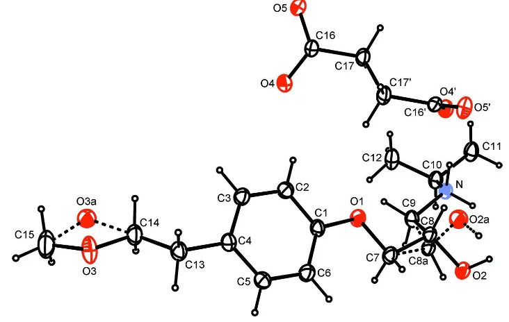
The structure of metoprolol succinate consists of cations formed by the N–protonated metoprolol molecule and of
succinate dianions, in 2:1 ratio. The asymmetric unit (Fig. 1) contains one metoprolol cation and the symmetry–
independent part of the succinate anion, the whole anion possessing two–fold rotational symmetry. Disorder affecting the
positions of the ether oxygen O3 and of the hydroxyl oxygen O2 was accounted for. The hydrogen atoms were included
in geometrically generated positions, although most of them, including the two ammonium H atoms, could be clearly
identified in difference Fourier maps. Consistent with a complete deprotonation of the dicarboxylic acid, the lengths of
the two carboxylate C—O bonds are similar. That formed by the O4 atom, which participates in two hydrogen bonds (see
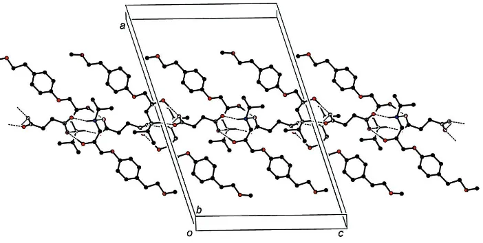
below), being only slightly larger (by 0.026 (2) Å) than the other one. In the structure, there are ribbons of hydrogen–
bonded ions parallel to the c axis (Fig. 2), characterized by the presence of centrosymmetric arrangements where pairs of
cations interact with pairs of carboxylate groups belonging to distinct anions. Contiguous arrangements of this type are
related to each other along the ribbon by the two–fold rotation axis and two of these contiguous arrangements form the
repeat motif in the c direction. There are no hydrogen–bond linkages between the ribbons. In detail, the metoprolol cation
forms hydrogen bonds to the two O atoms of a carboxylate group through its hydroxyl group (O2···O4i = 2.723 (2) Å, O2
—H2O···O4i = 179.4°; symmetry code (i): x, - y, -1/2 + z) and through an ammonium N—H bond (N···O5i = 2.745 (2) Å,
N—H1N···O5i = 162.3°). The same metoprolol cation is furthermore linked to the second anion in the centrosymmetric
arrangement along the ribbon, via the other N—H bond (N···O4ii = 2.796 (2) Å, N—H2N···O4ii = 169.7°; symmetry code
(ii): 1 - x, y, 3/2 - z). In this way, each metoprolol cation forms hydrogen bonds to two anions and each succinate anion
accepts hydrogen bonds from four cations, through its two carboxylate groups. No carbon atom of the phenyl group
deviates by more than 0.005 (1) Å from the best plane through the ring and the O1 and C13 atoms deviate respectively by
0.014 (2) Å and 0.009 (2) Å from it. The dihedral angle between the planes through the two parts of the anion, namely
atoms O4, O5, C16 and C17 and the symmetry–related ones, is 82.67 (5)° and the torsion angle through the carbon–
Samples of metoprolol succinate were kindly provided by SIMS (SIMS srl, Reggello Firenze, Italy). Crystals of the
compound, suitable for X-ray diffraction analysis, were obtained by slow evaporation from 3:1 methanol:octanol
solutions.
S3. Refinement
Hydrogen atoms were in geometrically generated positions, riding, and the constraint U(H) = 1.2Ueq(C,N) was applied on
the hydrogen temperature factors [U(H) = 1.5Ueq(C,O) for the H atoms of the methyl and hydroxyl groups]. It appears
that a 2.03 Å H···H contact involving the H2′ hydrogen of the disordered hydroxyl group, belonging to the fraction with
0.09 occupancy, whose position was (necessarily) geometrically generated, may be ignored, considering that it would be
easily released if the hydroxyl O—H bond were allowed to rotate.
A small number (12) of restraints were employed to ensure that the geometry and displacement parameters of the
[image:4.610.120.486.272.504.2]minor-component disordered atoms maintained chemically reasonable values.
Figure 1
A view of the two ions in the structure of the title compound. The asymmetric unit comprises one metoprolol cation and a
half succinate anion, as the latter lies in a site with two–fold rotational symmetry. Primed atoms are related by the
operation 1 - x, y, 3/2 - z. Displacement ellipsoids are drawn at the 30% probability level. Minor component disordered
atoms are denoted by labels with the trailing letter a and the bonds to which those atoms participate are denoted by
dashed lines. For the methyl and methylene groups affected by disorder only the hydrogen atoms belonging to the major
supporting information
sup-3
[image:5.610.133.484.70.247.2]Acta Cryst. (2009). E65, o1364–o1365
Figure 2
A view, approximately along b, of one of the ribbons, parallel to the c axis direction. Hydrogen bonds are denoted by
dashed lines. Only the hydrogen atoms involved in the formation of hydrogen bonds and only the major fractions in the
parts affected by disorder are shown.
{2-hydroxy-3-[4-(2-methoxyethyl)phenoxy]propyl}isopropylammonium hemisuccinate
Crystal data
C15H26NO3+·0.5C4H4O42− Mr = 326.40
Monoclinic, C2/c
Hall symbol: -C 2yc
a = 26.2630 (4) Å
b = 7.9396 (2) Å
c = 17.4629 (4) Å
β = 107.348 (2)°
V = 3475.68 (13) Å3 Z = 8
F(000) = 1416
Dx = 1.248 Mg m−3
Cu Kα radiation, λ = 1.54178 Å Cell parameters from 14350 reflections
θ = 5.0–72.4°
µ = 0.75 mm−1 T = 200 K
Elongated plate, colorless 0.60 × 0.20 × 0.06 mm
Data collection
Oxford Diffraction Xcalibur PX Ultra CCD diffractometer
Radiation source: fine-focus sealed tube Oxford Diffraction Enhance ULTRA assembly
monochromator
Detector resolution: 8.1241 pixels mm-1 ω scans
Absorption correction: multi-scan
(ABSPACK in CrysAlis PRO RED; Oxford Diffraction, 2006)
Tmin = 0.732, Tmax = 0.956
22961 measured reflections 3408 independent reflections 3108 reflections with I > 2σ(I)
Rint = 0.028
θmax = 72.7°, θmin = 5.3° h = −32→32
k = −9→9
l = −21→18
Refinement
Refinement on F2
Least-squares matrix: full
R[F2 > 2σ(F2)] = 0.045 wR(F2) = 0.124 S = 1.06 3408 reflections 226 parameters
12 restraints
Primary atom site location: structure-invariant direct methods
Secondary atom site location: difference Fourier map
w = 1/[σ2(F
o2) + (0.0619P)2 + 2.6692P]
where P = (Fo2 + 2Fc2)/3
(Δ/σ)max < 0.001
Δρmax = 0.21 e Å−3
Extinction correction: SHELXL97 (Sheldrick, 2008), Fc*=kFc[1+0.001xFc2λ3/sin(2θ)]-1/4
Extinction coefficient: 0.00076 (10)
Special details
Geometry. All e.s.d.'s (except the e.s.d. in the dihedral angle between two l.s. planes) are estimated using the full
covariance matrix. The cell e.s.d.'s are taken into account individually in the estimation of e.s.d.'s in distances, angles and torsion angles; correlations between e.s.d.'s in cell parameters are only used when they are defined by crystal symmetry. An approximate (isotropic) treatment of cell e.s.d.'s is used for estimating e.s.d.'s involving l.s. planes.
Refinement. Refinement of F2 against ALL reflections. The weighted R-factor wR and goodness of fit S are based on F2,
conventional R-factors R are based on F, with F set to zero for negative F2. The threshold expression of F2 > 2σ(F2) is
used only for calculating R-factors(gt) etc. and is not relevant to the choice of reflections for refinement. R-factors based on F2 are statistically about twice as large as those based on F, and R- factors based on ALL data will be even larger.
Fractional atomic coordinates and isotropic or equivalent isotropic displacement parameters (Å2)
x y z Uiso*/Ueq Occ. (<1)
C1 0.32938 (5) 0.08129 (18) 0.64968 (8) 0.0315 (3) C2 0.34141 (5) 0.01034 (19) 0.72575 (9) 0.0349 (3) H2 0.3768 −0.0250 0.7526 0.042* C3 0.30174 (6) −0.00870 (19) 0.76236 (9) 0.0350 (3) H3 0.3104 −0.0570 0.8144 0.042* C4 0.24918 (5) 0.04133 (18) 0.72460 (8) 0.0321 (3) C5 0.23807 (6) 0.1099 (2) 0.64854 (9) 0.0357 (3) H5 0.2026 0.1439 0.6213 0.043* C6 0.27752 (6) 0.1304 (2) 0.61068 (9) 0.0371 (3) H6 0.2689 0.1778 0.5584 0.044* O1 0.37140 (4) 0.09661 (14) 0.61815 (6) 0.0375 (3)
supporting information
sup-5
Acta Cryst. (2009). E65, o1364–o1365
H92′ 0.4532 0.2250 0.6267 0.042* 0.093 (3) N 0.50840 (4) 0.24135 (15) 0.56677 (7) 0.0311 (3)
H1N 0.5056 0.2456 0.5130 0.037* H2N 0.5178 0.1330 0.5839 0.037* C10 0.55297 (6) 0.3574 (2) 0.61095 (9) 0.0367 (3) H10 0.5452 0.4731 0.5877 0.044* C11 0.60366 (6) 0.2938 (2) 0.59633 (10) 0.0463 (4) H111 0.6133 0.1844 0.6226 0.069* H112 0.6326 0.3744 0.6184 0.069* H113 0.5979 0.2815 0.5385 0.069* C12 0.55708 (7) 0.3651 (3) 0.69955 (10) 0.0487 (4) H121 0.5605 0.2507 0.7216 0.073* H122 0.5249 0.4181 0.7061 0.073* H123 0.5885 0.4314 0.7281 0.073* C13 0.20539 (6) 0.0195 (2) 0.76350 (9) 0.0375 (3) H131 0.1726 0.0738 0.7291 0.045* H132 0.1979 −0.1023 0.7658 0.045*
C14 0.21768 (7) 0.0914 (2) 0.84700 (10) 0.0427 (4) 0.942 (5) H141 0.2478 0.0302 0.8844 0.051* 0.942 (5) H142 0.2273 0.2120 0.8472 0.051* 0.942 (5) O3 0.17048 (7) 0.0715 (2) 0.87015 (10) 0.0595 (6) 0.942 (5) C15 0.17273 (11) 0.1557 (3) 0.94139 (14) 0.0731 (7) 0.942 (5) H151 0.2006 0.1053 0.9859 0.110* 0.942 (5) H152 0.1382 0.1461 0.9519 0.110* 0.942 (5) H153 0.1809 0.2749 0.9363 0.110* 0.942 (5) C14′ 0.21768 (7) 0.0914 (2) 0.84700 (10) 0.0427 (4) 0.058 (5) H143 0.2570 0.0876 0.8696 0.051* 0.058 (5) H144 0.2080 0.2122 0.8398 0.051* 0.058 (5) O3′ 0.1975 (8) 0.0335 (14) 0.9084 (11) 0.038 (6)* 0.058 (5) C15′ 0.17273 (11) 0.1557 (3) 0.94139 (14) 0.0731 (7) 0.058 (5) H154 0.1401 0.1092 0.9494 0.110* 0.058 (5) H155 0.1636 0.2525 0.9050 0.110* 0.058 (5) H156 0.1969 0.1920 0.9931 0.110* 0.058 (5) C16 0.50084 (5) −0.18399 (18) 0.86196 (8) 0.0307 (3)
C17 0.52161 (6) −0.1855 (2) 0.78974 (8) 0.0380 (3) H171 0.5445 −0.0854 0.7923 0.046* H172 0.5441 −0.2867 0.7926 0.046* O4 0.46519 (4) −0.07642 (13) 0.86371 (6) 0.0372 (3) O5 0.52021 (4) −0.28659 (14) 0.91717 (6) 0.0430 (3)
Atomic displacement parameters (Å2)
U11 U22 U33 U12 U13 U23
O1 0.0293 (5) 0.0535 (6) 0.0340 (5) 0.0016 (4) 0.0157 (4) 0.0066 (4) C7 0.0311 (7) 0.0529 (9) 0.0300 (7) −0.0016 (6) 0.0136 (6) 0.0029 (6) C8 0.0310 (7) 0.0402 (11) 0.0265 (7) 0.0013 (7) 0.0125 (6) 0.0035 (7) O2 0.0367 (6) 0.0593 (8) 0.0300 (6) 0.0071 (5) 0.0157 (5) 0.0061 (5) C9 0.0323 (7) 0.0409 (8) 0.0359 (8) −0.0019 (6) 0.0170 (6) −0.0016 (6) C7′ 0.0311 (7) 0.0529 (9) 0.0300 (7) −0.0016 (6) 0.0136 (6) 0.0029 (6) C8′ 0.0310 (7) 0.0402 (11) 0.0265 (7) 0.0013 (7) 0.0125 (6) 0.0035 (7) C9′ 0.0323 (7) 0.0409 (8) 0.0359 (8) −0.0019 (6) 0.0170 (6) −0.0016 (6) N 0.0296 (6) 0.0377 (6) 0.0290 (6) −0.0030 (5) 0.0132 (5) −0.0003 (5) C10 0.0352 (7) 0.0433 (8) 0.0342 (7) −0.0086 (6) 0.0143 (6) −0.0051 (6) C11 0.0331 (8) 0.0720 (11) 0.0372 (8) −0.0087 (7) 0.0158 (6) −0.0074 (8) C12 0.0401 (8) 0.0724 (12) 0.0373 (9) −0.0115 (8) 0.0169 (7) −0.0167 (8) C13 0.0323 (7) 0.0452 (8) 0.0395 (8) −0.0027 (6) 0.0177 (6) 0.0009 (6) C14 0.0455 (8) 0.0484 (9) 0.0421 (8) 0.0002 (7) 0.0251 (7) 0.0009 (7) O3 0.0598 (11) 0.0758 (10) 0.0593 (11) −0.0063 (8) 0.0428 (9) −0.0093 (8) C15 0.1075 (18) 0.0667 (13) 0.0690 (14) 0.0119 (13) 0.0630 (14) 0.0016 (11) C14′ 0.0455 (8) 0.0484 (9) 0.0421 (8) 0.0002 (7) 0.0251 (7) 0.0009 (7) C15′ 0.1075 (18) 0.0667 (13) 0.0690 (14) 0.0119 (13) 0.0630 (14) 0.0016 (11) C16 0.0314 (7) 0.0360 (7) 0.0260 (7) −0.0034 (5) 0.0106 (5) −0.0038 (5) C17 0.0337 (7) 0.0535 (9) 0.0303 (7) 0.0025 (6) 0.0150 (6) 0.0017 (6) O4 0.0383 (5) 0.0404 (6) 0.0370 (6) 0.0025 (4) 0.0173 (4) −0.0002 (4) O5 0.0512 (6) 0.0502 (7) 0.0313 (5) 0.0110 (5) 0.0179 (5) 0.0078 (5)
Geometric parameters (Å, º)
supporting information
sup-7
Acta Cryst. (2009). E65, o1364–o1365
C9—H92 0.9900 C16—O4 1.2744 (17) C8′—O2′ 1.410 (9) C16—C17 1.5161 (18) C8′—H8′ 1.0000 C17—C17i 1.508 (3)
O2′—H2′ 0.8400 C17—H171 0.9900 N—C10 1.5086 (18) C17—H172 0.9900
O1—C1—C6 124.49 (13) C10—N—H2N 108.2 O1—C1—C2 115.91 (12) H1N—N—H2N 107.4 C6—C1—C2 119.60 (12) N—C10—C11 107.28 (12) C3—C2—C1 119.88 (13) N—C10—C12 110.72 (12) C3—C2—H2 120.1 C11—C10—C12 112.69 (13) C1—C2—H2 120.1 N—C10—H10 108.7 C2—C3—C4 121.64 (13) C11—C10—H10 108.7 C2—C3—H3 119.2 C12—C10—H10 108.7 C4—C3—H3 119.2 C10—C11—H111 109.5 C5—C4—C3 117.41 (12) C10—C11—H112 109.5 C5—C4—C13 120.40 (13) H111—C11—H112 109.5 C3—C4—C13 122.18 (13) C10—C11—H113 109.5 C4—C5—C6 121.80 (13) H111—C11—H113 109.5 C4—C5—H5 119.1 H112—C11—H113 109.5 C6—C5—H5 119.1 C10—C12—H121 109.5 C1—C6—C5 119.66 (13) C10—C12—H122 109.5 C1—C6—H6 120.2 H121—C12—H122 109.5 C5—C6—H6 120.2 C10—C12—H123 109.5 C1—O1—C7 117.42 (10) H121—C12—H123 109.5 O1—C7—C8 106.85 (12) H122—C12—H123 109.5 O1—C7—H71 110.4 C14—C13—C4 114.78 (12) C8—C7—H71 110.4 C14—C13—H131 108.6 O1—C7—H72 110.4 C4—C13—H131 108.6 C8—C7—H72 110.4 C14—C13—H132 108.6 H71—C7—H72 108.6 C4—C13—H132 108.6 O2—C8—C7 105.59 (12) H131—C13—H132 107.5 O2—C8—C9 111.85 (13) O3—C14—C13 106.20 (14) C7—C8—C9 110.43 (12) O3—C14—H141 110.5 O2—C8—H8 109.6 C13—C14—H141 110.5 C7—C8—H8 109.6 O3—C14—H142 110.5 C9—C8—H8 109.6 C13—C14—H142 110.5 N—C9—C8 110.19 (12) H141—C14—H142 108.7 N—C9—H91 109.6 C15—O3—C14 112.98 (17) C8—C9—H91 109.6 O5—C16—O4 123.48 (12) N—C9—H92 109.6 O5—C16—C17 118.19 (12) C8—C9—H92 109.6 O4—C16—C17 118.32 (12) H91—C9—H92 108.1 C17i—C17—C16 113.99 (15)
O2′—C8′—H8′ 113.4 C17i—C17—H171 108.8
C8′—O2′—H2′ 109.5 C16—C17—H171 108.8 C9—N—C10 116.31 (11) C17i—C17—H172 108.8
O1—C1—C2—C3 179.49 (13) O1—C7—C8—O2 173.83 (12) C6—C1—C2—C3 −0.8 (2) O1—C7—C8—C9 −65.09 (17) C1—C2—C3—C4 0.2 (2) O2—C8—C9—N −75.80 (16) C2—C3—C4—C5 0.5 (2) C7—C8—C9—N 166.91 (12) C2—C3—C4—C13 179.31 (14) C8—C9—N—C10 171.96 (12) C3—C4—C5—C6 −0.6 (2) C9—N—C10—C11 177.49 (12) C13—C4—C5—C6 −179.44 (14) C9—N—C10—C12 54.15 (17) O1—C1—C6—C5 −179.62 (14) C5—C4—C13—C14 −128.73 (16) C2—C1—C6—C5 0.7 (2) C3—C4—C13—C14 52.5 (2) C4—C5—C6—C1 0.0 (2) C4—C13—C14—O3 175.14 (14) C6—C1—O1—C7 1.5 (2) C13—C14—O3—C15 −170.64 (17) C2—C1—O1—C7 −178.87 (13) O5—C16—C17—C17i 132.10 (11)
C1—O1—C7—C8 −177.63 (12) O4—C16—C17—C17i −49.01 (14)
Symmetry code: (i) −x+1, y, −z+3/2.
Hydrogen-bond geometry (Å, º)
D—H···A D—H H···A D···A D—H···A
O2—H2O···O4ii 0.84 1.88 2.7231 (15) 179
N—H2N···O4i 0.92 1.89 2.7961 (16) 170
N—H1N···O5ii 0.92 1.85 2.7448 (15) 162