Penetrating eye injury in a dog: a case report
M. Lew
1, S. Lew
2, M. Drazek
1, A. Pomianowski
11Faculty of Veterinary Medicine, University of Warmia and Mazury, Olsztyn, Poland 2Faculty of Biology, University of Warmia and Mazury, Olsztyn, Poland
ABSTRACT: A four-year-old, male German Shepherd dog with severe pain in the left eye following a corneal perforation with a foreign body was examined. An ophthalmic examination revealed conjunctival hyperaemia and pancorneal dense oedema, preventing a diagnosis of deeper structures of the eye and lowered IOP. Vision testing was missing or impossible to detect. Ultrasonography showed a solid hyperechoic line protruding through the iris and lens into the vitreous and minor posterior lens displacement. The dog qualified for immediate surgical treat-ment. Intraoperative ophthalmic examination revealed a rupture of the anterior hyaloid membrane with vitreous herniation, posterior lens subluxation, lens capsule rupture and a torn iris. Partial iridectomy and intracapsular lens extraction (ICLE) was conducted. Slit-lamp biomicroscopy revealed iridodonesis and a gradual reduction of the corneal oedema, leading to complete transparency in the bottom two-thirds of the area on Day 12 after surgery. Direct and consensual PLR was sluggish and the dazzle reflex was positive. An electroretinographic examination confirmed normal activity of the retina.
Keywords: acute ocular trauma; intraocular foreign body; dog
Penetrating eye injuries, including corneal wounds and corneal and lens lacerations, are common in small animal ophthalmology practice, resulting from the cornea being scratched by a cat claw (Spiess et al. 1996; Paulsen and Kass 2012), a foreign body such as grass awn (Bussanich and Rootman 1981; Cullen and Grahn 2005), rose thorns (Dean 2004), fragments of plant material (Gelatt 1974; Crispin 1986; Gionfriddo and Chen 2011), porcupine quills (Williams and Wilcock 1988; Grahn et al. 1995; Sandmeyer et al. 2007), lead or air gun pel-lets (Slatter and Bryan 1972; Schmidt et al. 1975; Sansom and Labruyere 2012) and sticks (Martin 2010a). Perforating wounds require immedi-ate treatment (Gelatt and Gelatt 2001a; Hendrix 2007) due both to quite severe pain as a result of abundant sensory innervation (Barrett et al. 1991; Marfurt et al. 2001) and to a number of potential complications, such as secondary bacterial (Malar and Dubielzig 1995; Klatte et al. 2012; Bell et al. 2013) and fungal infections (Andrew 2003; Chew et al. 2010), as well as due to traumatic lens cap-sule rupture (Wilcock and Peiffer 1987; Davidson et al. 1991). The release of previously sequestered lens protein to the anterior chamber activates the
dynamic development of immune response, lead-ing to phacoclastic anterior uveitis or even panu-veitis (Bussanich and Rootman 1981; Davidson et al. 1991). Immediate treatment is important also in order to avoid the so called Septic Implantation Syndrome. Bell et al. (2013) pointed to an essential role of the inoculation of bacteria into the lens cor-tex resulting in secondary development of septic endophthalmitis often within no more than one week.
This case report describes the clinical presenta-tion, electroretinographic findings as well as sur-gical treatment in a German Shepherd dog with perforating corneal injury with traumatic lens rup-ture and vitreous hernia. To the best knowledge of the authors, no case of vision retention after such a multiple eye injury has ever been reported.
Case description
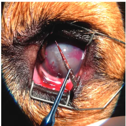
A four-year-old, male German Shepherd dog was referred to the ophthalmologic division of our clinic with severe pain in the left eye. The owners reported that the injury had occurred six to seven hours before. A clinical examination of the left eye (OS) revealed blepharospasm, epiphora, prolapse of the third eyelid and the presence of a foreign body (thorn) stuck in the medial part of the cor-nea (Figure 1). Digital slit-lamp biomicroscopy OS (Hawk Eye, Dioptrix, France) revealed moderate conjunctival hyperaemia and pancorneal dense oedema, preventing an examination of deeper structures of the eye, including the fundus. An ophthalmologic examination of the right eye (OD) performed with slit-lamp biomicroscopy, a port-able indirect ophthalmoscope (Omega 500, Heine Instruments, Germany) with a 20 D power lens (Volk Optical, USA) and direct ophthalmoscopy
[image:2.595.312.531.480.699.2](Beta 200S, Heine Instruments, Germany) revealed no anterior segment abnormalities. Vision testing, including menace response, dazzle reflex, direct and consensual pupillary light reflexes (PLR), were nor-mal for the OD and missing or impossible to detect in the OS. Further examination was performed un-der local anaesthesia induced with proxymetacaine hydrochloride (Alcaine, Alcon-Couvreur, Belgium) administered at a dose of two drops at 5-min in-tervals. Intraocular pressure (IOP) measured with an applanation tonometer (TonoPen XL, Reichert Technologies, USA) did not reveal any recording in the OS indicating a valve under 5 mmHg, which was below the measuring capacity of the device. IOP OD reached 22 mmHg. Fluorescein staining for corneal uptake was positive in the OS – around the foreign body – and negative in the OD. Ultrasonography (USG) was performed under premedication due to eye pain and severe blepharospasm. Atropine sul-phate was administered (Atropinum sulfuricum WZF, Polfa, Poland) at a dose of 0.04 mg/kg of body weight (b.w.) s.c., butorphanol (Dolorex, Intervet International B.V., Holland) at a dose of 0.2 mg/kg b.w. i.m. and medetomidine hydrochloride (Do- mitor, Orion Pharma Animal Health, Finland) at a dose of 20 µg/kg b.w. i.m. B-mode USG of the orbital and ocular structures was performed with a 10 MHz transducer (Ultrascan Imaging System, Alcon, USA). All scans were performed with a probe
[image:2.595.63.292.480.713.2]Figure 1. Foreign body (thorn) in the medial part of the cornea OS, moderate conjunctival hyperaemia and pan-corneal dense oedema
placed directly on the sclera and partially on the cornea, coated with 1.5% hypromellose (Goniovisc, HUB Pharmaceuticals, USA) as a coupling gel. An ophthalmic ultrasound OS showed a solid hyper-echoic line protruding through the iris and lens into the vitreous and a minor posterior lens displace-ment. The USG in the OD was unremarkable. The dog qualified for immediate surgical treatment. The total blood count and serum biochemical profile were within normal limits.
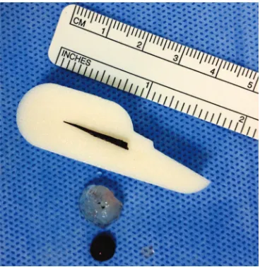
Surgical management. Before the surgery, the dog was anesthetised with propofol at a dose of 5 mg/kg b.w. i.v. (Scanofol, ScanVet, Poland). General anaesthesia was maintained with isoflu-rane (Aerisoflu-rane, Baxter Polska, Poland) at a con-centration of 1–1.5%. The left periocular skin and conjunctiva were flushed with povidone–iodine di-luted in saline in a 1 : 50 ratio. After a blepharostat was put in place, the cornea and conjunctiva were cleansed with sterile buffered-saline solution (BSS) to remove foreign material residues (AquaCrom, Croma-Pharma, Poland). The foreign body was re-moved with a vertical rotational movement with Bonn forceps. It was a thorn of 20 mm in length (Figures 2 and 3). The removal of the foreign body resulted in an opening in the structures that had been ruptured, including the anterior hyaloid membrane, posterior and anterior lens capsule, iris and the corneal wound. This resulted in a vitreous displacement into the anterior chamber, leakage of lens material from the damaged lens capsule, bleeding from the iridial vessels, aqueous humour outflow and the subsequent collapse of the
ante-rior chamber (Figure 2). Anteante-rior chamber per-ilimbal paracentesis was performed at 12 o’clock with a 3.2 mm slit-angled ophthalmic blade (Mani, Yamanashi, Japan). The accumulated tissue mate-rial originating from the torn iris and lens as well as hyphaema from the damaged iridial vessels were then flushed from the anterior and posterior cham-ber with a sterile BSS solution using an I/A cannula, Venturi pump system (Megatron S3 VIP, Geuder, Germany). High dispersive visceoclastics were used to restore the physiological depth of the anterior chamber (Eyefill HD, Croma-Pharma, Poland).
The restoration of partial transparency of the op-tical system in the anterior segment of the eyeball allowed for a more accurate evaluation of intraocu-lar injuries. These injuries included: a rupture of the anterior hyaloid membrane with Wieger’s ligament and herniation of the vitreous through the pupil into the anterior chamber, posterior cataractous lens sub-luxation with a typical aphakic crescent secondary to more than two-thirds zonular damage, posterior and anterior lens capsule rupture, traumatic iris tear at the pupillary margin and a puncture wound of the cornea.
[image:3.595.81.265.534.723.2]Partial iridectomy of the pupillary margin, which included a part of the torn iris, was performed (Figure 3). Bleeding from the iridial vessels was stopped with high-frequency bipolar wet-field co-agulation (Megatron S3 VIP, Geuder, Germany). Lack of sufficient visibility due to corneal oedema prevented a removal of the lens by phacoemulsifi-cation. It was decided to extend the incision of the cornea to approximately 150 degrees and perform intracapsular lens extraction (ICLE) (Figure 3).
[image:3.595.315.521.535.717.2]Figure 3. Removed thorn of 20 mm in length, a part of the torn iris and the lens material after ICLE
After lifting the cornea up with a viscoelastic and restoration of the anterior chamber, a paralimbal incision was closed with a single 9-0 nylon inter-rupted suture (Mani, Yamanashi, Japan), leaving a 2-mm opening. The next step was to remove the herniated vitreous from the pupil and the ante-rior chamber. A vitrectomy was performed with a Magnetic High-Speed drive combined with a double-bladed cutting head with the capacity of up to 4.000 cuts/min (Megatron S3 VIP, Geuder, Germany). After flushing the viceoclastics from the anterior chamber with an I/A cannula – Venturi pump system (Megatron S3 VIP, Geuder, Germany), the opening was sealed. The corneal wound was then closed with two single, interrupted sutures using 9-0 nylon.
Post-surgical management. Analgesic treat-ment was conducted with tramadol hydrochloride (Tramal 100, Polpharma S.A., Poland) at a dose of 4 mg/kg of b.w. i.m. for two days following the surgery. Topical ophthalmologic treatment was administered according to the following proce-dure: tobramycin 0.3% and dexamethasone 0.1% (Tobradex, Alcon-Couvreur, Belgium) q3h to both eyes (OU), phenylephrine hydrochloride 10% (Neo-Synephrine, Sanofi-Winthrop, USA) q12h OS, atropinum sulfuricum 1% (Polfa, Poland) q12h OS, dorzolamide hydrochloride (Trusopt, Merck Sharp&Dohme, France) q12h OS and dexpanthe-nol (Corneregel, Bausch and Lomb, Germany) q3h
[image:4.595.308.531.498.615.2]OS. The treatment procedure described above was continued for 14 days. Systemic treatment includ-ed a broad-spectrum antibiotic, i.e. amoxicillin (Betamox L.A., ScanVet, Poland), administered for 12 days and carprofen (Rimadyl, Pfizer Trading Polska, Poland) administered for three weeks. The measurements of IOP on Days 2, 5 and 12 after the surgery were 11, 8 and 7 mmHg OS and 18, 21 and 20 mmHg OD, respectively. Slit-lamp biomi-croscopy OS (Hawk Eye, Dioptrix, France) revealed iridodonesis and a gradual reduction of corneal oedema leading to complete transparency in the bottom two-thirds of the area on Day 12 after the surgery. Mild oedema was still found in the up-per one-third of the corneal surface and showed a progressive reduction (Figure 4). The results of slit-lamp biomicroscopy in the OD remained un-changed. Indirect ophthalmoscopy without a 20 D power lens and direct ophthalmoscopy in the OS did not reveal any lesions of the fundus. Single vitreous floaters/opacities were detected. The OD ophthalmoscopy results remained unchanged. A gonioscopic examination in the OS with a Koeppe lens with a 160° visual angle and 18 mm-diameter (Ocular Instruments, USA) revealed a deep ante-rior chamber and a wide iridocorneal angle. The OD was unremarkable. Direct and consensual PLR measured on Day 12 after the surgery was sluggish in the OS and normal in the OD. The dazzle reflex was positive in the OU. The menace response was
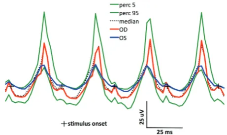
Figure 6.The dark-adapted, high-intensity (3 cd s/m2) flash
stimulated ERGs – a mixed rod-cone response. Normal baseline values of the implicit times and amplitudes in a control group of German Shepherd dogs with indicating the median (black, doted trace) and limits of normality using the 5th and 95th percentiles (green traces)
accord-ing to the protocol recommended by Ekesten et al. (2013). Calibration bars are denoted separately for each type of ERG response. Markers indicate the onset of the light stimulus (Figures 6, 7, 8)
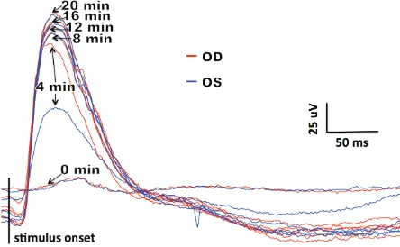
Figure 5. ERGs from the discussed case (four-year-old, male German Shepherd dog), obtained during the dynamic process of dark adaptation following low-inten-sity flash stimulation (0.03 cd s/m2), six times every 4 min
[image:4.595.64.286.516.653.2]difficult to note in the OS and was positive in the OD. Corneal sutures were removed on Day 12 after the surgery, immediately after electroretinographic examination.
Electroretinography. In order to verify the re-sults of vision tests and also due to a request sub-mitted by the owner, electroretinography (ERG) Easy VEP (Acrivet, Germany) was performed on Day 12 after the surgery. Atropine sulphate (Atropinum sulfuricum WZF, Polfa, Poland) at a dose of 0.04 mg/kg b.w. was used for premedica-tion and a combinapremedica-tion of xylazine (Vetaxyl, Vet-Agro, Poland) and ketamine (Vetketam, Vet-Vet-Agro, Poland) at a dose of 2 mg/kg b.w. and 5 mg/kg b.w., respectively, was used to induce general anaesthe-sia. The pupils were fully dilated with a tropicamide ophthalmic solution (Mydriacyl 0.5 %, Alcon Polska, Poland). An LED Flash 4W generator coupled with a Kooyman-Damhof electrode (Roland Consult, Germany) was used as a stimulator and was placed on the cornea covered with a 1.5% hypromellose so-lution (Goniovisc, HUB Pharmaceuticals, USA). A 0.3 mm stainless steel needle electrode was placed subcutaneously approximately 3 cm laterally from the temporal canthus as a reference electrode. A ground electrode was placed subcutaneously on the fronto-polar point (Fpz). The value of imped-ance between the electrodes was below 2 kOhm. Electroretinograms (ERGs) were performed under photopic and scotopic conditions. Rod function was evaluated during the dynamic process of dark adaptation following a standard flash stimulation (SF = 3 cd s/m2) reduced by two logarithmic units
(–20dB), six times every 4 min at 0, 4, 8, 12, 16 and 20 min, respectively (Figure 5). A single, mixed rod-cone response to SF was then recorded in dark adaptation (Figure 6). Cone-driven responses to SF after 10 min of light adaptation (Figure 7) were
ob-tained with a background light intensity of 30 cd/m2.
Eight responses were averaged in order to generate a result of the examination. A 31 Hz cone flicker response (Figure 8) was recorded under the same light conditions. The measurements of amplitudes and implicit times were taken according to the ca-nine ERG protocol recommended by the European College of Veterinary Ophthalmologists (ECVO) (Narfstom et al. 2002; Ekesten et al. 2013).
DISCUSSION AND CONCLUSIONS
[image:5.595.300.531.95.232.2]Corneal translucency is possible due to the co-existence of protective mechanisms against its ove-rhydration. The corneal epithelium protects the cornea against absorption of the aqueous humour via an active route through sodium-potassium ATPase pumps and passively by the tightness of cel-lular junctions called occluding junctions (Waring et al. 1982). In contrast to the well-regenerating epithelial cells, the regenerative capacity of the endothelial cells disappears with age (Gilger et al. 2007). In an adult dog, the mitotic activity of the endothelial cells is minimal and the replenishing of defects is achieved by enhancing the volume of cells and their migration (Befanis et al. 1981). Corneal epithelium mechanical injuries, which ex-ceed the repair capacity of endothelial cells result in permanent translucency defects. Perforation of the corneal stroma causes a release of hydrophilic glycosaminoglycans (GAG) which bind water both from the outside, such as in the lacrimal film, and from the inside originating from the aqueous hu-mour, causing corneal swelling (Gilger et al. 2007). In the present case, the damage to all protective mechanisms of the cornea caused by the thorn resulted in overhydration and pancorneal dense
[image:5.595.63.293.106.234.2]oedema within 6–7 h. If an ophthalmological exami-nation is performed a few hours after an injury, it is difficult to assess the depth of a corneal wound and the extent of intraocular damage (Hendrix 2007). A B-scan USG is generally recommended for ultra-sonographic diagnostics of the eye, and especially for imaging the lens, vitreous, chorioretinal layer and the sclera (Fielding 1992; Atta 1996). All changes in the vitreous cavity, such as foreign bodies and vitre-ous haemorrhage are diagnosed as echo-dense areas (Hendrix 2007). The correct volume and transpar-ency of the lens are regulated by the lens membrane and lens epithelial cells (LEC). Although the lens membrane is impermeable to albumins and globu-lins, it allows water and electrolytes to pass through. LEC with NA/K-ATPase pumps are located between the anterior lens capsule and the lens fibres (Mathias et al. 2010). A rupture of the lens capsule results in an inflow of Na+ ions to the interior of the lens,
which changes its osmolality and causes a second-ary inflow of water (Kinoshita 1974). Penetration of the aqueous humour into the lens fibres provokes their oedema, which results in a loss of transpar-ency within a few hours (Ofri 2008). A simultaneous inflow of neutrophils inside the lens and a release of lens proteins to the outside, leads to immune-mediate inflammation (Davidson et al. 1991; Gelatt and Gelatt 2001a), which is known as phacoclas-tic uveitis, perilenphacoclas-ticular fibroplasia or secondary glaucoma (Wilcock and Peiffer 1987). A traumatic lens capsule rupture mainly affects young animals and is located in the anterior and axial part of the lens (Wilcock and Peiffer 1987; Davidson et al. 1991; Davidson and Nelms 1999).
In the discussed case, uveitis could have been secondary to an auto-immune reaction and also a mechanical irritation caused by a posterior lens luxation (Curtis 1990). Secondary glaucoma and ocular hypertension may occur as a complica-tion of phacoclastic uveitis and lens subluxacomplica-tion as well as anterior hyaloid face damage. A pro-truded vitreous as well as a subluxated lens may cause a pupillary block, anterior displacement of the iris base and a narrowing of the ciliary cleft (Curtis et al. 1983; Bedford 1988). Vitreous dis-placement may additionally provoke obstruction at the trabecular meshwork level (Manning et al. 2006). These complications may cause a blockage of the aqueous outflow, a rapid IOP increase and quite severe pain (Curtis et al. 1983; Bedford 1988; Nasisse and Glover 1997). As discussed in this case,
posterior lens subluxation could have also caused retinal damage (Curtis et al. 1983; Bedford 1988). For these reasons, rapid removal of a displacement lens is commonly recommended (Glover et al. 1995; Pizzirani 1998).
The removal of a subluxated lens by phaco-emulsification (Curtis 1990; Santoro et al. 2003; Manning et al. 2006; Paulsen and Kass 2012) is less invasive than by ICLE (Glower et al. 1995; Nasisse and Glover 1997; Saroglu et al. 2007). In studies by Manning et al. (2006), long-term effects related to vision were better after lensectomy carried out with the phaco technique than after ICLE. In the present case, due to corneal oedema and a lack of sufficient visibility, it was decided to remove the subluxated lens by ICLE. A wide, 150-degree open-ing of the anterior chamber allowed for direct and precise visualisation of the surgical area.
In the case of perforating eye injuries, sudden decompression is the cause of uveal or iris prolapse that, together with fibrin, seal a defect (Hendrix 2007; Martin 2010a). In the present case, the thorn acted like a stopper that maintained relative sealing in the anterior chamber, lens and anterior vitre-ous face, which delayed and alleviated the induc-tion of an auto-immune response. This thorn also prevented a prolapse of the vitreous to the ante-rior chamber and reduced the risk of secondary retinal detachment. The removal of the foreign body resulted in the formation of a canal joining the vitreous with the external environment. The accumulation in the anterior chamber of a mass consisting of the vitreous, lens material and blood from the damaged iris, is suggestive of a poor prog-nosis (Hendrix 2007). The immediate flushing of the mass from the anterior chamber minimised the probability of all the above-mentioned complica-tions.
lac-eration with associated lens capsule disruption, it was demonstrated that the percentage of long-term complications, such as a loss of vision, was higher with surgical corneal and lens treatment than with pharmacological treatment. Obviously, this only applied to cases in which the range of corneal and lens damage allowed for conservative treatment. Minor lens capsule ruptures seal spontaneously as a result of the lens epithelial cells fibrous metapla-sia (Buschmann 1987; Davidson and Nelms 1999). In the case of disruptions of a lens capsule longer than 1.5 mm, an immediate lensectomy by phaco-emulsification is highly recommended due to the risk of phacoclastic uveitis (Davidson et al. 1991).
On Day 12 after the surgery, the observed direct and consensual PLR was sluggish in the OS. This could have been caused by a substantial intra-op-erative distension of the pupil that accompanied lensectomy by ICLE. In the studies by Yuguchi et al. (1999) with an iris retractor, it was observed that in patients with an intra-operatively distend-ed pupil up to a diameter of 5 mm, the PLR was diminished after the surgery. This phenomenon was explained by the permanent intra-operative damage of the sphincter muscle and a stronger post-operative inflammatory reaction (Yuguchi et al. 1999). Lensectomy by ICLE and anterior vitrec-tomy resulted in deepening of the anterior chamber and in iridodenesis as reported in the post-surgical ophthalmologic examination (Curtis 1990).
Anterior vitrectomy was the final stage of the described surgical procedure and was limited to the removal of the protruded vitreous from the anterior chamber and pupil up to the level of the anterior hyaloid face. This was to prevent the above-de-scribed papillary (Curtis et al. 1983; Bedford 1988; Gelatt and Gelatt 2001b) and potential trabecu-lar meshwork blockage (Manning et al. 2006). A similar pupillary block and a subsequent sudden increase in IOP up to 65 mmHg, was reported in the clinical case of a Boston Terrier puppy with a corneal perforation, iris prolapse and anterior lens capsule disruption (Denis 2002). In addition, the protruded vitreous extending through the pupil and incarcerated in a corneal wound contributes to-wards exacerbation of iritis, dyscoria and formation of an inflammatory membrane (Gelatt and Gelatt 2001b; Denis 2002). In the described case, a precise anterior vitrectomy resulted in a correct circulation of the aqueous humour and a lack of dramatic IOP increases. The OS ocular hypotension detected on
Days 2, 5 and 12 after the surgery was probably associated with anterior uveitis.
Penetrating eye injuries with lens and anterior hyaloid face damage caused by infectious agents such as foreign bodies may result in immune-mediated inflammation, hyphaema and vitreous liquefaction (Martin 2010b). Anterior uveitis, cat-aract and especially posterior lens capsule tears enhance this process (Gelatt and Gelatt 2001b). A small amount of blood originating from the torn iris could have remained in brief contact with the hyaloid, which also contributed to its liquefaction (Martin 2010b). Hemosiderin, as a product of blood decomposition, provokes macrophages to migrate inside the vitreal cavity, thereby contributing to a mild inflammation, which results in the forma-tion of a fibrinous membrane (Martin 2010b). The discussed processes could have been the cause of the single vitreous floaters/opacities observed on Day 12 after the surgery. The corneal oedema was progressively reduced and by Day 12 after the sur-gery it was limited to the upper one-third as a result of the 150-degree paralimbal corneal incision.
The above-described specific model of general anaesthesia for an ERG examination is routinely used in our clinic and is an element that is ap-plied to unify measurement conditions. The studies by Kommonen et al. (1988) and Jeong et al. (2009) showed that a combination of xylazine and ketamine resulted in the lowest changes in a-wave and b-wave implicit times and amplitudes. In addition, the combination of these anaesthet-ics does not induce lower rotation of the eyeball or myosis. The unification of ERG examinations requires that ERG analysis of the examined dogs is based on the normal values for the same breed and the same anaesthetic protocol (Narfstom et al. 2002). Normal baseline values of implicit time and amplitude in dark and light-adapted ERGs for a German Shepherd dog were established based on the clinic’s own data collected during screening examinations of eight healthy individuals of the same breed, which constituted a control group (Figures 6, 7, 8). In the discussed case, ERGs had implicit times in the OU and the amplitudes in the OD that approximated the normal baseline values, but the amplitudes in the OS were slightly lower (Figures 5, 6, 7, 8).
but also to restore the transparency of the optical system. The accompanying conservative treatment helped to reduce inflammatory processes, minimise complications in the retina and led to the preserva-tion of vision.
REFERENCES
Andrew SE (2003): Corneal fungal disease in small animals. Clinical Techniques in Small Animal Practice 18, 186–192. Atta HR (eds.) (1996): Ophthalmic Ultrasound, a Practical
Guide. 1st ed. Butterworth-Heinemann, Oxford. 164 pp. Barrett PM, Scagliotti RH, Merideth RE, Jackson PA, Alar-con FL (1991): Absolute corneal sensitivity and corneal trigeminal nerve anatomy in normal dogs. Progress in Veterinary and Comparative Ophthalmology 1, 245–254. Bedford PGC (1988): Primary lens luxation in the dog. In
Practice 10, 188–193.
Befanis P, Peiffer R, Brown D (1981): Endothelial repair of the canine cornea. American Journal of Veterinary Re-search 42, 590–595.
Bell CM, Pot SA, Dubielzig RR (2013): Septic implantation syndrome in dogs and cats: a distinct pattern of endoph-thalmitis with lenticular abscess. Veterinary Ophthalmol-ogy 16, 180–185.
Buschmann W (1987): Microsurgical treatment of lens cap-sule perforations. Part II: Clinical applications and re-sults. Ophthalmic Surgery 18, 276–282.
Bussanich MN, Rootman J (1981): Intraocular foreign body in a dog. Canadian Veterinary Journal 22, 207–210. Chew HF, Jungkind DL, Mah DY, Raber IM, Toll AD,
Tokarc-zyk MJ, Cohen EJ (2010): Post-traumatic fungal keratitis caused by Carpoligna species. Cornea 29, 449–452. Crispin S (1986): Removal of organic foreign bodies from
the canine anterior segment of the cornea. Canine Prac-tice 13, 12–14.
Cullen CL, Grahn BH (2005): Diagnostic ophthalmology. Right corneal foreign body, secondary ulcerative keratitis, and anterior uveitis. Canadian Veterinary Journal 46, 1054–1055.
Curtis R (1990): Lens luxation in the dog and cat. Veterinary Clinics of North America: Small Animal Practice 20, 755–773.
Curtis R, Barnett KC, Lewis SJ (1983): Clinical and patho-logical observations concerning the etiology of primary lens luxation in the dog. Veterinary Record 112, 238–246. Davidson MG, Nelms SR (1999): 23 Diseases of the lens and cataract formation. In: Gelatt KN (eds.): Veterinary Oph-thalmology. 3rd ed. Lippincott Williams & Wilkins,
Phil-adelphia. 797–825.
Davidson MG, Nasisse MP, Jamieson VE, English RV, Olivero DK (1991): Traumatic anterior lens capsule disruption. Journal of the American Animal Hospital Association 27, 410–414.
Dean E (2004): Eye traumas in dogs and cats. EMC – Vet-erinaire 5, 199–229.
Denis HM (2002): Anterior lens capsule disruption and suspected malignant glaucoma in a dog. Veterinary Oph-thalmology 5, 79–83.
Ekesten B, Komaromy AM, Ofri R, Petersen-Jones SM, Narfstrom K (2013): Guidelines for clinical electroreti-nography in the dog: 2013 update. Documenta Ophthal-mologica 127, 79–87.
Fielding JA (1992): Imaging the eye with ultrasound. British Journal of Hospital Medicine 47, 805–815.
Gelatt KN (1974): Organic corneal foreign bodies in the dog. Veterinary Medicine and Small Animal Clinician 69, 1423–1428.
Gelatt KN, Gelatt JP (2001a): 8. Surgery of the cornea and sclera. In: Gelatt KN, Gelatt JP (eds.): Small Animal Oph-thalmic Surgery. 1st ed. Butterworth-Heinemann, Oxford.
180–218.
Gelatt KN, Gelat JP (2001b): 12. Vitreoretinal surgery. In: Gelatt KN, Gelatt JP (eds.): Small Animal Ophthalmic Surgery. 1st ed. Butterworth-Heinemann, Oxford. 336–
361.
Gilger BC, Ollivier FJ, Bentley E (2007): 15 Diseases and surgery of the canine cornea and sclera. In: Gelatt KN (eds.): Veterinary ophthalmology. 4th ed. Blackwell
Pub-lishing, Oxford. 690–745.
Gionfriddo JR, Chen T (2011): A challenging case: A dog with a painful red eye. Veterinary Medicine 7, 346–350. Glover TL, Davidson MG, Nasisse MP, Olivero DK (1995):
The intracapsular extraction of displaced lenses in dogs: a retrospective study of 57 cases (1984–1990). Journal of the American Animal Hospital Association 31, 77–81. Grahn BH, Szentimrey D, Pharr JW, Farrow CS, Fowler D
(1995): Ocular and orbital porcupine quills in the dog: A review and case series. Canadian Veterinary Journal 36, 488–493.
Hendrix DVH (2007): 17 Diseases and surgery of the canine anterior uvea. In: Gelatt KN (eds.): Veterinary ophthal-mology. 4th ed. Blackwell Publishing, Oxford. 812–849.
Jeong MB, Narfstrom K, Park SA, Chae JM, Seo KM (2009): Comparison of the effects of three different combinations of general anesthetics on the electroretinogram of dogs. Documenta Ophthalmologica 119, 79–88.
Kinoshita JH (1974): Mechanisms initiating cataract forma-tion. Investigative Ophthalmology 13, 713–724.
Plesiomonas shigelloides following traumatic lamellar corneal laceration. Pediatric Infectious Disease Journal 31, 1200–1201.
Kommonen B, Karhunen U, Raitta C (1988): Effects of thio-pentone halothane-nitrous oxide anaesthesia compared to ketamine-xylazine anaesthesia on the DC recorded dog electroretinogram. Acta Veterinaria Scandinavica 29, 23–33.
Malar AJ, Dubielzig RR (1995): Delayed onset of endoph-thalmitis following lens capsule rupture. In: 25th Annual Meeting of the American College of Veterinary Ophthal-mologists, 46 pp.
Manning S, Renwick P, Heinrich CL, Cripps P (2006): The surgical management of lens dislocation in the dog: a retrospective study of 102 cases (155 eyes) 1994–2004. BSAVA Congress Clinical Research Abstracts.
Marfurt CF, Murphy CJ, Florczak JL (2001): Morphology and neurochemistry of canine corneal innervation. Investiga-tive Ophthalmology and Visual Science 42, 2242–2251. Martin CL (2010a): 10. Cornea and sclera. In: Martin CL
(ed.): Ophthalmic Disease in Veterinary Medicine. 2nd
ed. Manson Publishing, London. 241–288.
Martin CL (2010b): 14. Vitreous and ocula fundus In: Mar-tin CL (ed.): Ophthalmic Disease in Veterinary Medicine. 2nd ed. Manson Publishing, London. 401–460.
Mathias RT, White TW, Gong X (2010): Lens gap junctions in growth, differentiation, and homeostasis. Physiological Reviews 90, 179–206.
Narfstrom K, Ekesten B, Rosolen SG Spiess BM, Percicot CL, Ofri R (2002): Guidelines for clinical electroretinog-raphy in the dog. Documenta Ophthalmologica 105, 83–92.
Nasisse MP, Glover TL (1997): Surgery for lens instability. Veterinary Clinics of North America: Small Animal Prac-tice 27, 1175–1192.
Ofri R (2008): 13. Lens. In: Maggs DJ, Miller PE, Ofri R (eds.): Slatter’s Fundamentals of Veterinary Ophthalmol-ogy. 4th ed. Saunders, St. Louis. 258–276.
Paulsen ME, Kass PH (2012): Traumatic corneal laceration with associated lens capsule disruption: a retrospective
study of 77 clinical cases from 1999 to 2009. Veterinary Ophthalmology 15, 355–368.
Pizzirani S (1998): Lens dislocation. In: 4th European
FE-CAVA SCIVAC Congress Book, 343–344.
Sandmeyer LS, Bowen G, Grahn BH (2007): Diagnostic ophthalmology. Anterior uveitis, cataract, retinal detach-ment, and an intraocular foreign body. Canadian Veteri-nary Journal 48, 975–976.
Sansom J, Labruyere J (2012): Penetrating ocular gunshot injury in a Labrador Retriever. Veterinary Ophthalmology 15, 15–22.
Santoro S, Sannace C, Cascella MC, Lavermicocca N (2003): Subluxated lens: phacoemulsification with iris hooks. Journal of Cataract and Refractive Surgery 29, 2269–2273. Saroglu M, Erdikmen DO, Guzel O, Aydin D (2007): Luxa-tio lentis in dogs: a case report. Veterinarni medicina 52, 213–221.
Schmidt GM, Dice PF, Koch SA (1975): Intraocular lead foreign bodies in four canine eyes. Journal of Small Ani-mal Practice 16, 33–39.
Slatter DH, Bryan GM (1972): An unusual foreign body in the anterior chamber of a dog. Veterinary Medicine and Small Animal Clinician 67, 775–778.
Spiess BM, Fuhli MR, Bollinger J (1996): Eye injuries from cats claw at dog (in German). Schweizer Archiv fur Tier-heilkunde 138, 429–433.
Waring GO 3rd, Bourne WM, Edelhauser HF, Kenyon KR (1982): The corneal endothelium. Normal and pathologic structure and function. Ophthalmology 89, 531–590. Wilcock BP, Peiffer RL Jr (1987): The pathology of
lens-in-duced uveitis in dogs. Veterinary Pathology 24, 1549–1553. Williams MM, Wilcock BP (1988): Traumatic exogenous
uveitis. Veterinary and Comparative Orthopaedics and Traumatology 1, 35–37.
Yuguchi T, Oshika T, Sawaguchi S, Kaiya T (1999): Pupillary functions after cataract surgery using flexible iris retrac-tor in patients with small pupil. Japanese Journal of Oph-thalmology 43, 20–24.
Received: 2014–07–17 Accepted after corrections: 2015–03–07
Corresponding Author:
Marcin Lew, University of Warmia and Mazury, Faculty of Veterinary Medicine, Department of Surgery, 14 Oczapowskiego Street, 10-957 Olsztyn, Poland