Mesh repair of a large ventral hernia with interposition
of omentum in a calf: a case report
G. Giusto, C. Bellino, M. Casalone, V. Caramello, F. Comino, M. Gandini
Department of Veterinary Sciences, University of Turin, Grugliasco, Italy
ABSTRACT: A one-month-old, Piedmontese female calf was admitted to the Department of Veterinary Sciences, University of Turin, for repair of a large ventral hernia. A large ventral hernia, approximately 20 cm long and 15 cm large was noticed extending from 3 cm caudal to the umbilicus down to the pubis. At ultrasonography the hernia content was represented by small intestine and omentum and no adhesions to the hernial sac could be detected. The hernial sac was composed by skin only. Because of the large dimensions of the defect and the economic value of the animal, surgical correction was recommended to the owner. A prosthetic implant with a polypropylene mesh was elected due to the dimension of the abdominal defect. The implant was placed intra-abdominally with the interposition of the omentum between the mesh and the underlying viscera. Two and six months after sur-gery follow-ups were performed and a positive outcome was confirmed. This is the first report of ventral hernia repair in large animals that combines the use of a tension-free polypropylene mesh with the interposition of the omentum between the viscera and the mesh. This procedure is safe, cost-effective and not associated with major complications.
Keywords: bovine; soft tissue surgery; herniorraphy; adhesion; polypropylene
Case description
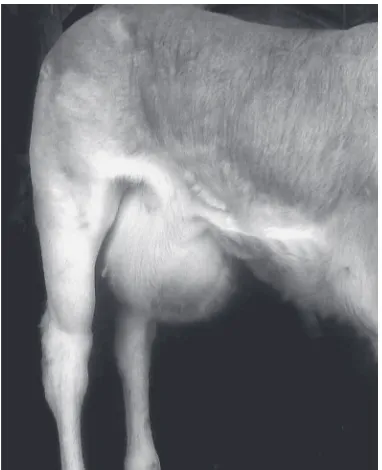
A one-month-old female Piedmontese calf, weighing 60 kg, was referred to the Department of Veterinary Science, University of Turin for evalu-ation and repair of a large ventral hernia. The calf was visited by the referring veterinarian two weeks prior to admission for sudden appearance of a ven-tral hernia 20 cm long and 15 cm wide, extending from 3 cm caudal to the umbilicus down to the pu-bis (Figure 1). Upon admission a complete clinical, haematological and ultrasonographical examina-tion was performed. Clinically, the presence of an uncomplicated hernia was confirmed, and other concurrent diseases ruled out. No alterations were detected in the haematological profile. At ultra-sonography the hernia content was represented by small intestine and omentum and no adhesions to the hernial sac were detected. The hernial sac was composed of skin only. The edges of the torn ab-dominal wall could be detected at the periphery of the sac. Because of the large dimensions of the her-nia and the economic value of the animal, surgical
correction was recommended to the owner. Food and water were withheld 12 h before the surgery. After clipping and aseptic preparation, an
[image:1.595.328.519.506.741.2]591
Veterinarni Medicina, 61, 2016 (10): 590–593 Case Report
doi: 10.17221/272/2015-VETMED
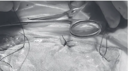
[image:2.595.307.534.96.221.2]venous catheter was inserted into the left jugular vein. The region around the first intercoccygeal space was clipped and after sterile skin preparation, the patient received epidural anaesthesia with 2% xylazine (0.05 mg/kg) and 2% lidocaine (0.2 mg/kg). After the loss of tone of the hind limbs, the calf was maintained in sternal position for 10 min and then placed in dorsal recumbency on a surgical table. The abdominal wall was clipped and a local inverted V-block was performed using 2% lidocaine cranial to the umbilicus to provide additional anal-gesia of the ventral abdominal wall. The abdomen was aseptically prepared and draped for surgery. Starting 3 cm caudally to the umbilicus, a 20 cm in-cision was made towards the pubis. To preserve the integrity of the mammary gland, the incision had a Y shape with the two branches extend laterally to the udder. Incision of the skin resulted in immedi-ate entry to the abdominal cavity. A full examina-tion of the abdominal cavity was performed and no other abnormalities were noticed. The torn edges of the abdominal wall were identified but were so con-tracted that they couldn’t be completely opposed, leaving a defect approximately 12 cm long and 7 cm wide. Caudally, the defect extended to the pelvic bones, leaving only a strip 3–5 mm wide of fascia that was not enough to safely anchor sutures under tension. For these reasons a tension-free mesh im-plantation was considered the best option. A 17 × 13 cm polypropylene mesh(Bard® Soft Mesh, Davol Inc., Cranston, RI, USA) was cut approximately in the shape of the defect. The omentum was reached and brought caudally to cover the viscera down to the pelvis. Several USP 1 nylonb (MonosofTM su-ture, Covidien, Segrate Milano, Italy) sutures were placed in the omentum along the perimeter of the hernia, leaving the strands approximately 10 cm long (Figure 2). The mesh was overlapped to the omentum and the strands passed, with removable needles, through the mesh and then through the fascia. Mosquito forceps were used to clamp the suture strands as they were applied to keep them in position before being tied. Particular care was taken to apply the mesh without any tension and wrinkles. When the mesh was in place, the nylon sutures were tied. Excess skin of the hernia sac was trimmed with scissors. A subcutaneous layer with polyglactin 910(Vicryl suture, Ethicon, Johnson & Johnson, Norderstedt, Germany) was made be-fore closing the skin with a simple continuous su-ture pattern with USP 1 nylon(MonosofTM suture,
Covidien, Segrate Milano, Italy). The duration of surgery was approximately 90 min. Recovery from sedation was uneventful and no postoperative com-plications or signs of pain were detected.
Clinical findings. The patient was discharged
on the same day. The calf was isolated in a sin-gle box for 10 days, checked daily by the referring veterinarian and was administered Penicillin G, 20 000 IU i.m. s.i.d. for three days and flunixin meglumine 1.1 mg/kg i.v.s.i.d. for two days. No complications were reported and weight gain was normal two and six months after surgery.
DISCUSSION AND CONCLUSIONS
Ventral hernia is a term that is used to describe a hernia through any part of the abdominal wall other than the umbilicus or inguinal canal (Tirgari 1980; Kawcak and Stashak 1995). Ventral and in-cisional hernias are common surgical problems in large animals and may occur due to midline or paramedian incision, or wherever the abdominal wall is severely traumatised (Wintzer 1962; Tirgari 1980; Kawcak and Stashak 1995). In young large animals the defect could be congenital or traumatic due to external manipulation during foal or calf delivery (Witte et al. 2008). Trauma was the most probable cause in our case, as the hernia appeared 15 days after the birth of the animal. Although small abdominal wall defects can be treated with good results, the outcome for larger defects is variable, both in humans (Sorour 2014) and large animals (Elce et al. 2005; Whitfield-Cargile et al. 2011).
and Stashak 1995). This condition may lead the surgeon to prefer a mesh implant, although it is de-manding in terms of surgical skills and time, more expensive and may lead to higher complication rated compared with suture repair (Williams et al. 2014).In horses similar results have been obtained with sutures and mesh implantation in ventral her-nia repair (Elce et al. 2005; Whitfield-Cargile et al. 2011), whereas in cattle mesh implantation has been associated with a higher complication rate in umbilical hernias (Kawcak and Stashak 1995). Nevertheless, simple repair with suture alone may not be effective in repairing large defects, and this could lead to recurrence of the hernia or muscle tearing (Tulleners and Fretz 1983; Elce et al. 2005; Whitfield-Cargile et al. 2011). In the case reported, the defect was large and a simple repair was not achievable due to the lack of sufficient tissue for safe anchoring of the sutures under tension. An intra-abdominal mesh repair was elected for, be-cause a retroperitoneal placement was not feasible, due to the absence of the peritoneum in the defect (Witte et al. 2008; Whitfield-Cargile et al. 2011).
The ideal mesh stimulates tissue grown from overlying fascia without the development of ad-hesions at the visceral mesh surface (Bernard et al. 2007). Synthetic materials of high tensile strength have been used to produce meshes for the repair of abdominal wall defects. The characteristic of these materials make them the best choice for use in large animals with large abdominal defects (Tulleners and Fretz 1983).
Generally, the prognosis of ventral hernia repair with a tension-free mesh implantation is associated with a fair-to-good prognosis even for defects up to 30 × 20 cm but complications such as wound infection, seromas, sinus formation, mesh extru-sion, and fistula formation may arise (Tulleners and Fretz 1983; Elce et al. 2005; Bernard et al. 2007; Whitfield-Cargile et al. 2011). Polypropylene mesh is one of the most commonly used prosthetic mate-rials for large ventral hernia repair in large animals (Tulleners and Fretz 1983; Finan et al. 2009). Its advantages include pliability, elasticity, inertness, strength, low rate of rejection, well-formed resist-ant tissue, and lower cost compared to expanded polytetrafluoroethylene mesh (Sorour 2014).The choice of a polypropylene mesh is also favoured by the structure of the mesh itself which is fined as a knitted mesh. This characteristic is de-sirable in closing large abdominal defects in horses
and cattle, as it tends to result in fewer wrinkles, particularly when the hernial sac is lacking and only subcutaneous tissue skin covers the mesh (Tulleners and Fretz 1983), as in the present case. Most post-operative problems result from adhesion of abdominal contents to the mesh, irritation of the intestine and subsequent rupture of the bowel, and eventually persistent drainage and infection of the mesh (Witte et al. 2008; Sorour 2014). Irreversible peritonitis following mesh implantation resulted in the death of four animals in the study of Tulleners and Fretz (1983). One of the horses included in that study, treated with the polypropylene mesh, began to suffer from small intestine rubbing against the mesh that led finally to leakage of ingesta, causing diffuse fatal peritonitis (Tulleners and Fretz 1983).
Using a double layer ePTFE mesh could prevent such issues due to the anti-adhesive characteristics of the mesh that allow its intraperitoneal place-ment; however, costs increase enormously (Caron and Mehler 2009). The same result can be obtained in cattle with the interposition of the omentum between the mesh and the viscera. This is virtually impossible to obtain in horses, because of the small dimensions of the omentum in this species. The interposition of omentum between the mesh and underlying intestine has been proposed as a protec-tive measure in humans and this procedure could be considered as an important step for preventing adhesion with a polypropylene mesh (Sorour 2014). It has not been reported as yet in large animals.
Placement of any permanent foreign material during surgery carries with it a high risk of infec-tion; therefore, asepsis should be an important con-sideration (Williams et al. 2014). An infection could lead to formation of a non-healing tissue, purulent persistent discharge from the incision and eventu-ally to a re-herniation (Witte et al. 2008; Whitfield-Cargile et al. 2011). This kind of surgery is carried out in humans with a higher level of asepsis that is not easily achieved in farm animal surgery un-der field conditions (Williams et al. 2014). Mesh implants require a high level of surgical skill and should be avoided under field conditions (Baird 2008).
593
Veterinarni Medicina, 61, 2016 (10): 590–593 Case Report
doi: 10.17221/272/2015-VETMED
to be trimmed during the process of disassembling the carcass of the animal; thus, any foreign material in this region should readily be detected. Another possibility would be to create a proper space in the animal’s passport to signal that a permanent implant has been used and that care must be taken during the slaughter process to carefully remove it.
To the best of our knowledge, this is the first report that combines the use of a tension-free polypropylene mesh with the interposition of the omentum between the viscera and the mesh in large animals. In our case this procedure was safe, cost-effective and was not associated with major com-plications. Therefore, this method may be a viable alternative to other methods of mesh hernia repair.
RefeReNCeS
Baird AN (2008): Umbilical surgery in calves. Veterinary Clin-ics of North America Food Animal Practice 24, 467–477. Bernard C, Polliand C, Mutelica L, Champault G (2007):
Repair of giant incisional abdominal wall hernias using open intraperitoneal mesh. Hernia 11, 315–320. Caron JP, Mehler SJ (2009): Laparoscopic mesh incisional
hernioplasty in five horses. Veterinary Surgery 38, 318–325. Elce YA, Kraus BM, Orsini JA (2005): Mesh hernioplasty
for repair of incisional hernias of the ventral body wall in large horses. Equine Veterinary Education 17, 252–256. Finan KR, Kilgore ML, Hawn MT (2009): Open suture ver-sus mesh repair of primary incisional hernias: a cost-utility analysis. Hernia 13, 173–182.
Kawcak CE, Stashak TS (1995): Predisposing factors, diag-nosis, and management of large abdominal wall defects in horses and cattle. Journal of American Veterinary Medical Association 206, 607–611.
Sorour MA (2014): Interposition of the omentum and/or the peritoneum in the emergency repair of large ventral hernias with polypropylene mesh. International Journal of Surgery 12, 578–586.
Tirgari M (1980): Ventral hernia in the sheep. Veterinary Record 5, 7–9.
Tulleners EP, Fretz PB (1983): Prosthetic repair of large ab-dominal wall defects in horses and food animals. Journal of American Veterinary Medical Association 182, 258–262. Whitfield-Cargile CM, Rakestraw PC, Hardy J, Cohen ND, Davis BE (2011): Comparison of primary closure of inci-sional hernias in horses with and without the use of pros-thetic mesh support. Equine Veterinary Journal 43, 69–75.
Williams HJ, Gillespie AV, Oultram JW, Cripps PJ, Holman AN (2014): Outcome of surgical treatment for umbilical swellings in bovine youngstock. Veterinary Record 174, 221–224.
Wintzer HJ (1962): Methods for surgical treatment of bo-vine abdominal hernias. Journal of American Veterinary Medical Association 141, 131–134.
Witte S, Rodgerson D, Hunt R, Spirito MA (2008): Trau-matic ventral herniation in foals as a complication of dystocia. Compendium Equine 3, 137–143.
Received: 2015–12–14 Accepted after corrections: 2016–07–20
Corresponding Author:
Marco Gandini, University of Turin, Department of Veterinary Sciences, Largo Paolo Braccini 2–5, Grugliasco (TO), Italy
The CENTAUR network aims at upgrading the standards of economically significant priority animal diseases control in the region with particular emphasis on transboundary animal diseases, animal health and consumer protection.
The CENTAUR is willing to achieve it through dissemination of scientific information, training, links with the international centres of excellence and cooperation. The important task is also to present the problems, personalities, institutions, and scientific achievement of the region. Efficient utilization of Internet, e-mail and improvement in English language proficiency is followed, too.
Under the CENTAUR network the CENTAUR NEWSLETTER FLASH INFORMATION (CNFI), an international electronic bulletin (ISSN 1213-368X), is published, providing subscribers with instant infor-mation in the form of e-mail messages relating to fields of interest which subscribers define themselves during the process of registration. CNFI covers global animal disease-related events and is distributed to the registered readers from all over the world. The number of subscribers has been growing rapi-dly and new registrations are always welcome. More than 1200 registered members of the CENTAUR network from 70 countries receive the e-mail information at present. The web page http://centaur.vri. cz is requently visited by colleagues from countries of all continents.
The forms of CNFI are as follows:
E-MAIL MESSAGES are distributed to field specific registered members. Sometimes identical infor-mation is distributed to more fields of interest. Therefore second mail with identical subject and time of dispatching should not be opened but immediately deleted.
CNFI BULLETIN: approximately 10 issues per year with general information for the CENTAUR network members are distributed to all registered addresses as an attachment to e-mail. This bulletin is also available for downloading from the CENTAUR web page http://centaur.vri.cz
CENTAUR network members are welcome as authors of original papers or reviews submitted for publication in an international peer reviewed jornal for veterinary medicine and biomedical sciences Veterinarni medicina, indexed is the Web of Science, Current Contents and other databases. Papers published in this journal are free in full text at http://vetmed.vri.cz
CENTAUR network members can request the Editor for search from the published papers if their intentions are oriented towards to contributions for CNFI or submission the manuscript for publication in the journal Veterinarni medicina.
CNFI subscription is free. Register your “fields of interest” according to the instructions available at http://centaur.vri.cz/default.asp?page=cent_reg.asp and you will receive instant confirmation of your choice by e-mail. To unsubscribe or change the selected fields of interest, send an e-mail to the CNFI editor <hruska@vri.cz>. Contributions, comments and requests of the subscribers are welcome.