Contents lists available at
ScienceDirect
Prostaglandins, Leukotrienes and Essential Fatty Acids
journal homepage:
www.elsevier.com/locate/plefa
Ischemia-modi
fi
ed albumin: Crosstalk between fatty acid and cobalt binding
James P.C. Coverdale
a,1, Kondwani G.H. Katundu
b,c,1, Amélie I.S. Sobczak
b, Swati Arya
b,
Claudia A. Blindauer
a,⁎, Alan J. Stewart
b,⁎aDepartment of Chemistry, University of Warwick, Coventry, United Kingdom
bSchool of Medicine, University of St Andrews, St Andrews, United Kingdom
cCollege of Medicine, University of Malawi, Blantyre, Malawi
A R T I C L E I N F O
Keywords:
Albumin cobalt binding assay Molecular diagnostics Free fatty acids Human serum albumin Myocardial ischemia
A B S T R A C T
Myocardial ischemia is difficult to diagnose effectively with still few well-defined biochemical markers for identification in advance, or in the absence of myocardial necrosis.“Ischemia-modified albumin”(IMA), a form of albumin displaying reduced cobalt-binding affinity, is significantly elevated in ischemic patients, and the albumin cobalt-binding (ACB) assay can measure its level indirectly. Elucidating the molecular mechanism underlying the identity of IMA and the ACB assay hinges on understanding metal-binding properties of albumin. Albumin binds most metal ions and harbours four primary metal binding sites: site A, site B, the N-terminal site (NTS), and the free thiol at Cys34. Previous efforts to clarify the identity of IMA and the causes for its reduced cobalt-binding capacity were focused on the NTS site, but the degree of N-terminal modification could not be correlated to the presence of ischemia. More recent work suggested that Co2+ions as used in the ACB assay bind
preferentially to site B, then to site A, andfinally to the NTS. This insight paved the way for a new consistent molecular basis of the ACB assay: albumin is also the main plasma carrier for free fatty acids (FFAs), and binding of a fatty acid to the high-affinity site FA2 results in conformational changes in albumin which prevent metal binding at site A and partially at site B. Thus, this review advances the hypothesis that high IMA levels in myocardial ischemia and many other conditions originate from high plasma FFA levels hampering the binding of Co2+to sites A and/or B. This is supported by biophysical studies and the co-association of a range of patho-logical conditions with positive ACB assays and high plasma FFA levels.
1. Introduction
Myocardial ischemia occurs due to restricted blood supply to the
muscular tissue of the heart (myocardium) resulting in insu
ffi
cient
oxygen supply. The main cause of this can be the partial or complete
blockage of a coronary artery, and a critical depletion of myocardial
oxygen leads to cell death, or infarction. Diagnosis of myocardial
ischemia typically includes exercise-electrocardiography stress tests,
coronary angiography, and imaging stress-echo tests
[1]
. While a
ple-thora of cardiac biomarkers have been described for detecting the
de-velopment of other acute coronary syndromes (ACS)
[2,3]
, there are
still few well-de
fi
ned biochemical markers for identi
fi
cation of
myo-cardial ischemia in advance, or in the absence of myomyo-cardial necrosis.
One of these biomarkers is based on albumin, the most abundant
protein in blood plasma. So-called
“
ischemia-modi
fi
ed albumin
”
(IMA)
is found to be signi
fi
cantly elevated in ischemic patients
[2,4
–
7]
, and
serves as a biomarker for early detection of myocardial ischemia before
the onset of irreversible cardiac injury
[6]
. IMA is solely characterised
by its reduced cobalt-binding a
ffi
nity, which can be measured indirectly
by the Food and Drug Administration-approved albumin cobalt-binding
(ACB) assay
[8,9]
.
In the commercially available ACB test, cobalt(II) chloride
(ap-proximately 1.5 mol equivalents per albumin molecule) is added to a
serum sample, to allow albumin-cobalt binding. Dithiothreitol (DTT), a
metal chelator that forms a coloured complex with Co
2+, is then added.
The resulting ill-de
fi
ned brown DTT-Co
2+product is measured by
ab-sorption spectrophotometry at 470 nm and compared to a serum-cobalt
blank without DTT present. The reduced cobalt-binding capacity of IMA
https://doi.org/10.1016/j.plefa.2018.07.014
Received 29 March 2018; Received in revised form 17 July 2018; Accepted 17 July 2018
Abbreviations:ACB, albumin cobalt-binding; ACS, acute coronary syndromes; ATCUN, amino terminal Cu(II) and Ni(II) binding motif; DTT, dithiothreitol; EPR, electron paramagnetic resonance; EXAFS, extended X-ray absorptionfine structure spectroscopy; FFAs, free fatty acids; HRG, histidine-rich glycoprotein; IMA, ischemia-modified albumin; ITC, isothermal titration calorimetry; NMR, nuclear magnetic resonance; NTS, N-terminal binding site on albumin
⁎Corresponding authors.
1These authors contributed equally.
E-mail addresses:c.blindauer@warwick.ac.uk(C.A. Blindauer),ajs21@st-andrews.ac.uk(A.J. Stewart).
0952-3278/ © 2018 The Authors. Published by Elsevier Ltd. This is an open access article under the CC BY license (http://creativecommons.org/licenses/BY/4.0/).
leaves more unbound Co
2+to complex with DTT, resulting in higher
absorbance readings
[10]
. The ACB test has an excellent negative
pre-dictive value,
i.e.
low IMA readings correspond well to the absence of
myocardial ischemia. However, a severe shortcoming is the high
in-cidence of false positives,
i.e.
high readings in the absence of ischemia.
After its
fi
rst description
[8]
, the molecular identity of IMA
re-mained elusive. Based on the general assumption that Co
2+would
preferentially bind to an N-terminal site
[11
–
13]
, e
ff
orts to elucidate
the molecular causes of reduced cobalt binding concentrated on this
site. It was hypothesized that ischemia causes the N-terminal end of the
albumin protein to undergo structural modi
fi
cations, hence that IMA
corresponded to N-terminally modi
fi
ed albumin
[13]
. The structural
modi
fi
cations proposed and investigated included cleavage of the
fi
rst
two residues and oxidation
[11]
, which were suggested to result from
free radical damage, exposure to free iron and copper, or disruption of
ion pumps
[8,14]
.
However, in-depth studies could not reveal a correlation between
N-terminal modi
fi
cations and ACB readings
[13,15]
; more recently, no
correlation was found between the ACB assay and an enzyme-linked
immunosorbent assay that speci
fi
cally detects N-terminal modi
fi
cation
of albumin in patients with either acute coronary syndrome or
non-ischemic chest pain
[16]
. Similarly, patients su
ff
ering from
acute-on-chronic liver failure have signi
fi
cantly elevated ACB assay readings, but
the same proportion of N-terminally modi
fi
ed albumin as healthy
in-dividuals
[17,18]
. In the light of such
fi
ndings, low plasma pH as a
result of acidosis, and altered plasma cysteine/cystine ratio as a
con-sequence of hypoxia or oxidative stress have also been suspected as
molecular causes of reduced cobalt binding
[19]
. The need to consider
the contribution of other plasma components to the Co-DTT complex
formation was also highlighted
[19]
. Indeed, a positive correlation has
been identi
fi
ed between the highly elevated serum levels of free fatty
acids (FFAs) in patients with acute ischemic myocardia and high levels
of IMA
[20]
. Following our discovery of FFA-mediated inhibition of
zinc binding to albumin
[21
–
24]
, we have demonstrated that the
con-formational changes that FFA-binding to albumin elicits in the protein
is su
ffi
cient to cause reduced cobalt binding capacity
[22,25]
. This
re-view will present essential background information on metal
ion-al-bumin interactions and discuss the molecular basis of FFA-mediated
inhibition of metal (in particular Co
2+) binding. It will also provide a
clinical perspective to highlight how conclusions from biochemical/
bioinorganic investigations are re
fl
ected in patient data.
2. Albumin
–
a carrier of essential and xenobiotic metal ions in
plasma
Albumin is a
∼
66 kDa protein containing 585 amino acids,
con-tributing to around 50% of the total protein concentration in blood
plasma, and up to 75% of the colloidal activity
[26]
. Albumin comprises
three homologous but structurally distinct domains, each divided into
two sub-domains
[27]
. One of its key roles in the body is to transport a
variety of small molecules, including cholesterol
[28]
, fatty acids
[29]
,
and pharmaceutical drugs
[30]
. Importantly, albumin also serves as an
important carrier of inorganic ions, including those required for regular
physiological function (Ca
2+, Cu
2+,Zn
2+)
[31]
, toxic metal ions (Cd
2+and Ni
2+)
[32,33]
, as well as metal-based therapeutics (Au
+and Pt
2+)
[34,35]
. Before considering cobalt binding in depth, we will brie
fl
y
summarise the interactions of albumin with other d-block metal ions,
with the exception of Cr
3+, Fe
3+, and Mn
2+, which are preferentially
transported by transferrin, another important metal ion transporter in
blood plasma. Whilst Fe
3+can, in principle, also bind to albumin, this
only occurs in cases of severe iron overload
[34]
.
2.1. Metal binding sites in serum albumins
[image:2.595.39.356.55.204.2]Though originally albumin was thought to transport ions in a
non-speci
fi
c
‘
sponge-like
’
manner
[30]
, four partially selective metal
binding sites have been identi
fi
ed, namely the N-terminal site (NTS),
sites A and B, and Cys34 (
Fig. 1
)
[34]
. Metal binding to such sites can be
studied using a variety of techniques. Stability constants for the binding
of d-block metals, including Zn
2+, Cu
2+, Ni
2+and Cd
2+, were
ori-ginally derived from equilibrium dialysis experiments
[36-39]
; more
recently, isothermal titration calorimetry (ITC) has provided valuable
thermodynamic data for metal ion binding
[40]
. Nevertheless, both of
these techniques only provide global binding constants
[34]
and need
to be complemented by techniques that address structural features. For
true transition metal ions such as Cu
2+and Co
2+, electronic
spectro-scopic methods such as circular dichroism allow metal binding to
al-bumin to be studied via transfer of chirality from metal-binding amino
acid residues to the d-d/charge-transfer bands of complexed metal ions,
providing insight into the geometry of metal-protein interactions
[41,42]
. The same ions have unpaired electrons, and can also be
in-vestigated using electron paramagnetic resonance (EPR) spectroscopy,
which provides insight into the chemical environment surrounding the
metal ion
[43,44]
. To obtain structural information on the binding of
diamagnetic d
10ions, such as Zn
2+and Cd
2+, that are largely silent in
the aforementioned spectroscopies, nuclear magnetic resonance (NMR)
methods have been employed, making use of either
partially-char-acterised
1H-resonances of metal-binding residues, or NMR-active
nu-clei such as the
111Cd or
113Cd isotopes of cadmium
[39,45
–
47]
. Further
information on the coordination mode, geometry and identi
fi
cation of
likely donor ligands has been gained using extended X-ray absorption
fi
ne structure spectroscopy (EXAFS)
[47]
. In addition, mass
spectro-metry has been used as a tool to detect crosslinking of His67 and His247
by platinum in site A
[48]
.
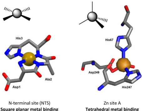
2.1.1. The N-terminal binding site (NTS)
two deprotonated backbone amide nitrogen atoms. This square planar
con
fi
guration of N-donor atoms (
Fig. 2
) is particularly suitable for Cu
2+and Ni
2+, which has led to the NTS being referred to by the acronym
‘
ATCUN
’
, for the Amino Terminal Cu(II) and Ni(II) binding motif
[42,50]
. The ATCUN motif is present in the majority of albumins from
di
ff
erent mammalian species, though porcine and canine albumins are
notable exceptions, as they lack His3
[34]
. Oligopeptide models of the
native ATCUN motif have been investigated extensively
[34]
. The NTS
motif is thought to have high conformational
fl
exibility in the absence
of bound metal, re
fl
ected in the crystal structures of albumin, all of
which lack de
fi
ned structures of the
fi
rst few N-terminal residues
[12]
.
Interestingly, the N-terminal X-X-His motif is not unique to albumin
–
many other proteins, such as the peptide hormone Hepcidin, can also
bind Ni
2+and Cu
2+ions via an ATCUN motif
[51]
.
Cu
2+binds preferentially to the NTS in albumin, occupying
ap-proximately 1
–
2% of the available NTS
–
equating to around 15% of
total copper in blood plasma
[34,52]
. Owing to the d
9electronic
con-fi
guration of Cu
2+, preference to form square planar complexes, and
high stability in the Irving-Williams series, Cu
2+is coordinated at the
NTS with 1 pM a
ffi
nity
[52]
, and binds preferentially over other metal
ions
[22]
. Cu
2+can also bind at other metal binding sites with
com-parable or even higher a
ffi
nities to those of Ni
2+and Zn
2+[41]
,
however its low relative concentration (10
–
20 µM total Cu
2+, and
sub-micromolar
‘
free
’
Cu
2+in plasma)
[53]
compared to albumin means
that, in practice, only the NTS is ever occupied by Cu
2+[52]
. Like
Cu
2+, Ni
2+binds to albumin preferentially at the NTS site
[33]
, with
micromolar a
ffi
nity
[34]
. Ni
2+is only present at nanomolar
con-centrations in plasma, however levels may be elevated under certain
pathological conditions (e.g.
stroke)
[54]
. Nearly all of plasma Ni
2+is
albumin-bound
[12,34]
. Binding of Ni
2+and Cu
2+can be modulated
by the redox state of Cys34
[43]
with higher metal a
ffi
nity in the
re-duced (free thiol) state.
Site A
–
the multi-metal binding site
Metal binding site A is located at the interface of domains I and II
[34]
(
Fig. 1
), and has been identi
fi
ed and characterised using
1H and
111/113Cd NMR spectroscopy
[39,45,46,55,56]
, circular dichroism,
site-directed mutagenesis
[56]
, EXAFS
[47]
and recently X-ray
crystal-lography
[57]
. As well as having a high nanomolar a
ffi
nity
[23,24,38,39,47]
for the d
10divalent cations Zn
2+and Cd
2+, site A can
also bind Cu
2+, Ni
2+and Co
2+–
hence it is also referred to as the
‘
multi-metal
’
binding site
[34,41,56]
. In fact, up to 90% of the total zinc
present in plasma (11.5
–
36.7 µM total Zn
2+in adults
[58]
) is bound to
albumin
[59,60]
; this amounts to
ca.
98% of exchangeable plasma zinc.
EXAFS, site-directed mutagenesis and molecular modelling initially
suggested that site A is formed by His67, His247, Asp249, and Asn99
[47]
, and so distorted trigonal bipyramidal coordination of Zn
2+was
proposed, with water (or chloride) as the
fi
fth ligand completing the
inner coordination sphere. However, the recent X-ray crystallographic
structures of human and equine albumins discounted participation of
Asn99 and showed site A to be essentially tetrahedral (
Fig. 2
), with the
fourth ligand being a water molecule
[57]
. Cu
2+coordination at site A
had also been suggested to be tetrahedral in geometry, as determined
by EPR and CD experiments
[61]
. The combination of amino acid
re-sidues bearing intermediate-to-hard N/O-donors
[60]
(HSAB principle)
provide a good coordination environment for metal ions with a small
ionic radius and moderate charge (e.g.
2+ cations). Notably though, the
a
ffi
nity of site A for Cu
2+is 4 orders of magnitude lower than that of
the NTS
[34]
, thus site A only becomes populated by Cu
2+when more
than one molar equivalent of Cu
2+is present. Finally, the comparison
of apo- and Zn-bound crystal structures of albumin has revealed high
structural similarity at site A. Thus, in marked contrast to the
fl
exible
NTS, site A is essentially
‘
pre-formed
’
for metal binding
[57,60]
. It is
important to note that site A is an inter-domain site, with His67 from
domain I, and His247 and Asp249 from domain II.
Site B
The other Cd
2+binding site (site B), which is distinct from site A
and the NTS and readily identi
fi
able using
111Cd or
113Cd NMR
(
Fig. 3
a), appears to bind Cd
2+with similar a
ffi
nity to the multi-metal
binding site A
[45,46]
. In contrast, site B's a
ffi
nity for zinc is markedly
less than that of site A. Based on NMR data, it is likely that only one
nitrogen donor ligand is involved at site B, suggesting this site to be
harder (HSAB principle) than site A. The location of site B has remained
elusive, but site-directed mutagenesis of His39Leu excluded His39 from
involvement in either site A or B
[47]
.
Cysteine-34
Albumin contains 17 disul
fi
de bonds, which contribute to the
structural stability of the protein. One free thiol residue (Cys34) is
lo-cated between helices 2 and 3 of subdomain IA (
Fig. 1
)
[34]
. Cys34 is
not involved in any intramolecular bridging, however it often forms
intermolecular disul
fi
des with small sulfur-containing molecules such
as cysteine and glutathione
[34]
. Under normal physiological
condi-tions, approximately 40% of albumin contains
‘
reduced
’
Cys34 (free
thiol)
[34]
. The restricted location of Cys34 in a crevice of albumin
helps to improve its speci
fi
city for binding metal ions that favour linear
coordination, including Hg
2+, Au
+, Ag
+and Pt
2+[34,35]
, but not
Cd
2+or Zn
2+[56]
.
Calcium binding sites
Albumin is an important transporter of Ca
2+in blood plasma. Many
reports suggest that this occurs in a non-speci
fi
c fashion, involving
various carboxylate side chains on the surface of albumin
[42,62]
,
while work by Majorek et al. detected three de
fi
ned Ca
2+binding sites
on bovine albumin
[63]
. It may be signi
fi
cant that one of the Ca
2+sites
detected by crystallography involves the key site A ligand, Asp248
(corresponding to Asp249 in human albumin) and indeed, Ca
2+ions
were found to interfere with the
113Cd signals for both sites A and B of
human albumin
[46]
. However, the a
ffi
nity of albumin for Ca
2+binding is relatively weak (K
dof 0.67 mM), with only around 45% of
Fig. 2.Contrasting geometries of metal binding sites on albumin. Left: squareplanar coordination of Cu2+or Ni2+at the NTS site; the structure shown is
derived from molecular modelling. The N-terminal amino group, two depro-tonated backbone amide N atoms and the N(delta) of the imidazole ring of His3 form a square plane around the central metal ion. Right: tetrahedral co-ordination of Zn2+at site A in human serum albumin (pdb5ijf). His67 uses its
the 2.4 mM of circulating Ca
2+bound to albumin
[63,64]
. Mg
2+,
which is also carried by albumin, is thought to bind to the same binding
sites as Ca
2+but with an even lower a
ffi
nity (K
d
of 10 mM)
[65]
.
2.2. Cobalt binding to albumin
Cobalt circulates in the blood as Co
2+and albumin is its principal
transporter in plasma
[34]
. While it is widely assumed that its binding
resembles that of Ni
2+and Cu
2+(d
8and d
9metal ions, respectively,
with preference for the formation of square planar/tetragonal
com-plexes), Co
2+(d
7) behaves in fact more like Zn
2+(d
10), has a similar
ionic radius (0.58 vs. 0.60 Å for Zn
2+[66]
), and shares a preference for
tetrahedral, penta-coordinate or octahedral geometry
[67]
. For
pre-cisely this reason, Co
2+has been used extensively as a spectroscopic
probe for Zn
2+in proteins
[68,69]
.
In total, three signi
fi
cant Co
2+binding sites have been identi
fi
ed on
albumins
–
the NTS, site A and site B
[42]
. Based on Co
2+perturbing
1H
NMR resonances for the three N-terminal residues
[11]
and Co
2+binding to an ATCUN peptide mimic
[13,49]
, it was assumed that the
primary cobalt-binding site was the NTS motif
[11,19,70]
. More recent
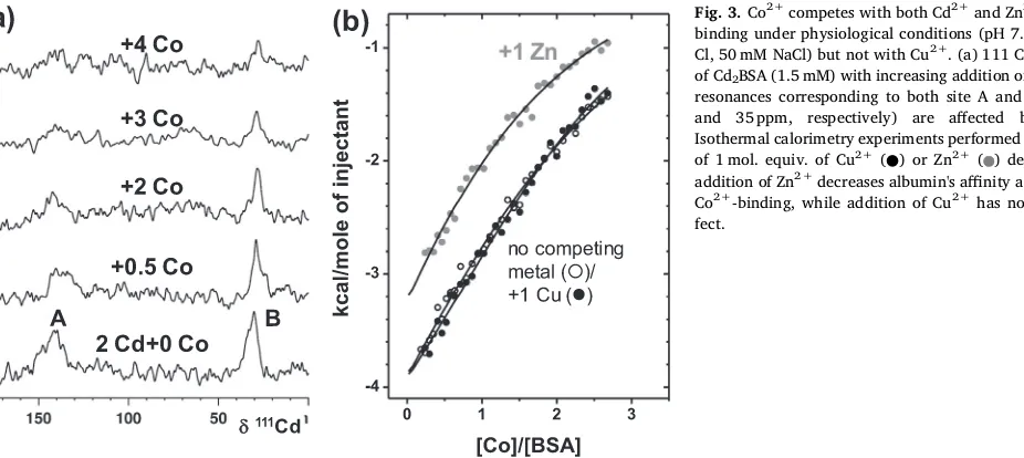
comprehensive studies on human albumin by Mothes and Faller
[71]
and Sokolowska et al.
[42]
, and on bovine albumin by our labs
[25]
have since rejected this claim. Competition with Cd
2+and Cu
2+monitored by electronic absorption spectroscopy strongly suggested
that sites A and B are the preferred Co
2+binding sites
[16,42,71]
.
Subsequently, ITC and spectroscopic studies identi
fi
ed site B as the
strongest cobalt binding site
[42]
. Co
2+binding to sites A and B was
also con
fi
rmed by
111Cd NMR spectroscopy for bovine albumin
(
Fig. 3
a), and competition with Zn
2+was evident from ITC (
Fig. 3
b)
[25]
. In contrast, blocking the NTS site with Cu
2+did not impart any
signi
fi
cant e
ff
ect on Co
2+binding
[25,71]
. It is important to note
however, that even though Co
2+and Zn
2+may be regarded as metal
ions with similar properties, the apparent binding constant for Co
2+binding
to
its
strongest
site
on
bovine
albumin
(log
K
app= 4.6 ± 0.3 × 10
4M
−1;
Fig. 5
b;
[25]
) and 9 ± 5 × 10
4M
−1for
human albumin
[42]
) were around one order of magnitude lower than
those determined for Zn
2+[25]
.
In summary, even though all three apparent binding constants for
Co
2+binding to human albumin lie between 9 ± 5 × 10
4M
−1and
0.9 ± 0.3 × 10
4M
−1[42]
, and hence the respective equilibria do
overlap, the NTS site is now known to have the weakest a
ffi
nity for
Co
2+[42]
. Most importantly, this weaker than anticipated binding of
Co
2+to the NTS means that the initially proposed molecular basis of
the ACB assay to assess the likelihood of myocardial infarction required
revision, since the original studies assumed that Co
2+binds exclusively
to the NTS
[8,19]
.
3. Free fatty acid binding to albumin and allosteric inhibition of
metal ion binding
Albumin has an unparalleled capacity to bind and transports a range
of organic molecules under physiological conditions
[72]
. Notable
among those transported are FFAs, important substrates in organismal
metabolism for which albumin is the main carrier
[73
–
76]
. FFAs are the
main source of energy for heart and skeletal muscle. Disturbances of the
levels and/or distribution of fatty acids in the body have been linked to
a spectrum of pathological disorders, including diabetes, cardiovascular
and neurological diseases, and cancer
[77]
. Owing to the abundance of
albumin in plasma, and the importance of fatty acids in metabolism and
disease progression, binding of FFAs to albumin has been studied
in-tensively in the past four decades
[73]
, in particular by X-ray
crystal-lography
[75,78,79]
and
13C NMR spectroscopy
[80,81]
.
Up to seven medium-to-long chain (C10-C18) fatty acid binding
sites (FA1-7) have been identi
fi
ed on albumin, spread over the three
domains (see
Table 1
and
Fig. 4
)
[75]
. The binding a
ffi
nities depend on
both the site and the FFA chain length. Four additional binding
loca-tions have been described for short-to-medium chain fatty acids
[82]
,
however for the purposes of this review we will focus on FA1-7 (
Fig. 4
).
In a normal physiological state, albumin circulates with between 0.1
–
2
equivalents of FFAs bound, however it pathologically can bind in excess
of 6 equivalents
[83]
. The seven identi
fi
ed binding sites can be broadly
split into two categories: the high-a
ffi
nity sites (FA2, FA4 and FA5) and
the low-a
ffi
nity sites (FA1, FA3, FA6 and FA7)
[83]
. The high-a
ffi
nity
site FA2 is close to metal-binding site A and therefore of particular
interest. This relatively hydrophobic site is, like metal site A, an
inter-domain site and is located between sub-inter-domains IA and IIA (
Fig. 4
)
[82]
. Compared to FFA-free albumin, accommodation of a fatty acid
molecule in site FA2 requires a change in the mutual arrangement of
domains I and II. While short-chain FFAs (<C8) were originally
thought to be too short to successfully dock in the FA2 site
[82]
, more
recent
1H and
111Cd NMR studies indicated that octanoate can bind to
this site. Molecular modelling suggested that the half-pocket in domain
II is su
ffi
cient to accommodate octanoate, and therefore does not
re-quire the domain-domain movement
[25]
.
[image:4.595.47.510.54.261.2]While metal site A is essentially
‘
pre-formed
’
for metal
(physiolo-gically Zn
2+) binding in FFA-free albumin
[60]
, this is not the case
when FA2 is occupied by a longer chain FFA (e.g.
myristic acid, C14), as
the distance between the metal-coordinating residues is too large after
Fig. 3.Co2+competes with both Cd2+and Zn2+for albuminbinding under physiological conditions (pH 7.4, 50 mM Tris-Cl, 50 mM NaCl) but not with Cu2+. (a) 111 Cd NMR spectra of Cd2BSA (1.5 mM) with increasing addition of Co2+. 111-Cd
resonances corresponding to both site A and B (∼140 ppm and 35 ppm, respectively) are affected by Co2+. (b) Isothermal calorimetry experiments performed in the presence of 1 mol. equiv. of Cu2+(●) or Zn2+( ) demonstrate that
addition of Zn2+decreases albumin's affinity and capacity for Co2+-binding, while addition of Cu2+has no significant
the conformational change
[21,24,56]
. This crucial discovery suggested
that FFA and zinc concentration(s) in blood plasma may be correlated
through an allosteric mechanism based on albumin
[21,22]
.
Competi-tion experiments monitored by ITC demonstrated that the zinc-binding
capacity of both bovine and human albumin for site A was dramatically
reduced
[23,25]
. Five equivalents of myristate were su
ffi
cient to
com-pletely inhibit Zn
2+coordination to site A in bovine albumin (
Fig. 5
a),
with site B also a
ff
ected more or less severely
[25]
. Importantly, FA2 is
one of the high a
ffi
nity sites, and will become signi
fi
cantly populated
already at 1 molar equivalent
[83,84]
. Indeed, the data in
Fig. 5
a
in-dicate that the largest e
ff
ect is seen between 0 and 2 molar equivalents.
The downstream implications of this allosteric switch for the fate of
plasma zinc are discussed elsewhere
[21
–
23]
.
Crucially, although the binding preferences of Zn
2+and Co
2+are
not identical, the presence of myristate also clearly reduced the binding
capacity of bovine albumin for Co
2+(
Fig. 5
b)
[25]
. The e
ff
ect on Co
2+binding is less severe than that on Zn
2+binding, with 5 molar
equivalents of myristate reducing binding by
ca.
50%
[25]
. This is in
agreement with the fact that Co
2+does not bind preferentially to site A,
but site B (which in BSA is a
ff
ected by FFA binding, but less severely)
[25]
, and can also bind to the NTS motif which is not expected to be
adversely a
ff
ected by the presence of FFA. Similarly to Zn
2+, it appears
that the bound metal ion must
fi
rst be removed from site A before fatty
acid binding can occur at FA2, identi
fi
ed by a reduction in the
exo-thermicity of the FFA-binding reaction (
Fig. 5
c)
[25]
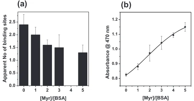
. The number of
apparent Co
2+binding sites in these experiments was in broad
agree-ment with other experiagree-mental data, although we note that the selected
experimental conditions did not allow to fully saturate all three binding
sites. The apparent number of binding sites in the absence of myristate
amounted to 2.4, and reduced to
ca. 1.3 sites, implying that (at least)
one binding site became non-functional (
Fig. 6
a). This is consistent with
an inhibition of cobalt-binding to site A, as a result of FFA binding to
the nearby FA2 site
[25]
. Most importantly, increasing the levels of FFA
in a mock ACB assay is su
ffi
cient to lead to increased formation of the
Co-DTT product, with concomitant higher absorbance readings
(
Fig. 6
b). The magnitude of the changes in absorbance at 470 nm is
broadly in line with e
ff
ects seen in clinical studies
[25]
. We next explore
whether this molecular mechanism may be re
fl
ected in clinical data.
4. Ischemia-modi
fi
ed albumin in disease states
As indicated previously, the diagnostic speci
fi
city of the ACB assay
is very low, resulting in a high proportion of false positives,
i.e.
high
readings despite the absence of ischemia
[15,85]
. This realisation has
motivated a large number of studies which found positive ACB readings
for a wide range of disease conditions including ACS
[20, 86]
, chronic
liver and kidney diseases
[87,88]
, infectious diseases such as malaria
[89]
, and pregnancy-related conditions such as pre-eclampsia
[90]
. In
addition, elevated IMA levels have been measured in metabolic
syn-drome
[91,92]
, diabetes
[93]
and obesity
[94,95]
while exercise and
trauma have also been investigated
[96,97]
. These conditions,
there-fore, should have a common feature that can explain elevated IMA
le-vels, and we propose that this common feature is elevated plasma FFAs.
The latter have been shown to independently in
fl
uence the ACB assay to
the same extent as ACS and other conditions
[20,25]
. Together with the
biochemical and biophysical studies detailed in
Section 3
, it is
com-pelling to suggest that IMA corresponds to albumins in which FA2 is
occupied. To further explore this hypothesis,
Table 2
compiles selected
conditions which are positive for the ACB assay and reports
quantita-tive data on serum FFAs drawn from the literature. Cobalt binding to
albumin is both speci
fi
c and proportional to the total serum albumin
concentration, and so many studies adjust for the total albumin level
[10]
.
[image:5.595.43.557.90.173.2] [image:5.595.37.360.597.739.2]ACSs are well-known to be associated with increased serum FFA
concentrations
[20,98]
. The pain and the stress associated with such
syndromes is thought to trigger a sympathetic discharge, with the
re-lease of catecholamines which activate hormone-sensitive tissue lipase
–
the enzyme which hydrolyses triglycerides and hence liberates FFAs
into the circulation
[99
–
101]
. This leads to elevated serum free fatty
acid concentrations within 1
–
2 hours from the onset of ACS, and the
degree of increase in FFA concentration has been positively associated
with serious ventricular arrhythmias
[102]
. Signi
fi
cantly, the ACB assay
values are also positively correlated to the severity of the ACS condition
[20,86,102]
. In addition, the IMA levels detected via the ACB assay
Table 1The location and characteristics of fatty acid binding sites FA1-7 of albumin. Particular attention is drawn to binding site FA2, since occupation of this site by FFAs causes an allosteric switch in metal binding at site A, owing to its close proximity; both are located between subdomains IA and IIA.
Site Affinity Physiologicala Subdomain Comments Reference
FA1 Low – IB Site is relatively accessible to solvent [79,82]
FA2 High Yes IA-IIA Allosteric switch affecting site A [21,82,83]
FA3 Low – IIB-IIIA Chain distorted in longer FFAs [78,82]
FA4 High Yes IIIA Inverted configuration for C18 FFAs [82,83,143]
FA5 High Yes IIIB C18 FFAs accommodated [82,83]
FA6 Low – IIA-IIB Absence of ligands for carboxylate [78,79,82]
FA7 Low – IIA Preference for shorter-chain FFAs [78,82]
a (Partially) Occupied under basal physiological conditions (pH 7.4, 0.5–2 mol. equiv. of FFA).
increase within minutes of the onset of ischemia, stay high for 6 to 12
hours before returning to normal level within 24 hours. This correlates
to similar changes in FFA levels, which return to normal after 24 to 48
hours after myocardial ischemia
[103]
, but is in contrast to
explana-tions invoking N-terminally modi
fi
ed albumin, as albumin has a
half-life of
ca.
20 days and so IMA should be detected for several days
fol-lowing ischemia
[104,105]
.
Higher FFA concentrations in plasma have been observed in several
non-communicable diseases which also result in a positive ACB assay
[22]
. In fatty liver disease for example, there is insulin resistance which
causes a withdrawal of the inhibition of dephosphorylation of
hormone-sensitive lipase activity to reduce fat hydrolysis
[106
–
108]
. Further to
this, the capacity of the liver to utilise and export FFAs is impaired,
leading to increased FFAs in the circulation
[87,109,110]
. Similarly,
chronic kidney disease is associated with raised FFA concentrations
arising from TNF-
α
-induced adipose tissue lipolysis as a consequence of
systemic in
fl
ammation
[111,112]
. In addition, when patients su
ff
ering
from metabolic syndrome are given high-fat diets, a signi
fi
cant increase
of their IMA/albumin ratio occurs
[113]
. It is therefore consistent with
our hypothesis that those disease states are associated with positive
ACB assays. Further conditions with positive ACB readings include
diabetes
[114]
, hypothyroidism
[115]
, intrauterine growth restriction
[116]
, chronic in
fl
ammation (rheumatoid arthritis
[117]
and
anky-losing spondylitis
[118]
), infection (sepsis
[119]
and malaria
[89]
),
exercise
[96]
and trauma
[97]
. All of these are also associated with high
FFA concentrations (see
Table 2
) through various physiological and
pathophysiological pathways
[120
–
125]
.
However, for some other conditions associated with high IMA levels
(psoriasis
[126]
and polycystic ovarian syndrome
[127]
) no variation in
FFA levels compared to healthy controls have been found. Yet some
speci
fi
c long-chain FFAs were measured at higher concentrations and
their increased binding a
ffi
nity for albumin may explain the observed
changes in albumin metal-binding capacities
[82]
. For other conditions
(obstructive sleep apnoea syndrome
[128]
, ovarian torsion
[129]
,
mo-thers bearing small-for-gestational-age foetuses
[116]
and preterm
ba-bies with respiratory distress syndrome
[130]
), FFA levels have not yet
been measured. Several other studies detected higher IMA levels in yet
more conditions (hyperemesis gravidarum
[131]
, perinatal asphyxia
[132]
, mild cognitive impairment
[133]
, pre-eclampsia
[90]
), however
they were not included in our analysis as
“
IMA levels
”
were measured
with an immunoassay (see next section) instead of the ACB assay.
5. Proposed alternatives to the ACB assay
An enzyme-linked immunosorbent assay has been developed as an
alternative to the ACB assay to speci
fi
cally detect N-terminal
mod-i
fi
cation of albumin. However, no correlation has been found between
the results of this assay and IMA levels measured via the ACB assay in
patients with either acute coronary syndrome or non-ischemic chest
pain
[16]
. This is consistent with metal binding sites A and B playing a
more important role in cobalt binding than the N-terminus.
[image:6.595.38.388.55.205.2]Other studies on human serum albumin have utilised Cu
2+and
Ni
2+instead of Co
2+to assess reduced metal binding. In some cases,
these studies were inspired by the originally proposed mechanism
in-volving binding to the NTS
[134
–
136]
. Even though Cu
2+and Ni
2+do
indeed preferentially bind to the N-terminus, these studies were
suc-cessful in demonstrating poor binding capacity of albumin for these
ions in coronary artery syndromes
–
similar to the ACB assay
[134
–
136]
. It should however be considered that site A is a potent
secondary binding site for these metal ions once the NTS is saturated, as
Fig. 5.Isothermal titration calorimetry experiments demonstrate the mutual modulation of metal and fatty acid binding to bovine albumin. The presence of the C14:0 fatty acid myristate (○, 0 mol. equiv.;●, 1 mol. equiv.;▽, 3 mol. equiv.; and★5 mol. equiv.) affects the binding capacity of albumin for Zn2+(a) and Co2+ (b) under near-physiological conditions (pH 7.4, 50 mM Tris-Cl, 50 mM NaCl). Co2+binding to albuminis not only weaker than that of Zn2+, but the effect of FFAs on Zn2+binding is also much more pronounced
than that of Co2+. (c) The presence of 1 mol. equiv. of
Zn2+( ) or Co2+(●) affects the energetics of fatty acid binding relative to the metal-free experiment (○), likely due to the need to remove the metal before the FFA can bind. Notably, again the effect for Zn2+is
larger than that for Co2+.
Fig. 6.Increasing FFA (myristate, C14:0) decreases the total Co2+binding capacity of BSA, (a) reflected in the number of apparent binding sites of albumin for Co2+
[image:6.595.137.458.551.719.2]explained in
Section 2 [34,137,138]
. Therefore, providing that such
tests employ an appropriate metal: albumin molar ratio (
≥
2), FFAs can
a
ff
ect the binding capacity of albumin for Cu
2+and Ni
2+binding to
site A (and site B) like for Zn
2+or Co
2+[8,22,135]
. Most recently, a
13C NMR-based protocol using
13C-methyl-labeled oleic acid (OA) as a
reporter molecule has also been developed to measure the amount of
long chain FFAs bound to albumin as an alternative to the ACB assay
that is not dependent on total albumin concentrations
[139]
.
6. Conclusion
Use of the ACB assay to measure IMA levels in multiple pathological
conditions has gained traction in recent years. The diagnostic value of
this test critically depends on understanding its molecular basis. In the
light of compelling evidence, there is now increasing recognition of the
fact that N-terminal modi
fi
cation is not a plausible explanation for
re-duced cobalt binding by albumin
[16,139,140]
. Nonetheless, the
FFA-based mechanism is not yet widely accepted either, with many recent
studies claiming that IMA corresponds to a marker for
“
oxidative
stress
”
. In principle, an altered redox balance may well a
ff
ect the
ill-de
fi
ned chemistry of complex formation between Co
2+and DTT, as
both agents are prone to oxidation. This alternative hypothesis which
does not require covalent modi
fi
cation of albumin may also be more
compatible with the timescales of increased and returned to normal
ACB readings. At present, corresponding quantitative data and
experi-ments to demonstrate the viability of this hypothesis are scarce, and it
leaves unclear the role of albumin in the readout, although the
possi-bility of ternary complex formation was raised
[19]
. The correlation
between ACB assay readings and FFA levels is clear (
Fig. 6
b), provides a
coherent explanation of the chemical identity of IMA, and is consistent
with all clinical observations. Serum FFA, in particular unbound FFA,
concentrations are useful biomarkers for early diagnosis of ACS
[141]
.
We suggest that the ACB asay
–
or indeed one of its variants using other
metal ions
–
may be re-purposed as a test for increased serum FFAs
[22,25,140,142]
. A comprehensive understanding of the chemical
species contributing to the overall readouts, including e
ff
ects of pH and
redox chemistry, should enable the design of a test with much better
speci
fi
city and diagnostic value.
Con
fl
ict of interest
There are no
fi
nancial or other relationships that might lead to a
con
fl
ict of interest for the authors.
Acknowledgements
This work was supported by the Leverhulme Trust (grant ref.
RPG-2017-214), BBSRC (grant ref. BB/J006467/1) and the British Heart
Foundation (grant refs. PG/15/9/31270 and FS/15/42/31556).
References
[1] E. Picano, A. Palinkas, R. Amyot, Diagnosis of myocardial ischemia in hyperten-sive patients, J. Hypertens. 19 (2001) 1177–1183.
[2] F.S. Apple, A.H. Wu, J. Mair, J. Ravkilde, M. Panteghini, J. Tate, F. Pagani, R.H. Christenson, M. Mockel, O. Danne, A.S. Jaffe, I. Committee on standardiza-tion of markers of cardiac damage of the, future biomarkers for detecstandardiza-tion of ischemia and risk stratification in acute coronary syndrome, Clin. Chem. 51 (2005) 810–824.
[3] P.O. Collinson, D.C. Gaze, Biomarkers of cardiovascular damage and dysfunc-tion–an overview, Heart Lung Circ. 16 (Suppl 3) (2007) S71–S82.
[4] P.O. Collinson, D.C. Gaze, Ischaemia-modified albumin: clinical utility and pitfalls in measurement, J. Clin. Pathol. 61 (2008) 1025–1028.
[5] D.C. Gaze, Ischemia modified albumin: a novel biomarker for the detection of cardiac ischemia, Drug Metab. Pharmacokinet. 24 (2009) 333–341. [6] E. Sbarouni, P. Georgiadou, V. Voudris, Ischemia modified albumin changes
-review and clinical implications, Clin. Chem. Lab. Med. 49 (2011) 177–184. [7] A.H. Wu, The ischemia-modified albumin biomarker for myocardial ischemia,
MLO Med. Lab. Obs. 35 (2003) 36–38 40.
[8] D. Bar-Or, E. Lau, J.V. Winkler, A novel assay for cobalt-albumin binding and its
potential as a marker for myocardial ischemia-a preliminary report, J. Emerg. Med. 19 (2000) 311–315.
[9] https://www.accessdata.fda.gov/cdrh_docs/pdf2/k023824.pdf, accessed 12/07/ 2018.
[10] I.M.I. Inc., Albumin cobalt binding (ACB) test (package insert). Reagent pack for Beckman Coulter Synchron LX-20.
[11] P.J. Sadler, A. Tucker, J.H. Viles, Involvement of a lysine residue in the N-terminal Ni2+and Cu2+binding site of serum albumins comparison with Co2+, Cd2+and
Al3+, Eur. J. Biochem. 220 (1994) 193–200.
[12] C. Harford, B. Sarkar, Amino terminal Cu(II)- and Ni(II)-binding (ATCUN) motif of proteins and peptides: metal binding, DNA cleavage, and other properties, Acc. Chem. Res. 30 (1997) 123–130.
[13] D. Bar-Or, G. Curtis, N. Rao, N. Bampos, E. Lau, Characterization of the Co2+and
Ni2+binding amino-acid residues of the N-terminus of human albumin, Eur. J.
Biochem. 268 (2001) 42–48.
[14] D. Roy, J. Quiles, D.C. Gaze, P. Collinson, J.C. Kaski, G.F. Baxter, Role of reactive oxygen species on the formation of the novel diagnostic marker ischaemia mod-ified albumin, Heart 92 (2006) 113–114.
[15] N.V. Bhagavan, E.M. Lai, P.A. Rios, J. Yang, A.M. Ortega-Lopez, H. Shinoda, S.A. Honda, C.N. Rios, C.E. Sugiyama, C.E. Ha, Evaluation of human serum al-bumin cobalt binding assay for the assessment of myocardial ischemia and myo-cardial infarction, Clin. Chem. 49 (2003) 581–585.
[16] B.J. Oh, M.H. Seo, H.S. Kim, Insignificant role of the N-terminal cobalt-binding site of albumin in the assessment of acute coronary syndrome: discrepancy between the albumin cobalt-binding assay and N-terminal-targeted immunoassay, Biomarkers 17 (2012) 394–401.
[17] M. Domenicali, M. Baldassarre, F.A. Giannone, M. Naldi, M. Mastroroberto, M. Biselli, M. Laggetta, D. Patrono, C. Bertucci, M. Bernardi, P. Caraceni, Posttranscriptional changes of serum albumin: clinical and prognostic significance in hospitalized patients with cirrhosis, Hepatology 60 (2014) 1851–1860. [18] R. Jalan, K. Schnurr, R.P. Mookerjee, S. Sen, L. Cheshire, S. Hodges, V. Muravsky,
R. Williams, G. Matthes, N.A. Davies, Alterations in the functional capacity of albumin in patients with decompensated cirrhosis is associated with increased mortality, Hepatology 50 (2009) 555–564.
[19] D. Bar-Or, L.T. Rael, R. Bar-Or, D.S. Slone, C.W. Mains, N.K.R. Rao, C.G. Curtis, The cobalt–albumin binding assay: Insights into its mode of action, Clin. Chim. Acta 387 (2008) 120–127.
[20] N.V. Bhagavan, J.S. Ha, J.H. Park, S.A. Honda, C.N. Rios, C. Sugiyama, G.K. Fujitani, I.K. Takeuchi, C.E. Ha, Utility of serum Fatty Acid concentrations as a marker for acute myocardial infarction and their potential role in the formation of ischemia-modified albumin: a pilot study, Clin. Chem. 55 (2009) 1588–1590. [21] J.P. Barnett, C.A. Blindauer, O. Kassaar, S. Khazaipoul, E.M. Martin, P.J. Sadler, A.J. Stewart, Allosteric modulation of zinc speciation by fatty acids, Biochim. Biophys. Acta Gen. Subj. 1830 (2013) 5456–5464.
[22] C.A. Blindauer, S. Khazaipoul, R. Yu, A.J. Stewart, Fatty acid-mediated inhibition of metal binding to the multi-metal site on serum albumin: implications for car-diovascular disease, Curr. Top. Med. Chem. 16 (2016) 3021–3032.
[23] O. Kassaar, U. Schwarz-Linek, C.A. Blindauer, A.J. Stewart, Plasma free fatty acid levels influence Zn2+-dependent histidine-rich glycoprotein–heparin interactions
via an allosteric switch on serum albumin, J. Thromb. Haemost. 13 (2015) 101–110.
[24] J. Lu, A.J. Stewart, D. Sleep, P.J. Sadler, T.J.T. Pinheiro, C.A. Blindauer, A mo-lecular mechanism for modulating plasma Zn speciation by fatty acids, J. Am. Chem. Soc. 134 (2012) 1454–1457.
[25] J. Lu, A.J. Stewart, P.J. Sadler, T.J.T. Pinheiro, C.A. Blindauer, Allosteric inhibi-tion of cobalt binding to albumin by fatty acids: implicainhibi-tions for the detecinhibi-tion of myocardial ischemia, J. Med. Chem. 55 (2012) 4425–4430.
[26] L.D. Lehman-McKeeman, Chapter 1 - biochemical and molecular basis of toxicity A2, in: W. Haschek, C.G. Rousseaux, M.A. Wallig (Eds.), Haschek and Rousseaux's Handbook of Toxicologic Pathology, Third ed., Academic Press, Boston, 2013, pp. 15–38.
[27] T. Topală, A. Bodoki, L. Oprean, R. Oprean, Bovine serum albumin interactions with metal complexes, Clujul Medical 87 (2014) 215–219.
[28] S. Sankaranarayanan, M.dela Llera-Moya, D. Drazul-Schrader, M.C. Phillips, G. Kellner-Weibel, G.H. Rothblat, Serum albumin acts as a shuttle to enhance cholesterol efflux from cells, J. Lipid Res. 54 (2013) 671–676.
[29] G.J. van der Vusse, Albumin as fatty acid transporter, Drug Metab. Pharmacokinet. 24 (2009) 300–307.
[30] M. Roche, P. Rondeau, N.R. Singh, E. Tarnus, E. Bourdon, The antioxidant prop-erties of serum albumin, FEBS Lett. 582 (2008) 1783–1787.
[31] S.H. Laurie, Transport and storage of metals, J. Inherit. Metab. Dis. 6 (1983) 9–14. [32] M. Nordberg, Cadmium toxicology, in: B. Caballero, P. Finglas, F. Toldra (Eds.),
Encyclopedia of Food Sciences and Nutrition, Second ed., Academic Press, Oxford, 2003, pp. 739–745.
[33] J.D. Glennon, B. Sarkar, Nickel(II) transport in human blood serum. Studies of nickel(II) binding to human albumin and to native-sequence peptide, and ternary-complex formation with L-histidine, Biochem. J. 203 (1982) 15–23.
[34] W. Bal, M. Sokołowska, E. Kurowska, P. Faller, Binding of transition metal ions to albumin: sites, affinities and rates, Biochim. Biophys. Acta Gen. Subj. 1830 (2013) 5444–5455.
[35] L. Messori, A. Merlino, Protein metalation by metal-based drugs: X-ray crystal-lography and mass spectrometry studies, Chem. Commun. 53 (2017) 11622–11633.
320–328.
[37] Y. Zhou, Y. Wang, X. Hu, J. Huang, Y. Hao, H. Liu, P. Shen, Equilibrium dialysis of metal-serum albumin. I. Successive stability constants of Zn(II)-serum albumin and the Zn2+-induced cross-linking self-association, Biophys. Chem. 51 (1994) 81–87. [38] J. Masuoka, P. Saltman, Zinc(II) and Copper(II) binding to serum-albumin - a
comparative-study of dog, bovine, and human albumin, J. Biol. Chem. 269 (1994) 25557–25561.
[39] W. Goumakos, J.P. Laussac, B. Sarkar, Binding of cadmium(II) and zinc(II) to human and dog serum albumins. An equilibrium dialysis and113Cd-NMR study,
Biochem. Cell Biol. 69 (1991) 809–820.
[40] D.E. Wilcox, Isothermal titration calorimetry of metal ions binding to proteins: an overview of recent studies, Inorg. Chim. Acta 361 (2008) 857–867.
[41] W. Bal, J. Christodoulou, P.J. Sadler, A. Tucker, Multi-metal binding site of serum albumin, J. Inorg. Biochem. 70 (1998) 33–39.
[42] M. Sokołowska, M. Wszelaka-Rylik, J. Poznański, W. Bal, Spectroscopic and thermodynamic determination of three distinct binding sites for Co(II) ions in human serum albumin, J. Inorg. Biochem. 103 (2009) 1005–1013.
[43] Y. Zhang, D.E. Wilcox, Thermodynamic and spectroscopic study of Cu(II) and Ni (II) binding to bovine serum albumin, J. Biol. Inorg. Chem. 7 (2002) 327–337. [44] L. Quintanar, L. Rivillas-Acevedo, Studying metal ion–protein interactions:
elec-tronic absorption, circular dichroism, and electron paramagnetic resonance, in: M.A. Williams, T. Daviter (Eds.), Protein-Ligand Interactions: Methods and Applications, Humana Press, Totowa, NJ, 2013, pp. 267–297.
[45] E.O. Martins, T. Drakenberg, Cadmium(II), zinc(II),and copper(II) ions binding to bovine serum albumin - a Cd-113 NMR study, Inorg. Chim. Acta 67 (1982) 71–74. [46] P.J. Sadler, J.H. Viles,1H and113Cd NMR investigations of Cd2+and Zn2+binding
sites on serum albumin: competition with Ca2+, Ni2+, Cu2+, and Zn2+, Inorg.
Chem. 35 (1996) 4490–4496.
[47] C.A. Blindauer, I. Harvey, K.E. Bunyan, A.J. Stewart, D. Sleep, D.J. Harrison, S. Berezenko, P.J. Sadler, Structure, properties, and engineering of the major zinc binding site on human albumin, J. Biol. Chem. 284 (2009) 23116–23124. [48] W. Hu, Q. Luo, K. Wu, X. Li, F. Wang, Y. Chen, X. Ma, J. Wang, J. Liu, S. Xiong,
P.J. Sadler, The anticancer drug cisplatin can cross-link the interdomain zinc site on human albumin, Chem. Commun. 47 (2011) 6006–6008.
[49] H. Lakusta, B. Sarkar, Equilibrium studies of zinc(II) and cobalt(II) binding to tripeptide analogues of the amino terminus of human serum albumin, J. Inorg. Biochem. 11 (1979) 303–315.
[50] C. Harford, B. Sarkar, Amino terminal Cu(II)- and Ni(II)-binding (ATCUN) motif of proteins and peptides: Metal binding, DNA cleavage, and other properties, Accounts Chem. Res. 30 (1997) 123–130.
[51] K. Kulprachakarn, Y.-L. Chen, X. Kong, M.C. Arno, R.C. Hider,
S. Srichairatanakool, S.S. Bansal, Copper(II) binding properties of hepcidin, J. Biol. Inorg. Chem. 21 (2016) 329–338.
[52] M. Rózga, M. Sokołowska, A.M. Protas, W. Bal, Human serum albumin coordinates Cu(II) at its N-terminal binding site with 1 pM affinity, J. Biol. Inorg. Chem. 12 (2007) 913–918.
[53] G.A. McMillin, J.J. Travis, J.W. Hunt, Direct measurement of free copper in serum or plasma ultrafiltrate, Am. J. Clin. Pathol. 131 (2009) 160–165.
[54] B.L. O'Dell, R.A. Sunde, Handbook of Nutritionally Essential Mineral Elements, CRC Press, 1997.
[55] I.M. Armitage, T. Drakenberg, B. Reilly, Use of113Cd NMR to probe the native
metal binding sites in metalloproteins: an overview, Met. Ions Life Sci. 11 (2013) 117–144.
[56] A.J. Stewart, C.A. Blindauer, S. Berezenko, D. Sleep, P.J. Sadler, Interdomain zinc site on human albumin, Proc. Natl. Acad. Sci. USA 100 (2003) 3701–3706. [57] K.B. Handing, I.G. Shabalin, O. Kassaar, S. Khazaipoul, C.A. Blindauer,
A.J. Stewart, M. Chruszcz, W. Minor, Circulatory zinc transport is controlled by distinct interdomain sites on mammalian albumins, Chem. Sci. 7 (2016) 6635–6648.
[58] W. Hussain, A. Mumtaz, F. Yasmeen, S.Q. Khan, T. Butt, Reference range of zinc in adult population (20-29 years) of Lahore, Pakistan, Pak. J. Med. Sci. 30 (2014) 545–548.
[59] D.C. Chilvers, J.B. Dawson, M.-H. Bahreyni-Toosi, A. Hodgkinson, Identification and determination of copper-and zinc-protein complexes in blood plasma after chromatographic separation on DEAE-Sepharose CL-6B, Analyst 109 (1984) 871–876.
[60] J. Lu, AlanJ. Stewart, PeterJ. Sadler, TeresaJ.T. Pinheiro, ClaudiaA. Blindauer, Albumin as a zinc carrier: properties of its high-affinity zinc-binding site, Biochem. Soc. Trans. 36 (2008) 1317–1321.
[61] M. Valko, H. Morris, M. Mazúr, J. Telser, E.J.L. McInnes, F.E. Mabbs, High-affinity binding site for copper(II) in human and dog serum albumins (an EPR study), J. Phys. Chem. B. 103 (1999) 5591–5597.
[62] D.J. Eatough, T.E. Jensen, L.D. Hansen, H.F. Loken, S.J. Rehfeld, The binding of Ca2+and Mg2+to human serum albumin: a calorimetric study, Thermochim. Acta
25 (1978) 289–297.
[63] K.A. Majorek, P.J. Porebski, A. Dayal, M.D. Zimmerman, K. Jablonska, A.J. Stewart, M. Chruszcz, W. Minor, Structural and immunologic characterization of bovine, horse, and rabbit serum albumins, Mol. Immunol. 52 (2012) 174–182. [64] U. Kragh-Hansen, H. Vorum, Quantitative analyses of the interaction between
calcium ions and human serum albumin, Clin. Chem. 39 (1993) 202–208. [65] K.O. Pedersen, Binding of calcium to serum albumin. III. Influence of ionic
strength and ionic medium, Scand. J. Clin. Lab. Invest. 29 (1972) 427–432. [66] R. Shannon, Revised effective ionic radii and systematic studies of interatomic
distances in halides and chalcogenides, Acta Cryst. 32 (1976) 751–767. [67] F.A. Cotton, D.M.L. Goodgame, M. Goodgame, The electronic structures of
tetra-hedral cobalt(II) complexes, J. Am. Chem. Soc. 83 (1961) 4690–4699.
[68] W. Maret, B.L. Vallee, Cobalt as probe and label of proteins, Methods Enzymol. 226 (1993) 52–71.
[69] B. Bennett, EPR of cobalt-substituted zinc enzymes, in: G. Hanson, L. Berliner (Eds.), Metals in Biology: Applications of High-Resolution EPR to Metalloenzymes, Springer New York, New York, NY, 2010, pp. 345–370.
[70] H. Liang, J. Huang, C.-Q. Tu, M. Zhang, Y.-Q. Zhou, P.-W. Shen, The subsequent effect of interaction between Co2+and human serum albumin or bovine serum
albumin, J. Inorg. Biochem. 85 (2001) 167–171.
[71] E. Mothes, P. Faller, Evidence that the principal CoII-binding site in human serum albumin is not at the N-terminus: implication on the albumin cobalt binding test for detecting myocardial ischemia, Biochemistry 46 (2007) 2267–2274. [72] M. Fasano, S. Curry, E. Terreno, M. Galliano, G. Fanali, P. Narciso, S. Notari,
P. Ascenzi, The extraordinary ligand binding properties of human serum albumin, IUBMB Life 57 (2005) 787–796.
[73] A.A. Spector, Fatty acid binding to plasma albumin, J. Lipid Res. 16 (1975) 165–179.
[74] A. Stahl, R.E. Gimeno, L.A. Tartaglia, H.F. Lodish, Fatty acid transport proteins: a current view of a growing family, Trends Endocrinol. Metab. 12 (2001) 266–273. [75] S. Curry, Plasma albumin as a fatty acid carrier, Advances in Molecular and Cell
Biology, Elsevier, 2003, pp. 29–46.
[76] C.E. Ha, N.V. Bhagavan, Novel insights into the pleiotropic effects of human serum albumin in health and disease, Biochim. Biophys. Acta 1830 (2013) 5486–5493. [77] N. Kaur, V. Chugh, A.K. Gupta, Essential fatty acids as functional components of
foods- a review, J. Food Sci. Technol. 51 (2014) 2289–2303.
[78] S. Curry, H. Mandelkow, P. Brick, N. Franks, Crystal structure of human serum albumin complexed with fatty acid reveals an asymmetric distribution of binding sites, Nat. Struct. Biol. 5 (1998) 827.
[79] S. Curry, P. Brick, N.P. Franks, Fatty acid binding to human serum albumin: new insights from crystallographic studies, Biochim. Biophys. Acta Mol. Cell. Biol. Lipids 1441 (1999) 131–140.
[80] J.A. Hamilton, NMR reveals molecular interactions and dynamics of fatty acid binding to albumin, Biochim. Biophys. Acta 1830 (2013) 5418–5426. [81] D.P. Cistola, D.M. Small, J.A. Hamilton, C-13 NMR-studies of saturated fatty-acids
bound to bovine serum albumin. 1. Thefilling of individual fatty-acid binding sites, J. Biol. Chem. 262 (1987) 10971–10979.
[82] A.A. Bhattacharya, T. Grüne, S. Curry, Crystallographic analysis reveals common modes of binding of medium and long-chain fatty acids to human serum albumin, J. Mol. Biol. 303 (2000) 721–732.
[83] J.R. Simard, P.A. Zunszain, C.-E. Ha, J.S. Yang, N.V. Bhagavan, I. Petitpas, S. Curry, J.A. Hamilton, Locating high-affinity fatty acid-binding sites on albumin by x-ray crystallography and NMR spectroscopy, Proc. Natl. Acad. Sci. U.S.A. 102 (2005) 17958–17963.
[84] J.R. Simard, P.A. Zunszain, J.A. Hamilton, S. Curry, Location of high and low affinity fatty acid binding sites on human serum albumin revealed by nmr drug-competition analysis, J. Mol. Biol. 361 (2006) 336–351.
[85] R.H. Christenson, S.H. Duh, W.R. Sanhai, A.H. Wu, V. Holtman, P. Painter, E. Branham, F.S. Apple, M. Murakami, D.L. Morris, Characteristics of an Albumin Cobalt Binding Test for assessment of acute coronary syndrome patients: a mul-ticenter study, Clin. Chem. 47 (2001) 464–470.
[86] P. Gurumurthy, S.K. Borra, R.K. Yeruva, D. Victor, S. Babu, K.M. Cherian, Estimation of ischemia modified albumin (IMA) levels in patients with acute coronary syndrome, Indian J. Clin. Biochem. 29 (2014) 367–371.
[87] J. Zhang, Y. Zhao, C. Xu, Y. Hong, H. Lu, J. Wu, Y. Chen, Association between serum free fatty acid levels and nonalcoholic fatty liver disease: a cross-sectional study, Sci. Rep. 4 (2014) 5832.
[88] A. Kiyici, I. Mehmetoglu, H. Karaoglan, H. Atalay, Y. Solak, S. Turk, Ischemia-modified albumin levels in patients with end-stage renal disease patients on he-modialysis: does albumin analysis method affect albumin-adjusted ischemia-modified albumin levels? J. Clin. Lab. Anal. 24 (2010) 273–277.
[89] K. Ghosh, M.G. Muddeshwar, M. Lokhande, K. Ghosh, Albumin cobalt binding or ischaemia modified albumin: a test of great prognostic value in malaria, Mediterr. J. Hematol. Infect. Dis. 9 (2017) e2017041.
[90] S. Vyakaranam, A.V. Bhongir, D. Patlolla, R. Chintapally, Maternal serum ischemia modified albumin as a marker for hypertensive disorders of pregnancy: a pilot study, Int. J. Reprod. Contracept. Obstet. Gynecol. 4 (2015) 611–616. [91] E. Zurawska-Plaksej, E. Grzebyk, D. Marciniak, A. Szymanska-Chabowska,
A. Piwowar, Oxidatively modified forms of albumin in patients with risk factors of metabolic syndrome, J. Endocrinol. Invest. 37 (2014) 819–827.
[92] M.G.V. Gottlieb, I.B. da Cruz, M.M. Duarte, R.N. Moresco, M. Wiehe, C.H. Schwanke, L.C. Bodanese, Associations among metabolic syndrome, ischemia, inflammatory, oxidatives, and lipids biomarkers, J. Clin. Endocrinol. Metab. 95 (2010) 586–591.
[93] A. Piwowar, M. Knapik-Kordecka, M. Warwas, Ischemia-modified albumin level in type 2 diabetes mellitus - Preliminary report, Disease markers 24 (2008) 311–317. [94] I. Mehmetoglu, S. Kurban, F.H. Yerlikaya, H. Polat, Obesity is an independent
determinant of ischemia-modified albumin, Obesity facts 5 (2012) 700–709. [95] S.J. Piva, M.M. Duarte, I.B. Da Cruz, A.C. Coelho, A.P. Moreira, R. Tonello,
S.C. Garcia, R.N. Moresco, Ischemia-modified albumin as an oxidative stress bio-marker in obesity, Clin. Biochem. 44 (2011) 345–347.
[96] T. Çolak, B. Bamaç, S. Çolak, C. Duman, B. Bayazit, S. Öztürk, B. Meriç, A. Özbek, F. Yildiz, The influence of a single bout of wrestling exercise on serum levels of ischemia-modified albumin, J. Exerc. Sci. Fit. 8 (2010) 67–72.
[97] M. Can, S. Demirtas, O. Polat, A. Yildiz, Evaluation of effects of ischaemia on the albumin cobalt binding (ACB) assay in patients exposed to trauma, Emerg. Med. J. 23 (2006) 537–539.
![Fig. 1. Location of the three metal binding sites that havebeen successfully identified on human serum albumin,PDB:5IJF [60]](https://thumb-us.123doks.com/thumbv2/123dok_us/8754171.390047/2.595.39.356.55.204/location-metal-binding-sites-havebeen-successfully-identied-albumin.webp)