TDP-43 regulates early-phase insulin secretion
via CaV1.2-mediated exocytosis in islets
Kunihiko Araki, … , Gen Sobue, Masahisa Katsuno
J Clin Invest.
2019;
129(9)
:3578-3593.
https://doi.org/10.1172/JCI124481
.
Graphical abstract
Research Article
Metabolism
Neuroscience
Find the latest version:
Introduction
Amyotrophic lateral sclerosis (ALS) is a selective motor neu-ron disorder leading to death in 3–5 years after the onset, due to rapid progression of muscle atrophy, including atrophy of the respiratory and bulbar muscles. TAR DNA-binding protein of 43 kDa (TDP-43) pathology is seen in 90%–95% of sporadic ALS subjects (1–4) as well as in familial ALS linked to TARDBP or
C9orf72 (5, 6). Several lines of evidence indicate that the loss of
nuclear TDP-43 results in motor neuron degeneration in vitro and in vivo (7–10), while the cytoplasmic aggregation of TDP-43 is also cytotoxic (3, 4, 11, 12).
In addition to their neuromuscular symptoms, ALS patients develop nonneuronal comorbidities (13). In particular, glucose intolerance and dyslipidemia are frequently reported in patients with ALS (13). Normal TDP-43 protein is abundant and
ubiqui-tous in both neuronal and nonneuronal tissues, and it is highly expressed in the pancreas (14), suggesting the possibility that TDP-43–related pathology underlies the nonneuronal manifesta-tion of ALS. However, the molecular mechanism underlying the extra-neural pathology of ALS is poorly understood (15–18).
The purpose of this study was to elucidate the mechanism of impaired insulin dynamics in ALS patients, with a particular focus on TDP-43 pathology in the β cells of pancreatic islets. Here we demonstrated decreased early-phase insulin secretion in patients with ALS by performing an oral glucose tolerance test (OGTT), together with the loss of nuclear TDP-43 in the pancreatic islets of autopsied cases. We further investigated the insulin secretory mechanism in Tardbp knocked-down MIN6 cells in vitro and β cell–specific Tardbp knockout mice in vivo. The results pro-vide the novel insight that loss of TDP-43 inhibits exocytosis via the CaV1.2 calcium channel and reduces the early phase of glucose-induced insulin secretion.
Results
Patients with ALS have reduced early-phase insulin secretion and nuclear loss of TDP-43 in the islets. To explore the extra-neural
pathogenesis, we analyzed plasma glucose levels and serum insu-lin using an OGTT in subjects with ALS (Table 1). Plasma glucose TAR DNA-binding protein 43 kDa (TDP-43), encoded by TARDBP, is an RNA-binding protein, the nuclear depletion of which is
the histopathological hallmark of amyotrophic lateral sclerosis (ALS), a fatal neurodegenerative disorder affecting both upper and lower motor neurons. Besides motor symptoms, patients with ALS often develop nonneuronal signs including glucose intolerance, but the underlying pathomechanism is still controversial, i.e., whether it is impaired insulin secretion and/or insulin resistance. Here, we showed that ALS subjects reduced early-phase insulin secretion and that the nuclear localization of TDP-43 was lost in the islets of autopsied ALS pancreas. Loss of TDP-43 inhibited exocytosis by downregulating CaV1.2 calcium channels, thereby reducing early-phase insulin secretion in a cultured β cell line (MIN6) and β cell–specific Tardbp– knockout mice. Overexpression of CaV1.2 restored early-phase insulin secretion in Tardbp–knocked-down MIN6 cells. Our findings suggest that TDP-43 regulates cellular exocytosis mediated by L-type voltage–dependent calcium channels and, thus, plays an important role in the early phase of insulin secretion by pancreatic islets. Thus, nuclear loss of TDP-43 is implicated in not only the selective loss of motor neurons, but also in glucose intolerance due to impaired insulin secretion at an early stage of ALS.
TDP-43 regulates early-phase insulin secretion via
CaV1.2-mediated exocytosis in islets
Kunihiko Araki,
1Amane Araki,
1Daiyu Honda,
1Takako Izumoto,
2Atsushi Hashizume,
1Yasuhiro Hijikata,
1Shinichiro Yamada,
1Yohei Iguchi,
1Akitoshi Hara,
3Kazuhiro Ikumi,
1Kaori Kawai,
1Shinsuke Ishigaki,
1Yoko Nakamichi,
4Shin Tsunekawa,
2Yusuke Seino,
5Akiko Yamamoto,
6Yasunori Takayama,
7Shihomi Hidaka,
5Makoto Tominaga,
7Mica Ohara-Imaizumi,
4Atsushi Suzuki,
5Hiroshi Ishiguro,
6Atsushi Enomoto,
3Mari Yoshida,
8Hiroshi Arima,
2Shin-ichi Muramatsu,
9,10Gen Sobue,
11and Masahisa Katsuno
11Department of Neurology, 2Department of Endocrinology and Diabetes, and 3Department of Pathology, Nagoya University Graduate School of Medicine, Nagoya, Aichi, Japan. 4Department of Biochemistry,
Kyorin University School of Medicine, Mitaka, Tokyo, Japan. 5Department of Endocrinology and Metabolism, Fujita Health University, Toyoake, Aichi, Japan. 6Department of Human Nutrition, Nagoya
University Graduate School of Medicine, Nagoya, Aichi, Japan. 7Division of Cell Signaling, National Institute for Physiological Sciences, Okazaki, Aichi, Japan. 8Department of Neuropathology, Institute for
Medical Science of Aging, Aichi Medical University, Nagakute, Aichi, Japan. 9Division of Neurological Gene Therapy, Jichi Medical University, Shimotsuke, Tochigi, Japan. 10Center for Gene & Cell Therapy, The
Institute of Medical Science, The University of Tokyo, Tokyo, Japan. 11Brain and Mind Research Center, Nagoya University Graduate School of Medicine, Nagoya, Aichi, Japan.
Authorship note: KA and AA contributed equally to this work.
Conflict of interest: SM owns equity in Gene Therapy Research Institution, a gene ther-apy company that commercializes the use of AAV vectors for gene therther-apy applications. Copyright: © 2019, American Society for Clinical Investigation.
Submitted: August 29, 2018; Accepted: June 4, 2019; Published: July 29, 2019. Reference information: J Clin Invest. 2019;129(9):3578–3593.
The Journal of Clinical Investigation
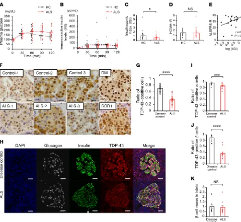
R E S E A R C H A R T I C L Ecantly different between the controls and ALS patients (Figure 1, H-K). These results indicate a possible role for nuclear TDP-43 depletion in the islets in the decreased insulin secretion of patients with sporadic ALS.
Insulin secretion is impaired in Tardbp knocked-down MIN6 cells. We examined the hypothesis that nuclear TDP-43 regulates
early-phase insulin secretion in MIN6 cells by performing in vitro knockdown experiments. The efficiency of Tardbp siRNA knock-down (set1, T1; set2, T2) was confirmed by quantitative real-time polymerase chain reaction (qRT-PCR) and immunoblotting (Fig-ure 2, A and B). Both sets of Tardbp siRNA significantly decreased glucose-induced insulin secretion in MIN6 cells (Figure 2C). The siRNA-resistant form of TDP-43 (a plasmid of mutant [mut]-mT-DP-43) recovered glucose-induced early-phase insulin secretion (Figure 2, D and E). Total internal reflection fluorescence (TIRF) imaging was used to confirm the decrease of early-phase insu-lin secretion due to Tardbp knockdown. First, we confirmed the transfection of control or T1 siRNA into MIN6 cells with mCherry (Figure 2F). We then labeled the insulin granules with GFP, and then analyzed the real-time imaging of the insulin-GFP sig-nal, which was released from the mCherry-positive MIN6 cells with glucose stimulation. The results showed that insulin-GFP signaling was decreased in Tardbp knocked-down MIN6 cells, which revealed an impaired insulin secretion (Supplemental Videos 1 and 2). Quantitative analysis revealed that the glucose- stimulated, first-phase insulin secretion was inhibited in Tardbp knocked-down MIN6 cells (Figure 2, G–I), although the number of insulin granules morphologically docked to the plasma mem-brane was not affected (Figure 2, J and K). KCL-induced insulin secretion was also significantly decreased in Tardbp knocked-down cells (Figure 2L). Collectively, these findings demonstrate the dysfunction in the first phase of insulin secretion in Tardbp knocked-down MIN6 cells.
Loss of TDP-43 reduces insulin secretion by downregulating CaV1.2 calcium channel expression in MIN6 cells. To identify the
gene expression changes that mediate Tardbp knockdown– induced impairment of early-phase insulin secretion, we per-formed microarray analysis on the MIN6 cells treated with control siRNA or T1 siRNA against Tardbp. In this analysis, we identified genes with altered expression: the expression of 338 genes was increased (>120%; Supplemental Table 1), while the expression of 611 genes was decreased (<80%; Supplemental Table 2), in the T1 siRNA–treated MIN6 cells compared with control siRNA–treated MIN6 cells (Gene Expression Omnibus database, accession num-ber GSE125424). These contained 2 voltage-dependent calcium channels, Cacna1c and Cacna2d1 (Table 2). We confirmed that T1 siRNA decreased Cacna1c and Cacna2d1 mRNA levels as well as CaV1.2 and Ca2d1 protein levels (Figure 3, A and B). However, T2 siRNA against Tardbp did not decrease the mRNA lev-els of Cacna2d1, indicating an off-target effect (Figure 3, A and B). Cotransfection of Tardbp siRNA and a CaV1.2 plasmid recovered glucose-induced insulin secretion (Figure 3, C and D), confirming that TDP-43 regulates early-phase insulin secretion via CaV1.2. In situ hybridization showed that human CACNA1C mRNA staining was substantially attenuated in the islets of patients with sporadic ALS (Figure 3E). As CACNA1D and CACNA1A are abundantly expressed in islets, we also examined the mRNA staining of those measured at 120 minutes after glucose load was significantly
higher in ALS subjects (173 ± 59 mg/dL) than in healthy control subjects (130 ± 25 mg/dL). The immunoreactive insulin level (IRI) measured at 30 minutes was significantly lower in ALS subjects (41 ± 16 μU/mL) than in healthy control subjects (63 ± 48 μU/ mL). (Figure 1, A and B). The basal insulin secretion level (fast-ing endogenous insulin secretion) was preserved in ALS subjects (Figure 1B). By contrast, the insulinogenic index (IGI), an index of early-phase insulin secretion, was lower in ALS subjects than in controls (Figure 1C). There were no significant differences in the homeostasis model assessment for insulin resistance (HOMA-IR), appendicular lean soft tissue mass, or creatinine among the groups (Figure 1D and Table 1). The IGI was positively correlated with the revised ALS functional rating scale (ALSFRS-R) (Figure 1E). Taken together, these results suggest that early-phase insu-lin secretion is decreased in the early stage of ALS and that this impairment of insulin dynamics occurs in parallel with motor dysfunction. To examine whether TDP-43 pathology is associated with the decreased insulin secretion in patients with ALS, we per-formed immunohistochemistry analysis of autopsied pancreas. We observed a substantial loss of nuclear TDP-43 in the islets of sporadic ALS subjects: the number of TDP-43–positive islet cells in ALS subjects (positive rate of 34% ± 2.6%) was decreased to half of that in controls (positive rate of 70% ± 2.0%) (Figure 1, F and G). Nuclear TDP-43 staining was preserved in the postmor-tem islets of subjects with diabetes mellitus and mutant superox-ide dismutase 1–linked (SOD1-linked) familial ALS, suggesting that the pancreatic TDP-43 pathology is specific to sporadic ALS (Figure 1F). The loss of nuclear TDP-43 was prominent in the β cells compared with α cells, though the β cell mass was not
signifi-Table 1. Baseline clinical and hematological features of healthy control and ALS subjects
Factors HC (n = 24) ALS (n = 25) P
Sex, M / F 16 / 8 18 / 7 NS
Age at examination, years 59.8 ± 7.5 63.6 ± 7.8 NS
ALSFRS-R 47.6±0.74A 36.7±9.0 <0.001
Grip power, kg 36.8 ± 9.1 19.1 ± 9.5 <0.001
Tongue pressure, kPa 37.5 ± 9.0 22.2 ± 12.1 <0.001
Body mass index, kg/m2 22.5 ± 2.6 21.6 ± 3.7 NS
ALST mass lean, ×103 kg 43.8 ± 8.2 40.8± 9.6 NS
Systolic blood pressure, mmHg 125.9 ± 17.6 125.4 ± 12.3 NS Diastolic blood pressure, mmHg 78.2 ± 9.7 75.5 ± 10.6 NS
% FVC, % 109.3 ± 15.6 90.6 ± 27.1B 0.005
Total protein, mg/dL 7.3 ± 0.3 7.0 ± 0.5 0.003
Albumin, mg/dL 4.6 ± 0.4 4.2 ± 0.5 0.003
Creatinine, mg/dL 0.77 ± 0.14 0.64± 0.27 NS
Total cholesterol, mg/dL 208 ± 44 197 ± 25 NS
Hemoglobin, g/dL 14.5 ± 1.5 13.6 ± 1.2 0.021
Hemoglobin A1c, % 5.7 ± 0.3 5.7 ± 0.5 NS
Values are mean ± SD. AFifteen healthy control subjects were included. BOne subject was excluded because he could not expire into the
[image:3.585.42.288.94.289.2]demonstrate that the dysfunction in the first phase of insulin secre-tion in Tardbp knocked-down MIN6 cells was due to the inhibisecre-tion of CaV1.2 and eventual impairment of calcium influx.
TDP-43 regulates the transcriptional activity of CaV1.2 calcium channel. As TDP-43 is a nuclear protein that has a variety of roles
in RNA metabolism, we investigated how TDP-43 controls the mRNA expression of Cacna1c. RNA immunoprecipitation anal-ysis showed that mouse WT TDP-43 binds to Cacna1c (Figure 5, A and B), and that Tardbp knockdown reduces mature Cacna1c genes. CACNA1D mRNA staining was attenuated but CACNA1A
[image:4.585.43.522.54.496.2]expression was enhanced in the islets of patients with sporadic ALS (Figure 3F). In Fura-2 imaging, Ca2+ influx was decreased in response to 30 mmol/L KCl and 50 mmol/L glucose in Tardbp knocked-down MIN6 cells (Figure 4, A–H). Furthermore, in whole-cell patch-clamp recordings of the Ca2+ current measurements, the voltage-dependent calcium channel inward currents were sig-nificantly smaller in T1 siRNA–treated MIN6 cells than in control siRNA–treated MIN6 cells (Figure 4I). Collectively, these findings
The Journal of Clinical Investigation
R E S E A R C H A R T I C L EFigure 2. Endogenous TDP-43 depletion suppresses the first phase of insulin secretion in MIN6 cells. (A) mRNA expression levels of Tardbp, encoding TDP-43 (control siRNA, Control; Tardbp siRNA, T1 or T2) (n = 7 each, 1-way ANOVA). (B) Immunoblotting of MIN6 cells transfected with control, T1, or T2 (con-trol, n = 10; T1, n = 5; T2, n = 7, 1-way ANOVA). (C) The insulin assay with low (2.8 mmol/L) and high glucose (16.7 mmol/L) (control, n = 12; T1, n = 12; T2, n = 7, 1-way ANOVA). (D) TDP-43 rescue experiments with mock plasmid or a siRNA-resistant form of murine TDP-43 (mut-mTDP) (n = 6 each, 1-way ANOVA). (E) The insulin assay with low and high glucose. mut-mTDP-43 recovered the glucose-induced insulin secretion (n = 8 each, 1-way ANOVA). (F) T1 siRNA against
Tardbp effectively reduced the protein level of TDP-43 (green). Scale bars: 5 μm. (G and H) Histogram showing the number of fusion events from GFP-tagged granules in control siRNA (G) and T1 siRNA–treated MIN6 cells (H) (per 200 μm2) at 1-minute intervals after glucose stimulation and measured by TIRF
microscopy (control, n = 11 cells; T1, n = 15 cells, unpaired t test). (I) Quantitative analysis of the total number of exocytotic events detected during the first phase (0–7 minutes) or second phase (>7 minutes). (J) TIRF imaging of insulin granules morphologically docked to the plasma membrane. Scale bar: 10 μm. (K) Number of docked insulin granules (per 200 μm2, control, n = 20; T1, n = 23 cells, unpaired t test). (L) The insulin assay with low glucose, high glucose,
luciferase assays showed that Tardbp knock-down reduces the promoter activity of both mouse Cacna1c and human CACNA1C (Fig-ure 5, G and H). In silico analysis showed that the TDP-43 protein bound to the promoter region of CACNA1C according to TARDBP chromatin immunoprecipitation sequencing (ChIP-seq) data (ENCODE phase3 data base, accession number ENCSR753GIA, https:/ /www.encodeproject.org/experiments/ ENCSR753GIA/). These results indicate that TDP-43 depletion downregulates CaV1.2 in MIN6 cells by decreasing gene tran-scripts and not by increasing RNA processing.
Early-phase insulin secretion and CaV1.2 expression are reduced in pancreas-specific Tardbp knockout mice. To confirm the role of
TDP-43 in early-phase insulin secretion in vivo, we created an AAV-mediated β cell–specific Tardbp knockout (AAV-KO) mouse mRNA levels in MIN6 cells (Figure 5, C and D). To confirm the
[image:6.585.34.358.85.146.2]nonsense-mediated decay of TDP-43 as RNA binding protein, we performed RNA stability. The results showed that there was no difference in mRNA stability of Cacna1c between the MIN6 cells treated with control siRNA and those treated with T1 Tardbp siRNA (Figure 5E). Instead, the amount of premature RNA was reduced in Tardbp knocked-down MIN6 cells (Figure 5F). Furthermore,
Table 2. Gene expression changes in Tardbp knocked-down MIN6 cells
Probe set ID P Fold change Gene symbol Gene title
1435730_at 0.010184 0.817997 Cacna1c calcium channel, voltage-dependent, L-type, alpha 1C subunit 1433643_at 0.018122 0.722274 Cacna2d1 calcium channel, voltage-dependent,
alpha2/delta subunit 1
Figure 3. CaV1.2 downregulation mediates impairment of the first phase of insulin secretion in Tardbp knockdown MIN6 cells. (A) mRNA expression levels of Cacna1c and Cacna2d1 that encode CaV1.2 and Ca2d1, respectively (control siRNA, Control; Tardbp siRNA, T1 or T2) (Cacna1c, n = 6; Cacna2d1,
[image:6.585.83.482.279.663.2]The Journal of Clinical Investigation
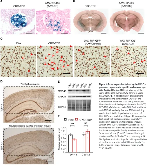
R E S E A R C H A R T I C L Eby intraperitoneally administrating AAV8 carrying Cre recombi-nase driven by rat insulin promoter (AAV-RIP-Cre) to Tardbpfl/fl mice. TDP-43 expression was attenuated in the islets of AAV-KO mice by about 30%, but not in Tardbpfl/fl mice treated with AAV-Control (Figure 6, A and B). A loss of nuclear TDP-43 was seen in the β cells but not in the α cells, though the β cell mass was not significantly different between AAV-Control and AAV-KO (Figure 6, B–E). IPGTT showed that the plasma glucose at 120 minutes was higher in AAV-KO mice than in control groups (Fig-ure 6F). Insulin secretion at 15 minutes of IGTT was decreased in AAV-KO mice (Figure 6G). In situ hybridization showed that
Cacna1c mRNA staining was substantially attenuated in the islets
of AAV-KO mice (Supplemental Figure 1, A and B). We also infected primary islets extracted from Tardbpfl/fl mice with AAV-RIP-Cre. In those primary islets, the mRNA levels of Tardbp and Cacna1c were reduced by 30% and 37%, respectively, but Cacna2d1 was not downregulated (Figure 6H). The islets infected with AAV-RIP-Cre had a significantly decreased glucose-induced insulin secre-tion compared with the control AAV-infected islets (Figure 6, I–K). Similarly, Tardbpfl/fl mice were mated with RIP-Cre transgenic mice to produce another type of β cell–specific Tardbp knockout
[image:7.585.40.443.56.445.2](CKO-TDP) mouse. The CKO-TDP mice showed glucose intoler-ance at 120 minutes with impaired insulin secretion at 15 minutes (Figure 7, A–G). The severity of glucose intolerance was greater in CKO-TDP than in AAV-KO, reflecting the fact that the effi-cacy of Tardbp knockdown is higher in CKO-TDP mice (Figure 7, F and G). To examine the time course of the insulin secretion response to 16.7 mmol/L glucose, perfusion experiments were performed. In CKO-TDP mice, the amount of secreted insulin (AUCinsulin) after glucose stimulation (from 5 to 10 minutes) was significantly less than that of Tardbpfl/fl mice (Figure 7, H and I). The Cacna1c mRNA levels were also reduced in the islets of CKO-TDP (Supplemental Figure 2, A and B). By contrast, nuclear TDP-43 staining was preserved in the islets of NOD mice, an ani-mal model of diabetes mellitus (Supplemental Figure 2C). Cre recombinase driven by the rat insulin promoter has been shown to be expressed in the hypothalamus in addition to the islet (19). In the present study, Cre recombinase was barely expressed in the hypothalamus of AAV-KO mice, but it was strongly expressed in the hypothalamus of CKO-TDP mice (Figure 8, A–C). This differ-ence appears to underlie the observation that CKO-TDP mice had a greater impairment of glucose tolerance than AAV-KO mice.
Figure 4. Fura-2 imaging and whole-cell patch-clamp recordings of Tardbp knocked-down MIN6 cells. (A and B) Fura-2 imaging of control (A) or T1 (B) siRNA–treated MIN6 cells with KCL stimulation (control,
The CaV1.2 protein was not downregulated in the hippocampus of neuron-specific Tardbp knockout mice (Figure 8, D–F). Taken together, these findings suggest that TDP-43 regulates early- phase insulin secretion from islet β cells in vivo.
Discussion
TDP-43 is a nuclear protein which possesses a variety of functions in RNA metabolism (transcription, splicing, and transport), syn-apse (synaptic vesicle transport, neurotransmitter secretion, and synaptic transmission), development, and cell morphology (3, 4, 11, 12). TDP-43 is normally localized in the nucleus and histopa-thology of ALS is characterized by the loss of TDP-43 from the nucleus as well as the presence of cytoplasmic protein aggregates containing TDP-43. Neuro-specific depletion of TDP-43 leads to a progressive neurodegenerative phenotype similar to human ALS, suggesting that TDP-43 is required for the maintenance of neuronal integrity (3, 8–10). However, the precise pathomecha-nism of neurodegeneration due to loss of TDP-43 is elusive. Here we revealed a novel function of TDP-43 as a regulator of insulin secretion. Insulin secretion consists of 2 phases, early and late, which are regulated differently. Early-phase insulin secretion is instigated by glucose uptake via glucose transporters and facil-itated by the activation of potassium channels followed by Ca2+ influx via calcium channels. This process releases the insulin pooled in vesicles via exocytosis. Our results indicate that TDP-43 facilitates insulin secretion via the transcriptional regulation of the L-type calcium channel CaV1.2, which is required for early- phase insulin release and the maintenance of systemic glucose tolerance (20, 21). The plasma glucose values during the IPGTT
in β cell–specific CaV1.2 knockout mice were shown to be similar to those of our β cell–specific Tardbp knockout mice (20). Thus, the present findings suggest that TDP-43 is a novel regulator of insulin secretion, providing a molecular basis for insulin intoler-ance in early-stage ALS subjects.
TDP-43 is a global regulator of gene expression involved in regulation of transcription, as well as a DNA- and RNA-binding protein involved in RNA processing (4, 11, 22). The TDP-43 exonic targets include RNA metabolism, synapse, development, cell morphology, and cell signaling, among others (22). Particularly, there is increasing evidence that TDP-43 controls synaptic protein expression (11, 22), although the precise mechanism has yet to be elucidated. TDP-43 overexpression impaired presynaptic integrity by downregulating soluble N-ethylmaleimide–sensitive factor (NSF) attachment receptors (SNARE) proteins including synapsin I and synaptotagmin, resulting in altered glutamate metabolism together with dysregulated excitatory-inhibitory neurotransmit-ter balance and synchrony (23). The pathogenic mutant TDP-43 upregulates HDAC6 with Bruchpilot deacetylase, leading to syn-aptic and locomotion defects in flies (24). The present study thus provides what we believe is a novel insight into the function of TDP-43 in vesicle exocytosis.
[image:8.585.45.529.57.259.2]L-type calcium channels play an important role not only in the pancreas but also in the nervous system (25, 26). The asso-ciation of TDP-43 with CaV1.2 is shown by the basis of cross- linking, immunoprecipitation, and high-throughput sequenc-ing (CLIP-seq or HITS-CLIP) experiments in the mouse brain (11). In Drosophila, lack of neuronal TDP-43 results in locomo-tion deficits, which are restored by the exogenous expression of
The Journal of Clinical Investigation
R E S E A R C H A R T I C L Etion by exogenous CaV1.2 strongly indicate the primary roles of calcium channels in the regulation of insulin secretion by TDP-43. This view is further supported by physiology experi-ments using MIN6 cells.
Glucose intolerance in patients with ALS has been repeat-edly reported, but the pathophysiologic mechanisms have been elusive (13, 15–18, 28, 29). Glucose intolerance may result from insulin resistance with a marked loss of skeletal muscle (17, 18, 29) and/or impaired insulin secretion (15, 16, 28). Our results suggest that the loss of nuclear TDP-43 underlies the impaired early-phase secretion of insulin in early-stage ALS subjects. a voltage-gated calcium channel (26). Furthermore, L-type
cal-cium channel agonists ameliorate motor impairment in zebra-fish larvae bearing a TDP-43 mutation (25). Given that subtle synaptic dysfunction has been postulated to be a primary event in the initial pathophysiology of ALS (9, 27), L-type calcium channels may also be affected by the loss of TDP-43 in motor neurons, though further work is needed to verify this hypothe-sis. Although the possibility that there is a CaV1.2-independent pathway in the TDP-43–mediated regulation of insulin secre-tion cannot be excluded, the decreased Ca2+ entry upon KCL stimulation by Tardbp knockdown and rescue of insulin
secre-Figure 7. Insulin secretion is suppressed in Tardbp flox mouse mated with RIP-Cre mouse (CKO-TDP). (A) Immunofluorescence of the islets in Tardbpfl/fl
and CKO-TDP mice. Scale bars: 10 μm. (B) Ratio of TDP-43–positive total cells (n = 8 each, unpaired t test). (C) Ratio of TDP-43-positive α cells (n = 10 each, unpaired t test). (D) Ratio of TDP-43–positive β cells (n = 10 each, unpaired t test). (E) β cell mass in islets (n = 12 each, unpaired t test). (F) IPGTT showed that plasma glucose measured at 120 minutes was significantly higher in CKO-TDP mice than in Tardbpfl/fl mice (n = 10 each, P < 0.0001, ANOVA). (G) Insulin
measurement at 0 minutes (fasting) and 15 minutes after intraperitoneal glucose load (n = 10 each, unpaired t test). (H) Insulin secretion in perfused pancreas in response to high glucose (16.7 mmol/L) (n = 4 each). (I) The amounts of secreted insulin of Tardbpfl/fl and CKO-TDP mice after glucose stimulation were
[image:10.585.37.531.62.504.2]The Journal of Clinical Investigation
R E S E A R C H A R T I C L EMethods
ALS patients and oral glucose tolerance test. We enrolled 25
consecu-tive Japanese ALS patients without diabetes mellitus or family history of ALS (Table 1). All patients were diagnosed as definite, probable, or probable laboratory-supported ALS under the El Escorial and the revised Airlie House diagnostic criteria. We compared these ALS patients with 24 age- and sex-matched Japanese volunteers without Our observation that the IGI, an index of insulin secretion,
[image:11.585.42.549.60.634.2]correlated with motor function measured with the ALSFRS-R further implies that motor neuronal damage occurs in parallel with β cell dysfunction in patients with ALS. This view is likely supported by the fact that islet β cells and motor neurons share key molecules, such as islet1 and HB9, during their develop-ment (30, 31).
Figure 8. Brain expression driven by the RIP-Cre promoter in pancreatic-specific and neuron-spe-cific Tardbp KO mice. (A) X-gal staining of the islets of the CKO-TDP and AAV-KO mice. Scale bar, 20 μm. (B) X-gal staining of brain sections including the hypothalamus in CKO-TDP and AAV-KO mice. Scale bars: 500 μm. (C) Immuno-histochemistry of the hypothalamus in Tardbpfl/fl,
CKO-TDP, AAV-Control, and AAV-KO mice with an anti–TDP-43 antibody. Nuclear TDP-43 staining was lost only in the hypothalamic neurons of CKO-TDP mice, Scale bars: 20 μm. (D) Immunohis-tochemistry of the hippocampus in Tardbpfl/fl
and neuron-specific Tardbp knockout mouse with an anti–TDP-43 antibody. Nuclear TDP-43 staining was lost in the neuron of cortices and CA1 in neuron-specific Tardbp knockout mouse. Scale bars: 20 μm. (E and F) Immunoblotting of cortices and CA1 in Tardbpfl/fl and neuron-specific Tardbp knockout mice, together with quantitation
Japan). The cells were cultured in Dulbecco’s modified Eagle’s medium containing 10% (vol/vol) FBS (10270, Life Technologies) with 5% penicillin/streptomycin (15140-122, Wako) and β-mercaptoethanol (M6250, Sigma-Aldrich) at 37°C and 5% CO2. MIN6 cells seeded at a density of 3.0 × 10⁵ cells on a 12-well plate were electroporated with 100 pmol siRNA (Control, TDP-43 set1, and TDP-43 set2) using the NEON Transfection System (electroporation: 1200 V pulse, 20 ms width; Thermo Fisher Scientific) according the manufacturer’s procedure, unless otherwise mentioned. Oligonucleotide siRNA duplexes were synthesized by Sigma-Aldrich with the following sequences: control-siRNA, 5′-GAAUCAGAUGCACAUGAGUTT-3′;
Tardbp siRNA-set1-T1, 5′-GAACGAUGAACCCAUUGAATT-3′; and Tardbp siRNA-set2-T2, 5′-GUUCUUAUGGUUCAGGUCATT-3′.
Unless otherwise mentioned, set1 siRNA (T1) was used for Tardbp knockdown throughout the experiments. The siRNA-resistant form of the TDP-43 gene (mut-mTDP-43) was generated as described else-where (7). In the TDP-43 and CaV1.2 rescue experiments, MIN6 cells seeded at a density of 3.0 × 10⁵ cells on a 12-well plate were electro-porated with 100 pmol control siRNA, 2 μg mock plasmid, 100 pmol T1-siRNA, and either 2 μg mut-mTDP-43 or 50 ng v5-His-CaV1.2 plasmid (as appropriate), using the NEON Transfection System as described above. The pCDNA6/V5-His-CaV1.2 (v5-His-CaV1.2) con-struct was purchased from Addgene (plasmid 26572). At 48 hours after transfection, MIN6 cells were collected and used for qRT-PCR, immunoblotting, and measurement of insulin secretion.
RNA extraction and qRT-PCR. mRNA levels were measured by
qRT-PCR as previously described (38, 39). RNA was prepared from MIN6 cells using an RNeasy Mini Kit (74106, Qiagen) according to the manufacturer’s instructions. Total RNA (500 ng) was used as a tem-plate for reverse transcription using ImProm-II Reverse Transcrip-tase (A3800, Promega). qRT-PCR was performed using the primers listed in Table 3 and KAPA SYBR FAST qPCR Master mix (KK4602, KAPA Biosystems) according to the manufacturer’s instructions. Data were shown as the ratio of the mRNA level to that of Gapdh.
Immunoblotting. MIN6 cells were scraped from the culture dish
and homogenized with RIPA buffer (182-02451, Wako) containing 1% protease inhibitors (4693116001, Roche) and phosphatase inhibitors (1862495, Thermo Fisher Scientific). Homogenates were incubated on ice for 20 minutes and centrifuged at 13,000g for 10 minutes at 4°C. The lysates were mixed with 4 times NuPAGE LDS sample buffer (NP0008, Novex) and heated at 95°C for 5 minutes. After denatur-ation, each cell lysate was separated by SDS-PAGE (5%–20% gradient gel) and analyzed by immunoblotting with ECL Plus detection reagents (NEL121001EA, PerkinElmer) using the following primary antibod-ies: anti–TDP-43 rabbit polyclonal (1:3000, 10782-2-AP; Proteintech), anti-GAPDH mouse monoclonal (1:1000; M171-3, MBL International Corporation), anti-CaV1.2 rabbit polyclonal (1:2000; ACC-003, Alo-mone Labs), anti-Ca2d1 (E10) mouse monoclonal (1:100, sc-271697; Santa Cruz Biotechnology), and anti–v5-Tag (D3H8Q) rabbit poly-clonal (1:1000, 13151; Cell Signaling Technology). Band intensity was quantified by Multi Gauge 3.0 software (Fujifilm).
Insulin secretion in MIN6 cells following glucose stimulation. MIN6
cells were cultured at a density of 3.0 × 10⁵ cells on a 12-well plate for 48 hours after electroporation (MPK10025, Thermo Fisher Scientific) as described above. The cells were preincubated with a 2.8 mmol/L (low glucose), Krebs-Ringer Modified Buffer (KRB) containing 10 mM HEPES (pH 7.4), 110 mM NaCl, 4.4 mM KCl, 1.45 mM KH2PO4, 1.2 neurological disease and diabetes mellitus. All patients were followed
in Nagoya University Hospital. The data were collected from May 2013 and August 2014.
All patients underwent a 75-gram OGTT after a 12-hour over-night fast. The plasma glucose and immunoreactive insulin levels were measured before glucose loading and every 30 minutes until 120 minutes after glucose loading. OGTT was performed via feeding tubes in the 2 ALS patients with gastrostomy tubes and orally in the remaining 23 ALS patients and in 24 healthy controls. In an effort to examine glucose metabolic pathology, we calculated several indices of insulin secretion and resistance by measuring C-peptide immuno-reactivity, fasting and GTT plasma glucose, and IRI. The IGI, a mea-sure of early-phase insulin secretion, defined as the ratio of the incre-ment of insulin to that of plasma glucose 30 minutes after a glucose load, was calculated with the formula: IGI = [(IRI30 – IRI0) / (PG30 – PG0)], where IRI0 is fasting plasma insulin (μU/mL); IRI30 is insulin 30 minutes after glucose load (μU/mL); PG0 is fasting plasma glucose (mg/dL); and PG30 is plasma glucose 30 minutes after glucose load (mg/dL) (32, 33). Insulin secretory insufficiency was defined as an IGI <0.4 (34). The serum C-peptide immunoreactivity index (CPI), an index of fasting endogenous insulin secretion, was calculated by the formula: CPI = [fasting serum C-peptide levels (ng/mL) × 100 / PG0 (mg/dL)] (35). The HOMA-IR, which primarily reflects hepatic insulin resistance, was calculated by the formula: HOMA-IR = [PG0 × IRI0 / 405] (36). The measured biochemical parameters included serum creatinine and appendicular lean soft tissue (ALST) mass as indices of skeletal muscle mass (37).
Immunocytochemistry for human autopsy samples. We analyzed
autopsied pancreatic islet specimens from patients with ALS and from age-matched disease control subjects. Pathological diagnoses of the disease controls were hepatic failure, Parkinson’s disease, and diabetes mellitus. The ALS pancreatic islets were excised at autopsy and fixed immediately in a 10% buffered formalin solution. Sections (3 μm) were deparaffinized, heated in a microwave for 15 minutes in 10 mM citrate buffer (pH 6.0), and incubated overnight with an anti–TDP-43 rabbit polyclonal antibody (1:6000; 10782-2-AP, Pro-teintech). Subsequent staining procedures were performed using the DAKO EnVision+ HRP System (Dako) and photographed with an opti-cal microscope (BZ-X710; Keyence) as previously described (38, 39).
Immunofluorescence for human autopsy samples. The ALS pancreatic
islets were excised at autopsy and fixed immediately in a 10% buff-ered formalin solution. Sections (3 μm) were deparaffinized, heated in a microwave for 15 minutes in 10 mM citrate buffer (pH 6.0), and incubated overnight with the following primary antibodies: anti– TDP-43 rabbit polyclonal (1:1000; 10782-2-AP, Proteintech), anti- insulin pig (1:200; ab7842, Abcam), and anti-glucagon mouse (1:500; ab10988, Abcam). After washing, the samples were incubated with Alexa-488–conjugated donkey anti-pig IgG (1:1000; A11073, Invitro-gen), Alexa-488–conjugated donkey anti-mouse IgG (1:1000; R37114, Invitrogen), Alexa-546–conjugated donkey anti-rabbit IgG (1:1000; A10040, Invitrogen), and Alexa-647–conjugated donkey anti-mouse IgG (1:1000; A31571, Invitrogen) for 1 hour, mounted using ProLong Gold with DAPI (P36935, Thermo Fisher Scientific), then imaged with a laser confocal microscope (LSM880; Zeiss).
Cell culture and transfection. MIN6 β cells, a line derived from
The Journal of Clinical Investigation
R E S E A R C H A R T I C L Ecopy, MIN6 cells were incubated for 20 minutes at 37°C in KRB. The cells were then transferred to the stage of a TIRF microscope and glucose stimulation was achieved by the addition of 40 mM glucose in KRB into the chamber (final concentration 22 mM glucose).
To evaluate the number of docked insulin granules by TIRF microscopy, MIN6 cells transfected with mCherry and control or
Tardbp siRNA were cultured on high-refractive-index glass, fixed,
permeabilized with 4% paraformaldehyde/0.1% Triton X-100, and processed for immunohistochemistry. The cells were labeled with an anti–insulin antibody (I2018, Sigma-Aldrich) and processed with goat anti-mouse IgG conjugated to Alexa Fluor 488 (A-11001, Invitrogen). For the TIRF imaging of Alexa Fluor 488–labeled insulin granules in cells expressing mCherry, we used the 488-nm and 561-nm laser line for excitation and an image splitter (W-view Gemini; Hamamatsu Photonics) that divided the green and red components of the images with a 565-nm dichroic mirror (Q565; Chroma Technology Corpora-tion), passing the green component through a 530 nm ± 15 nm band-pass filter (HQ530/30 m; Chroma Technology Corporation) and the red component through a 630 nm ± 25 nm bandpass filter (HQ630/50 m; Chroma Technology Corporation) (43).
Measurement of intracellular Ca2+ concentration. Intracellular free
Ca2+ concentration ([Ca2+]i) was estimated by microfluorometry in MIN6 cells loaded with Fura-2 AM (4987481523647, Dojindo Labora-tories) as previously described (44). MIN6 cells, electroporated with control siRNA or Tardbp siRNA (T1), were incubated for 60 minutes at room temperature with the acetoxymethyl ester Fura-2 (10 μM) in the standard KRB solution (2.8 mmol/L). The Fura-2–loaded MIN6 cells were attached to glass coverslips, which were then covered with 500 μL KRB for 2–4 minutes, KCL (30 mmol/L) for 1 minute, and KRB for 2–4 minutes at 37°C on the stage of an inverted microscope (Olympus IX). Alternatively, the experiment was performed with 500 μL KRB for 2–4 minutes and 50 mM glucose for 7 minutes at 37°C. Microfluo-rometry was performed on 10–20 MIN6 cells illuminated alternately at 340 and 380 nm. Fluorescence intensities (F340 and F380) were mea-sured at 510 nm. Changes in [Ca2+]i are presented as changes in the F340/F380 fluorescence ratio.
Whole-cell voltage-clamp recordings. MIN6 cells were transfected
with 500 pmol/2 mL siRNA using Lipofectamine 3000 (L3000015, Invitrogen). The cells were used 48–72 hours after transfection. The bath solution contained 140 mM NaCl, 5 mM KCl, 5 mM EGTA, 6.9 mM BaCl2, (approximately 2 mM free Ba2+ calculated by the MAXC program, Stanford University), 2 mM MgCl2, 10 mM glucose, and 10 mM HEPES, pH 7.40, adjusted with NaOH. The pipette solution con-tained 140 mM NMDG-Cl, 5 mM EGTA, and 10 mM HEPES, pH 7.30, adjusted with NMDG. Pipette resistances were 3 ± 1 MΩ. The currents were recorded with step pulses from –60 mV to +20 mV in 20-mV increments from a –90 mV holding potential using an Axopatch 200B amplifier (Molecular Devices), filtered at 5 kHz with a low-pass filter, and digitized with Digidata 1440A (Axon Instruments). Data were acquired with pCLAMP 10 (Axon Instruments).
Immunoprecipitation. The pcDNA3.1/V5-His-mouse TDP-43
(GenBank accession number NM_145556, v5-mTDP-43-WT) con-struct was produced as previously described (7). Total lysates of MIN6 cells transfected with 2 μg v5-mTDP-43-WT by electroporation (1.0 × 10⁶ cells, 1200 V, 20 ms, 2 pulses) on a 6-well plate were mixed with 2 μg rabbit IgG as control or 2 μg anti–v5-Tag (D3H8Q) rabbit poly-clonal antibody (13151, Cell Signaling Technology), which binds to mM MgSO4, 2.3 mM calcium gluconate, 4.8 mM NaHCO3, 4 mM
glu-cose, and 0.3% bovine serum albumin for 30 minutes, and stimulated with 16.7 mmol/L (high glucose) glucose and 30 mmol/L KCL for 30 minutes. Insulin levels in the supernatant (release) and acid ethanol extraction (content) were measured using an Insulin Ultra-sensitive Assay kit (62IN2PEG, CisBio). The amount of secreted insulin was calculated as a ratio of release to content.
Microarray analysis of MIN6 cells transfected with control and Tardbp siRNA. We analyzed alterations in gene expression in MIN6
cells transfected with control and Tardbp siRNA, using GeneChip Mouse Genome 430 2.0 array (Affymetrix). Total RNA was isolated from the MIN6 cells treated by control or Tardbp T1 siRNA accord-ing to the manufacturer’s instructions (RNeasy, Qiagen). The cDNA preparation, hybridization process, and microarray data analysis were performed at RIKEN Genesis. Biotinylated cRNA was prepared using GeneChip 3′IVT PLUS Reagent Kit (Thermo Fisher Scientific) accord-ing to the manufacturer’s standard protocol from 100 ng total RNA. Following fragmentation, 12.5 μg cRNA was hybridized for 16 hours at 45°C on a GeneChip Mouse Genome 430 2.0 Array. GeneChips were washed and stained in the GeneChip Fluidics Station 450. GeneChips were scanned using the GeneChip Scanner 3000 7G. The data were analyzed with Microarray Suite version 5.0 (MAS 5.0) using Affymet-rix Expression Console Software (Thermo Fisher Scientific) according to the default analysis settings. The criteria used to detect the differ-ences in gene expression were a 1.2-fold change in MIN6 cells treated with control siRNA compared with Tardbp siRNA. The microarray pro-filing data were deposited in the NCBI’s Gene Expression Omnibus database (GSE125424).
TIRF imaging for insulin secretion by MIN6 cells transfected with control or Tardbp siRNA. MIN6 cells were plated on fibronectin-
coated high-refractive-index glass for imaging by TIRF microscopy or on fibronectin-coated normal coverslips for immunostaining (40). After 18–24 hours, the cells were transfected with 50 nM various vectors (insulin-GFP, ref. 41), mCherry (42), and siRNAs (control or
Tardbp siRNAs) using Lipofectamine 2000 (11668-019, Invitrogen)
in the presence of serum, according to the manufacturer’s instruc-tions. The experiments were performed 3 days after transfection. The Olympus total internal reflection system was used with a high- aperture objective lens (Apo ×100 OHR, numerical aperture 1.65; Olympus) essentially as previously described (43). For real-time imag-ing of the insulin granules labeled by insulin-GFP under TIRF micros-Table 3. Primer sequences
Gene
symbol Forward primer Reverse primer
Tardbp CCGCATGTCAGCCAAATACAAG ACCAGAATTGGCTCCAACAACAG
Gapdh TGTGTCCGTCGTGGATCTGA TTGCTGTTGAAGTCGCAGGAG
Actin GGCTGTATTCCCCTCCATCG CCAGTTGGTAACAATGCCATGT
Cacna1c TCCTCATCGTCATTGGGAGC AGATGCGGGAGTTCTCCTCT
Cacna2d1 TCTTCAGCCAAAGAACCCCA ATTGACAGGCGTCCATGTGT
Exon30-32 TCCTCATCGTCATTGGGAGC AGATGCGGGAGTTCTCCTCT Exon2 CCCGGATGTACTGAGGATGC CAATGCTTATGCACGCCCTC
Cacna1c Exon47 TGCCTCACTGTTCTCGTGAC GCTCTGAGGCTTATCCCGAC
[image:13.585.41.288.84.225.2]In silico analysis. We extracted the TARDBP ChIP-seq data from the
NCBI Gene Expression Omnibus database (ENCSR753GIA, ENCODE phase3, https://www.encodeproject.org/experiments/ENCSR753GIA/). Using the UCSC genome browser, we confirmed the presence of a binding site for TADBP protein in CACNA1C promoter region (human
CACNA1C promoter, ID S71093, LightSwitch Assay; Active Motif). Animals. All mice (background C57BL/6J) were maintained at 25°C
in a light-controlled (12-hour light-dark cycle) environment (3–5 mice per cage) with food and water provided ad libitum. Tardbpfl/fl mice were generated using the Cre/LoxP recombination system as previously described (9). RIP-Cre transgenic mice (B6.Cg-Tg[Ins2-cre]25Mgn/J, 3573) were purchased from The Jackson Laboratory. NOD female 10-week-old mice (NOD/Shi JIC) were purchased from CLEA-Japan.
Recombinant AAV production. Modified pBluescript II
KS/RIP-Cre-ER (DM 265) was purchased from Addgene (plasmid 15029). The AAV vector plasmids contained an expression cassette consisting of a RIP, Cre recombinase, and poly A tail (RIP-Cre), and RIP-GFP between the inverted terminal repeats of the AAV3 genome. The recombinant AAV vectors were produced by transient transfection of HEK293 cells with the vector plasmid, an AAV8 vp expression plasmid, and an adenoviral helper plasmid (pHelper, Agilent Technologies), as previously described (45). The recombinant viruses were purified by isolation from 2 sequential continuous CsCl gradients, and viral titers were determined by qPCR.
Pancreas-specific Tardbp knockout mice. Male Tardbpfl/fl mice (8–10
weeks old) were injected intraperitoneally with normal saline, 4.0 × 10¹¹ vector genomes (vg)/mouse AAV8-RIP-GFP (AAV-Control), or 4.0 × 10¹¹ vg/mouse AAV8-RIP-Cre (AAV-KO) as Tardbp knockout mice. The IPGTT and insulin test were performed 2 weeks after injection. RIP-Cre mice were purchased from The Jackson Laboratory (strain B6.Cg-Tg[Ins2-cre]25Mgn/J, 3573). Pancreas-specific Tardbp knock-out mice were generated by mating Tardbpfl/fl mice with RIP-Cre mice (CKO-TDP). In Tardbpfl/fl and CKO-TDP mice without AAV injection, the IPGTT and insulin test were performed in 6-week-old male mice.
Insulin and glucose tolerance tests in mice. After fasting for 16 hours
(17:00–9:00), 2.0 g/kg bodyweight D-glucose was injected intraper-itoneally into the mice. Plasma glucose levels were measured after 0, 15, 30, 60, and 120 minutes using the Glucogard G Sensor (Arkray). Plasma insulin levels were measured after 0 and 15 minutes using a mouse insulin enzyme-linked immunosorbent assay kit (Morinaga Institute of Biological Science).
Immunofluorescence for islets. Pancreatic islets were excised from
mice with perfusion fixation using 4% paraformaldehyde, and fixed immediately in a 10% buffered formalin solution. Sections (3 μm) were deparaffinized and heated in a microwave for 15 minutes in 10 mM citrate buffer (pH 6.0). The following primary and secondary anti-bodies were the same as used for human samples: anti–TDP-43 rabbit polyclonal, anti-insulin pig, and anti-glucagon mouse.
Pancreatic islet isolation. Pancreatic islets were isolated from
Tardbpfl/fl mice at 8–10 weeks of age by modifying a previously
described procedure (46). Briefly, the pancreas of an anesthetized mouse was distended via the pancreatic duct with 3 ml RPMI 1640 (11875119, Thermo Fisher Scientific) containing 1.5 mg/mL colla-genase (C7657, Sigma-Aldrich). The distended pancreas was then excised and digested in a water bath at 37°C for 11.5 minutes, and then shaken manually 5 times in sets of 7. The digest was washed 3 times with 25 mL RPMI 1640 containing 10% (vol/vol) FBS (10270, Life WT mTDP-43, and rotated at 4°C for 1 hour. Protein G Dynabeads
(10004D, Invitrogen) were added to the mixture and further incubated at 4°C for 2 hours. The beads were washed 4 times with RIPA buffer (182-02451, Wako) containing 1% protease inhibitors (4693116001, Roche) and phosphatase inhibitors (1862495, Thermo Fisher Scientific). The beads were mixed directly with 2× NuPAGE LDS-PAGE sample buffer (NP0008, Novex) containing β -mercap-toethanol, heated at 70°C for 10 minutes, and analyzed by immuno-blotting using an anti–TDP-43 rabbit polyclonal antibody (1:3000, 10782-2-AP; Proteintech) and GAPDH mouse monoclonal anti-body (1:1000; M171-3, MBL International Corporation).
Ribonucleoprotein immunoprecipitation. Ribonucleoprotein
immu-noprecipitation was performed using the RIP Assay Kit (RN1001, MBL International Corporation) according to the manufacturer’s instruc-tions. RNA concentrations were measured with the NanoDrop system (Thermo Fisher Scientific). RNA obtained from immunoprecipitates was reverse transcribed into first-strand cDNA using ImProm-II Reverse Transcriptase (A3800, Promega). qRT-PCR was performed using the primers listed in Table 3 with KAPA SYBR FAST qPCR Master mix (KK4602, KAPA Biosystems).
Luciferase assay for mouse Cacna1c promoter. To analyze the
puta-tive promoter activity of mouse Cacna1c (CaV1.2, gene ID 12288), a series of reporter plasmid constructs were made using the backbone of the pGL3-Basic reporter vector and pRL-TK vector (E2231, Promega). To clone the 5′-flanking region of mouse Cacna1c as a putative pro-moter region, the following 2 primers were used: 5′-GTAGAGCTC-CACAGTGCCA-3′, which contained a 10-bp SacI DNA fragment (–1542 to –1533), and 5′-GGACTCGAGTGTCTGGTTG-3′, which contained a 10-bp XhoI Cacna1c gene fragment (+257 to +248). The
Cacna1c promoter was amplified by PCR from a BAC clone
(RP23-123O15, ID 638275; Advanced GenoTechs), and contained a 1799-bp
SacI/XhoI insertion (–1542 to +257). Amplified DNA was ligated into
the SacI and XhoI sites of the pGL3-Basic-reporter vector (Promega), generating a 1799-bp fragment (–1542 to +257) of the CaV1.2 promot-er in pGL3-Basic. The construct was vpromot-erified by sequencing. MIN6 cells seeded at 9.0 × 10³ cells on a 96-well plate were transfected with 100 pmol siRNA (Control, T1), 10 ng pGL3-Basic-CaV1.2 pro-moter, and 5 ng pRL/SV40 (Promega) using the NEON Transfection System (Thermo Fisher Scientific) as described above. At 48 hours after transfection, luciferase activity was measured using the Dual- Luciferase Reporter Assay System (E1910, Promega) according to the manufacturer’s instructions.
Human CACNA1C promoter reporter assay. HEK293T cells seeded
at 9.0 × 10³ cells on a 96-well plate were transfected with 100 pmol siRNA (Control, T1) and 50 ng GoClone plasmid DNA (human
CACNA1C promoter, ID S71093) using Lipofectamine 2000
(11668-019, Invitrogen) according to the manufacturer’s instructions. At 48 hours after transfection, luciferase activity was measured using the LightSwitch Assay Reagent (LS010, SwitchGear Genomics) according to the manufacturer’s instructions.
RNA stability. Actinomycin D (final concentration 10 μg/mL), a
The Journal of Clinical Investigation
R E S E A R C H A R T I C L Estat and stained with the same method as for the brains (MIR 2600; Mirus Bio LLC).
Neuron-specific Tardbp knockout mice. We crossed Tardbpfl/fl
mice with CamkII-Cre mice (49) (gift from Tsuyoshi Miyakawa, Fujita Health University, Toyoake, Japan) and generated Tardbpfl/fl/ CamKII-Cre+ mice as neuron-specific Tardbp knockout mice. The brains were excised from the neuron-specific Tardbp knockout mice at 12 weeks of age with perfusion fixation using 4% paraformalde-hyde, and fixed immediately in a 10% buffered formalin solution. Sections (3 μm) were deparaffinized and heated in a microwave for 15 minutes in 10 mM citrate buffer (pH 6.0). The anti–TDP-43 rab-bit polyclonal was the same as for human samples. Horizontal slices of the hippocampus were excised and homogenized in RIPA buffer (182-02451, Wako) containing 1% protease inhibitors (4693116001, Roche) and phosphatase inhibitors (1862495, Thermo Fisher Scien-tific) with a Nippi Biomasher II homogenizer. Homogenates were incubated on ice for 20 minutes and centrifuged at 13,000g for 10 minutes at 4°C. The lysates were mixed with 4× NuPAGE LDS sam-ple buffer (NP0008, Novex) and heated at 95°C for 5 minutes. After denaturation, each cell lysate was separated by SDS-PAGE (5%–20% gradient gel) and analyzed by immunoblotting with ECL Plus detec-tion reagents (NEL104001EA, PerkinElmer) using the following primary antibodies: anti–TDP-43 rabbit polyclonal (1:3000; 10782-2-AP, Proteintech), GAPDH mouse monoclonal (1:1000; MBL Inter-national Corporation), and anti-CaV1.2 rabbit polyclonal (1:2000; ACC-003, Alomone Labs). Band intensity was quantified by Multi Gauge 3.0 software (Fujifilm).
Statistics. For qRT-PCR and immunoblotting in vitro, the data
were obtained from at least 3 independent experiments and are pre-sented as relative value to the control. For the IPGTT and insulin test in vivo, at least 3 animals were in each group. Statistical analy-sis throughout the study was performed by applying a 2-tailed Stu-dent’s t test for 2-group comparison and 1-way ANOVA with Turkey post hoc test for multi-group comparison. In all experiments and for clinical studies, graph and other data are expressed as mean ± SEM. Significance was defined at P less than 0.05 using the SPSS 23.0J sta-tistical software package. In clinical studies, intergroup differences in categorical and continuous variables were assessed using unpaired t test, and correlation coefficients were assessed using the Pearson cor-relation. For experiments using cell cultures and mice, the number of experiments or animals is stated in the figure legends.
Study approval. This human study adhered to the ethics guidelines
for human genome/gene analysis research and those for medical and health research involving human subjects endorsed by the Japanese government, and was approved by the ethics review committees of Nagoya University Graduate School of Medicine. All participants pro-vided written informed consent. Experimental procedures involving human subjects were conducted according to the principles expressed in the Declaration of Helsinki. Animal protocols were approved by the IACUC at Nagoya University.
Author contributions
MK, KA, and AA contributed to the study concept and design. KA, AA, DH, TI, A Hashizume, YH, SY, YI, A Hara, KI, KK, SI, YN, ST, YS, AY, YT, SH, MT, MOI, AS, HI, AE, MY, HA, SM, GS, and MK conducted experiments and acquired data. KA, AA, MI, and MK drafted the manuscript and figures.
Technologies), and then filtered through a tea strainer. The filtered tissue was centrifuged (1000g for 2 minutes), and the pellet resus-pended with 10 mL Ficoll-Paque PLUS (17144002, GE Healthcare) in a 50-mL conical tube. Next, it was overlayed slowly with 10 mL RPMI 1640 containing 10% (vol/vol) FBS, which was centrifuged (1000g for 20 minutes at 4°C). Isolated islets appeared at the intermediate phase and were moved on the cell culture dish. The islets were isolated by hand-picking using a pipette with RPMI 1640 containing 10% (vol/ vol) FBS, and incubated for 2 hours in 5% CO2 incubator at 37°C.
Infection of isolated islets by AAV. The isolated islets were cultured
with 1.2 × 10¹¹ vg/mL AAV8-RIP-GFP or AAV8-RIP-Cre for 36 hours. Then mRNA levels were measured with qRT-PCR as described above for MIN6 cells, and the insulin secretion of infected islets was mea-sured with an Insulin Ultra-Sensitive Assay kit (62IN2PEG, CisBio) as described above for MIN6 cells. The 10 isolated islets were prein-cubated on 96-well plate with a 2.8 mmol/L glucose (low glucose) for 30 minutes, and stimulated with 16.7 mmol/L glucose (high glucose) for 30 minutes.
Perfusion experiments of Tardbpfl/fl and CKO-TDP mice. Perfusion
experiments were performed as previously described (47, 48). Briefly, 10- to 12-week-old male mice were fasted overnight (16 hours). The perfusion protocol began with a 10-minute equilibration period with the same buffer used in initial step (1–5 minutes; 2.8 mM glucose) and glucose loading step (5–25 minutes, 16.7 mM glucose) (Figure 6). The insulin levels in the perfusate were measured with an Insulin Ultra-Sensitive Assay kit (62IN2PEG, CisBio).
In situ hybridization. We purchased 4 probes against mouse Cacna1c (NM_009781.4, bp4846–6605, ACD445451), human CACNA1C (NM_199460.3, bp501–2144, ACD460061), human CACNA1D (NM_000720.3, bp544-1957, ACD544881), and human CACNA1A (NM_023035.2, bp646-1855, made-to-order ACD320269)
from Advanced Cell Diagnostics, and applied them to 3-μm sections of paraffin-embedded pancreas samples according to the RNAscope Sample Preparation and Pretreatment Guide for FFPE Tissue Part 1 (catalog 320511). We used the single-plex RNAscope assay according to the RNAscope 2.5 HD Detection Kit (BROWN) User Manual Part 2 (catalog 320497), containing a number of channel 1 probes against mouse Cacna1c, human CACNA1C, human CACNA1D, and human
CACNA1A molecules detected with DAB. A semiquantitative scoring
scale was used according to the manufacturer’s guidelines to evaluate the staining results: 0, no staining or less than 1 dot per 10 cells; 1, 1–3 dots per cell; 2, 4–9 dots per cell with no or very few dot clusters; and 3, 10–15 dots per cell and fewer than 10% dots are in clusters. Scoring was performed at ×60 magnification.
X-gal staining for Cre recombinase driven by the RIP promoter.
Cryo-Committee of CNS Degenerative Diseases, Research on Policy Planning and Evaluation for Rare and Intractable Diseases, Health, Labor and Welfare Sciences Research Grants, the Minis-try of Health, Labor and Welfare, Japan (to MY), a grant from the Naito Foundation (to MK), and a grant from the Hori Sciences & Arts Foundation (to MK).
Address correspondence to: Masahisa Katsuno, Department of Neurology, Nagoya University Graduate School of Medicine, 65 Tsurumai-cho, Showa-ku, Nagoya 466-8550 Japan. Phone: 81.52.744.2385; Email: ka2no@med.nagoya-u.ac.jp.
Acknowledgments
We thank Mika Ito and Naomi Takino (Jichi Medical Universi-ty) for their help with the production of the AAV vectors. This work was supported by Grants-in-Aid (KAKENHI) from the Ministry of Education, Culture, Sports, Science, and Technology (MEXT) of Japan (grants 16K09742, 16K15480, 17H04195, and 17K08547 to MK; grants 16K09392 and 16K09393 to HI). This work was partly supported by grants from the Japan Agency of Medical Research and Development (AMED) (grants JP17dm0107105h0002 and JP16kk0205009h0001 to MY). This work was also supported by Grants-in-Aid from the Research
1. Arai T, et al. TDP-43 is a component of ubiquitin- positive tau-negative inclusions in frontotem-poral lobar degeneration and amyotrophic lateral sclerosis. Biochem Biophys Res Commun. 2006;351(3):602–611.
2. Neumann M, et al. Ubiquitinated TDP-43 in fron-totemporal lobar degeneration and amyotrophic lateral sclerosis. Science. 2006;314(5796):130–133. 3. Wegorzewska I, Baloh RH. TDP-43-based animal models of neurodegeneration: new insights into ALS pathology and pathophysiology. Neurodegener Dis. 2011;8(4):262–274.
4. Scotter EL, Chen HJ, Shaw CE. TDP-43 prote-inopathy and ALS: insights into disease mecha-nisms and therapeutic targets. Neurotherapeutics. 2015;12(2):352–363.
5. DeJesus-Hernandez M, et al. Expanded GGGGCC hexanucleotide repeat in noncoding region of C9ORF72 causes chromosome 9p-linked FTD and ALS. Neuron. 2011;72(2):245–256. 6. Renton AE, et al. A hexanucleotide repeat
expan-sion in C9ORF72 is the cause of chromosome 9p21-linked ALS-FTD. Neuron. 2011;72(2):257–268. 7. Iguchi Y, et al. TDP-43 depletion induces
neuronal cell damage through dysregula-tion of Rho family GTPases. J Biol Chem. 2009;284(33):22059–22066.
8. Wu LS, Cheng WC, Shen CK. Targeted depletion of TDP-43 expression in the spinal cord motor neurons leads to the development of amyo-trophic lateral sclerosis-like phenotypes in mice. J Biol Chem. 2012;287(33):27335–27344. 9. Iguchi Y, et al. Loss of TDP-43 causes
age-depen-dent progressive motor neuron degeneration. Brain. 2013;136(Pt 5):1371–1382.
10. Yang C, et al. Partial loss of TDP-43 function causes phenotypes of amyotrophic lateral sclerosis. Proc Natl Acad Sci U S A. 2014;111(12):E1121–E1129. 11. Polymenidou M, et al. Long pre-mRNA depletion
and RNA missplicing contribute to neuronal vulnerability from loss of TDP-43. Nat Neurosci. 2011;14(4):459–468.
12. Taylor JP, Brown RH, Cleveland DW. Decod-ing ALS: from genes to mechanism. Nature. 2016;539(7628):197–206.
13. Dupuis L, Pradat PF, Ludolph AC, Loeffler JP. Energy metabolism in amyotrophic lateral sclero-sis. Lancet Neurol. 2011;10(1):75–82.
14. Buratti E, Dörk T, Zuccato E, Pagani F, Romano M, Baralle FE. Nuclear factor TDP-43 and SR proteins promote in vitro and in vivo CFTR exon 9 skipping. EMBO J. 2001;20(7):1774–1784. 15. Quick DT, Greer M. Pancreatic dysfunction in
patients with amyotrophic lateral sclerosis. Neurology. 1967;17(2):112–116.
16. Goto F, Kitamura A, Koto A, Kataoka K, Atsuji H. Abnormal insulin secretion in amyotrophic lateral sclerosis. J Neurol Sci. 1972;16(2):201–207. 17. Reyes ET, Perurena OH, Festoff BW, Jorgensen
R, Moore WV. Insulin resistance in amyotrophic lateral sclerosis. J Neurol Sci. 1984;63(3):317–324. 18. Pradat PF, et al. Impaired glucose tolerance in
patients with amyotrophic lateral sclerosis. Amyotroph Lateral Scler. 2010;11(1-2):166–171. 19. Song J, Xu Y, Hu X, Choi B, Tong Q. Brain
expres-sion of Cre recombinase driven by pancreas- specific promoters. Genesis. 2010;48(11):628–634. 20. Schulla V, et al. Impaired insulin secretion
and glucose tolerance in beta cell-selective Ca(v)1.2 Ca2+ channel null mice. EMBO J. 2003;22(15):3844–3854.
21. Taylor JT, Huang L, Keyser BM, Zhuang H, Clark-son CW, Li M. Role of high-voltage-activated cium channels in glucose-regulated beta-cell cal-cium homeostasis and insulin release. Am J Physiol Endocrinol Metab. 2005;289(5):E900–E908. 22. Sephton CF, et al. Identification of neuronal RNA
targets of TDP-43-containing ribonucleoprotein complexes. J Biol Chem. 2011;286(2):1204–1215. 23. Heyburn L, Moussa CE. TDP-43 overexpression impairs presynaptic integrity. Neural Regen Res. 2016;11(12):1910–1911.
24. Miskiewicz K, et al. HDAC6 is a Bruchpilot deacetylase that facilitates neurotransmitter release. Cell Rep. 2014;8(1):94–102.
25. Armstrong GA, Drapeau P. Calcium channel ago-nists protect against neuromuscular dysfunction in a genetic model of TDP-43 mutation in ALS. J Neurosci. 2013;33(4):1741–1752.
26. Chang JC, Hazelett DJ, Stewart JA, Morton DB. Motor neuron expression of the voltage-gated calcium channel cacophony restores locomotion defects in a Drosophila, TDP-43 loss of function model of ALS. Brain Res. 2014;1584:39–51. 27. van Zundert B, Izaurieta P, Fritz E, Alvarez FJ.
Early pathogenesis in the adult-onset neurode-generative disease amyotrophic lateral sclerosis. J Cell Biochem. 2012;113(11):3301–3312. 28. Saffer D, Morley J, Bill PL. Carbohydrate
metabo-lism in motor neurone disease. J Neurol Neurosurg Psychiatry. 1977;40(6):533–537.
29. Perurena OH, Festoff BW. Reduction in insulin receptors in amyotrophic lateral sclerosis cor-relates with reduced insulin sensitivity. Neurology. 1987;37(8):1375–1379.
30. Nakano T, Windrem M, Zappavigna V, Goldman
SA. Identification of a conserved 125 base-pair Hb9 enhancer that specifies gene expression to spinal motor neurons. Dev Biol. 2005;283(2):474–485. 31. Lee H, et al. Slit and Semaphorin signaling
gov-erned by Islet transcription factors positions motor neuron somata within the neural tube. Exp Neurol. 2015;269:17–27.
32. Phillips DI, Clark PM, Hales CN, Osmond C. Understanding oral glucose tolerance: compar-ison of glucose or insulin measurements during the oral glucose tolerance test with specific measurements of insulin resistance and insulin secretion. Diabet Med. 1994;11(3):286–292. 33. Matsumoto K, et al. Glucose tolerance, insulin
secretion, and insulin sensitivity in nonobese and obese Japanese subjects. Diabetes Care. 1997;20(10):1562–1568.
34. Matsuda A, Kuzuya T. The prevalence of low insu-lin responders to oral glucose load among groups with various patterns of family history of diabetes. Diabet Med. 1996;13(9 Suppl 6):S59–S62. 35. Funakoshi S, et al. Utility of indices using C-peptide
levels for indication of insulin therapy to achieve good glycemic control in Japanese patients with type 2 diabetes. J Diabetes Investig. 2011;2(4):297–303. 36. Matthews DR, Hosker JP, Rudenski AS, Naylor
BA, Treacher DF, Turner RC. Homeostasis model assessment: insulin resistance and beta-cell function from fasting plasma glucose and insulin concentrations in man. Diabetologia. 1985;28(7):412–419.
37. Hijikata Y, et al. Impaired muscle uptake of cre-atine in spinal and bulbar muscular atrophy. Ann Clin Transl Neurol. 2016;3(7):537–546. 38. Udagawa T, et al. FUS regulates AMPA receptor
function and FTLD/ALS-associated behaviour via GluA1 mRNA stabilization. Nat Commun. 2015;6:7098.
39. Yokoi S, et al. 3’UTR length-dependent con-trol of SynGAP isoform α2 mRNA by FUS and ELAV-like proteins promotes dendritic spine maturation and cognitive function. Cell Rep. 2017;20(13):3071–3084.
40. Ohara-Imaizumi M, et al. ELKS, a protein struc-turally related to the active zone-associated pro-tein CAST, is expressed in pancreatic beta cells and functions in insulin exocytosis: interaction of ELKS with exocytotic machinery analyzed by total internal reflection fluorescence microscopy. Mol Biol Cell. 2005;16(7):3289–3300.
The Journal of Clinical Investigation
R E S E A R C H A R T I C L Ewave microscopy: distinct behavior of granule motion in biphasic insulin release. J Biol Chem. 2002;277(6):3805–3808.
42. Aoyagi K, Ohara-Imaizumi M, Nishiwaki C, Nakamichi Y, Nagamatsu S. Insulin/phospho-inositide 3-kinase pathway accelerates the glucose-induced first-phase insulin secretion through TrpV2 recruitment in pancreatic β-cells. Biochem J. 2010;432(2):375–386.
43. Ohara-Imaizumi M, et al. Imaging analysis reveals mechanistic differences between first- and second-phase insulin exocytosis. J Cell Biol.
2007;177(4):695–705.
44. Yamamoto A, et al. Ethanol induces fluid hyper-secretion from guinea-pig pancreatic duct cells. J Physiol (Lond). 2003;551(Pt 3):917–926. 45. Li XG, et al. Viral-mediated temporally controlled
dopamine production in a rat model of Parkinson disease. Mol Ther. 2006;13(1):160–166. 46. Rhodes CJ, Halban PA. Newly synthesized
proin-sulin/insulin and stored insulin are released from pancreatic B cells predominantly via a regulated, rather than a constitutive, pathway. J Cell Biol. 1987;105(1):145–153.
47. Miki T, et al. Distinct effects of glucose-dependent insulinotropic polypeptide and glucagon-like peptide-1 on insulin secretion and gut motility. Diabetes. 2005;54(4):1056–1063.
48. Shibasaki T, et al. Essential role of Epac2/ Rap1 signaling in regulation of insulin granule dynamics by cAMP. Proc Natl Acad Sci U S A. 2007;104(49):19333–19338.