R E S E A R C H
Open Access
Critical role of NLRP3-caspase-1 pathway in
age-dependent isoflurane-induced
microglial inflammatory response and
cognitive impairment
Zhi Wang
1†, Shiyu Meng
1†, Lin Cao
1†, Ying Chen
1, Zhiyi Zuo
1,2,3*and Shuling Peng
1*Abstract
Background:Elderly patients are more likely to suffer from postoperative cognitive dysfunction (POCD) after surgery and anesthesia. Except for declined organ function, the particular pathogenesis of POCD in elderly patients remains unknown. This study is carried out to determine the critical role of the NOD-like receptor protein 3 (NLRP3)-caspase-1 pathway in isoflurane-induced cognitive impairment.
Methods:Young (6–8 months old) and aged (14 months old) healthy male C57BL/6 mice were exposed to 1.5% isoflurane for 2 h. Some mice received intraperitoneal injection of Ac-YVAD-cmk (8 mg/kg), a specific inhibitor of caspase-1, 30 min before the isoflurane exposure. Morris water maze test was carried out 1 week after the isoflurane anesthesia. Brain tissues were harvested 24 h after the isoflurane anesthesia. Western blotting was carried out to detect the expression of NLRP3, interleukin (IL)-1β, and IL-18 in the hippocampus. Mouse microglial cell line BV-2 and primary microglial cultures were primed by lipopolysaccharide for 30 min before being exposed to isoflurane. NLRP3 was downregulated by RNA interference.
Results:Compared to young mice, aged mice had an increased expression of NLRP3 in the hippocampus. Isoflurane induced cognitive impairment and hippocampal inflammation in aged mice but not in young mice. These effects were attenuated by Ac-YVAD-cmk pretreatment (P< 0.05). Isoflurane activated NLRP3-caspase-1 pathway and increased the secretion of IL-18 and IL-1βin cells pretreated with lipopolysaccharide but not in cells without pretreatment.
Downregulation of NLRP3 attenuated the activation of NLRP3 inflammasome by isoflurane.
Conclusions:NLRP3 priming status in aged mouse brain may be involved in isoflurane-induced hippocampal inflammation and cognitive impairment.
Keywords:Aging, Isoflurane, Neuroinflammation, NOD-like receptor protein 3 inflammasome, Postoperative cognitive dysfunction
Background
Human aging has been viewed as functional decline and reduced reserve in multiple systems [1,2]. Elderly patients are more likely to suffer from postoperative cognitive dys-function (POCD), which includes memory, concentration, or attention impairment, and is associated with increased
mortality [3,4]. Although POCD is a very significant clin-ical syndrome, its etiology and mechanism remain largely unknown.
Neuroinflammation has been suggested to play a critical role in the development of POCD [4]. Clinical and experimental evidence has shown that anti-inflammation treatment could reduce POCD [5–7]. Age-related inflam-mation is characterized by a chronic mild inflaminflam-mation called “inflammaging” [1, 8], which negatively impacts central nervous system (CNS) function [9]. In aged brain, microglia develop a“priming”phenotype and an overactive
* Correspondence:[email protected];[email protected] †Equal contributors
1Department of Anesthesiology, Sun Yat-Sen Memorial Hospital, Sun Yat-Sen University, Guangzhou 510289, Guangdong, China
Full list of author information is available at the end of the article
inflammatory status, which consequently impairs cognitive function [7,10–12].
Volatile anesthetics, such as isoflurane, have been shown to induce inflammatory response and increase caspase-3 activation in the aged rat brain [13, 14]. Our previous study has shown that lidocaine attenuates cognitive impairment after isoflurane anesthesia in aged rat by anti-neuroinflammation [15]. In addition, isoflurane does not influence the cognitive function of interleukin (IL)-1β-de-ficient mice [15]. Notably, young animals develop little neuroinflammation or cognitive impairment after isoflurane exposure [16]. These results suggest the critical role of neu-roinflammation in isoflurane-induced cognitive impairment in aged animals.
Known as one of the innate immune sensors, the NOD-like receptor protein 3 (NLRP3) inflammasome composes of NLRP3, apoptosis-associated speck-like protein containing a caspase recruitment domain (ASC), and caspase-1. This inflammasome regulates caspase-1 activation that controls the maturation and secretion of IL-1β and IL-18 [17–19]. Compared to young mice, canonical NLRP3 inflammasome activation is linked to inflammation-induced cognitive function decline and neuropathological changes with aging [20]. Ablation of each component of NLRP3 inflammasome protects cogni-tive function from age-related neuroinflammation [20] and neurodegeneration, such as in Alzheimer’s disease [21, 22]. NLRP3 inflammasome activation has also been suggested to be the mechanism for the neurotoxicity and cognitive impairment induced by polybrominated diphe-nyl ethers, materials used widely in industry [23]. Interest-ingly, canonical NLRP3 inflammasome activation takes two steps [24]. First, priming by up-regulation of NLRP3 is likely via nuclear factor-κB (NF-κB) pathway to increase
NLRP3 transcription. The deubiquitination of NLRP3 protein allows the protein to be assembled with the com-ponents of NLRP3 inflammasome, which completes the priming process [25]. The second step involves activating the NLRP3 inflammasome by diverse “danger signals” to induce caspase-1 activation. Notably, NLRP3 inflamma-some can be activated by damage-associated molecular patterns (DAMPs) and reactive oxygen species [25, 26]. However, the processes of the priming and activation are largely unknown (Additional file1).
Based on the above discussion, we hypothesize that aging-associated NLRP3 priming makes the aged brain vulnerable for cognitive dysfunction after isoflurane expos-ure. To test our hypothesis, we performed animal and cell culture studies.
Methods Ethics statement
All animal experiments were performed in accordance with current Chinese regulations and standards regarding
the use of laboratory animals, and approved by the animal ethics committee of Sun Yat-Sen University.
Animal groups
Six to 8-month-old male C57BL/6 mice were randomly assigned to two groups: (1) group YC (young mice not being exposed to isoflurane or any drugs), or (2) group Y-ISO (young mice exposed to isoflurane). Fourteen-month old male C57BL/6 mice were randomly assigned to four groups: (1) group AC (aged mice not being exposed to isoflurane or any drugs), (2) group A-ISO (aged mice exposed to isoflurane), (3) group A-ISO-cmk (Ac-YVAD-cmk administered before aged mice were exposed to isoflurane), or (4) group A-ISO-PBS (solvent administered before aged mice were exposed to isoflurane). Each group had 21 mice: 15 mice were subjected to Morris water maze test and 6 mice were sacrificed for brain tissue preparation.
Animal treatment
The mice received isoflurane anesthesia in a gas-tight chamber prefilled with 1.5% isoflurane in 100% O2. The chamber was continuously gassed with 1.5% isoflurane in 100% O2at 1.5 L/min. Mice were in this chamber for 2 h [16]. Isoflurane concentration was continuously monitored by sampling the exhaust gases with a Datex-Ohmeda ULT-SV analyzer (Madison, USA). After anesthesia, the mice were supplied with 100% O2for emergency.
Ac-YVAD-cmk (Sigma-Aldrich, Darmstadt, Germany), an inhibitor of NLRP3-caspase-1, was first dissolved in dimethylsulphoxide (DMSO) and then diluted with phosphate-buffered saline (PBS). Group A-ISO-cmk or group A-ISO-PBS received intraperitoneal administration of 8 mg/kg Ac-YVAD-cmk or its solvent (PBS + DMSO), respectively, 30 min before isoflurane anesthesia [27]. The mice in the group YC and AC received 100% O2for 2 h in an identical chamber. Mouse temperature was maintained at 37 ± 0.5 °C by an animal heating pad during the anesthesia procedure.
Morris water maze (MWM)
day, and mice were allowed to swim for 120 s to test their memory (platform-crossing times and target quadrant traveling time). The escape latency to reach the platform during the 5 days of training as well as the target quadrant traveling time, platform-crossing times, and the average swimming speed on the sixth day were used to reflect the performance of mice in MWM. Relative average escape latency was the average escape latency during the 5 days of training. The final result of this parameter was calculated in the following way. First, the average escape latency in the 5 days of training was calculated for each mouse. These data were then normalized by the corresponding average escape latency of the group YC (for all groups of young mice) or group AC (for all groups of aged mice).
Cell culture and treatment
The BV-2 mouse microglial cell line was purchased from Bio-platform (Guangzhou, China). The cells were grown in DMEM/F-12 containing 10% fetal bovine serum (FBS), 10μg/mL streptomycin, and 10 U/mL penicillin at 37 °C under 5% CO2and 95% air. BV-2 cells from passages 2 to 4 were used in experiments.
To prepare primary microglial cultures, 1- to 3-day old C57BL/6 mice were sacrificed. The whole brain was harvested on ice and digested in 0.125% trypsin and 12. 5 kU/mL DNase I at 37 °C for 20 min. Pooled cells pre-pared from two mice were planted in one 75 cm2 cell culture flasks for 14 days. The floating microglial cells over the mixed glial cultures were collected and seeded in 6-well plates. The purity of the primary microglial cultures was > 95% as determined by flow cytometry staining for CD11b.
BV-2 cells and primary microglial cultures were incu-bated with lipopolysaccharide (LPS) or control culture medium. The cells were subsequently exposed to or were not exposed to isoflurane in 5% CO2and 95% air for 6 h in a gas-tight chamber at 37 °C. During isoflurane exposure, the isoflurane concentration was continuously monitored by a Datex-Ohmeda ULT-SV analyzer. Four treatment groups were studied: control, ISO, LPS (NLRP3-primed), LPS (NLRP3-primed) + ISO.
Western blotting
After transcardial perfusion with ice-cold PBS under deep anesthesia, the hippocampus was harvested at 24 h after the isoflurane exposure for the determination of NLRP3, IL-1β, and IL-18 expression. The BV-2 cells were har-vested immediately after the isoflurane treatment. Briefly, hippocampus were removed immediately on ice and frozen in liquid nitrogen. Frozen tissues were lysed in Strong RIPA lysis buffer (Beyotime, Shanghai, China) with 1% Protease Inhibitor Cocktail (Biotool, Houston, USA) for 30 min on ice. The solution was centrifuged for 20 min at 12000 rpm at 4 °C, and the supernatant was used for western blotting. BV-2 cells and primary
microglial cultures were harvested immediately after isoflurane exposure. Cells were lysed in RIPA lysis buffer (Beyotime, Shanghai, China) with 1% Protease Inhibitor Cocktail for 30 min on ice. The solution was centrifuged for 20 min at 12000 rpm at 4 °C and the supernatant was used for western blotting.
Proteins from lysed tissue and cells were separated by SDS-PAGE and transferred to polyvinylidene difluoride membranes (Millipore, USA). The membrane was blocked in 5% skim milk dissolved in Tris-buffered saline contain-ing 0.1% Tween-20 (TBST) for 1 h and incubated over-night at 4 °C with specific antibody against NLRP3 (1: 1000, Cell Signaling Technology, USA), IL-1β(1:1000, Cell Signaling Technology, USA), IL-18 (1:1000, Abcam, UK), caspase-1 p45 (1:500, Santa Cruz, CA), caspase-1 p20 (1: 500, Santa Cruz, CA), ASC (1:500, Santa Cruz, CA), cleaved caspase-3 (1:1000, Abcam, UK), Iba-1 (1:1000, Abcam, UK), or glyceraldehyde-3-phosphate dehydrogen-ase (GAPDH) (1:10000, Abcam, UK), respectively. The membranes were then incubated with an HRP-conjugated secondary antibody. The volumes of protein bands were measured, and the values of protein bands were normal-ized by these of GAPDH from the same samples.
RNA isolation and real-time PCR assay
Total RNA was extracted with TRIzol reagent (TaKaRa, Japan). Reverse transcription was performed with Prime-Script RT reagent Kit (TaKaRa, Japan) according to the manufacturer’s instructions. For real-time PCR analysis, the resultant cDNA products were amplified using SYBR Green qPCR Master Mix (Biotool, USA) in triplicates. Primer sequences of the genes analyzed were NLRP3 5′
-ATTACCCGCCCGAGAAAGG-3′and 5′-TCGCAGCAA
AGATCCACACAG-3′; GAPDH 5′ –GGTGAAGGTCG
GTGTGAACG-3′ and 5′-CTCGCTCCTGGAAGATG
GTG-3′.
Enzyme-linked immunosorbent assay (ELISA)
The IL-1βsecreted into cell supernatants was quantitated by ELISA (Biolegend, San Diego, CA, USA) according to the manufacturer’s instruction. The absorbance was mea-sured at 450 and 570 nm using a micro-plate reader.
Cell viability
reader. The data were collected from three independent experiments.
RNA interference assay
RNA oligo nucleotides prepared by GenePharma (Shanghai, China) were transfected into BV-2 cells using lipofectamine RNAiMax (Invitrogen, Carlsbad, USA) according to the manufacturer’s instructions. The NLRP3 siRNA sequences
used were 5′-GGCGAGACCUCUGGGAAAATT-3′ and
5′-UUUUCCCAGAGGUCUCGCCTT-3′.
Immunoprecipitation
BV-2 cells were used in immunoprecipitation. Total protein lysate from BV-2 cells was prepared in immuno-precipitation buffer. Five hundred microgram protein was mixed with 2.5 μg anti-NLRP3 antibody, or IgG. The mixture was incubated on a rotating shaker at 4 °C for 8 h. Beads (Santa Cruz, CA, USA) were added to the mixture and incubated at 4 °C overnight. The beads were collected by centrifugation at 2500 rpm for 5 min at 4 °C, and then washed 3–5 times by immunoprecipitation buffer. Sample loading buffer at 5× was added to the beads before being boiled for 10 min. The supernatant was collected and used in western blotting.
Statistical analysis
Values were presented as the mean ± SD or in individual value. Statistical analysis was performed with SPSS 22.0 (SPSS Inc., Chicago, IL, USA). Statistical comparisons among groups were analyzed by one-way analysis of vari-ance (ANOVA), and post hoc comparisons were conducted by the Bonferroni test or by Dunnett method if the homo-geneity of variance was not met. Escape latency during the training of water maze was analyzed by two-way repeated measures ANOVA for the comparisons between different groups. Platform crossing times were analyzed by ANOVA on ranks. Results were considered statistically significant at
P< 0.05.
Results
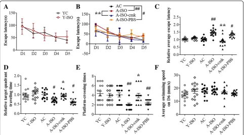
Isoflurane induced age-related cognitive impairment No animal died or was apneic during the anesthesia or the intended observation period after the anesthesia. In MWM, the escape latency of each training day and the relative average escape latency of five training days were used to indicate the learning ability. Significant differences in escape latency during the whole training phase were observed between group AC and group ISO, group A-ISO-cmk and group A-ISO, group A-A-ISO-cmk and group A-ISO-PBS (Fig.1a, b). The relative average escape latency of group A-ISO in five training days was significantly longer than group AC. This increase was attenuated by the pretreatment with the caspase-1 inhibitor Ac-YVAD-cmk (Fig.1c). In the probe trail, target quadrant traveling
time and the number of platform-crossing indicate the memory activities. Compared to group AC, the time in the target quadrant and frequency to cross the platform were significantly decreased in group ISO and group A-ISO-PBS. This decrease was reversed by Ac-YVAD-cmk pretreatment (Fig. 1d, e). No significant difference was observed in the average swimming speed on the sixth day among the groups (Fig.1f). Different from the situation in aged mice, isoflurane did not have an effect on the performance of young mice in MWM (Fig.1a–f). These results indicate that 1.5% isoflurane anesthesia for 2 h leads to cognitive impairment in the aged mice and that this impairment can be attenuated by Ac-YVAD-cmk pretreatment.
Isoflurane induced NLRP3 inflammasome activation in aged brain
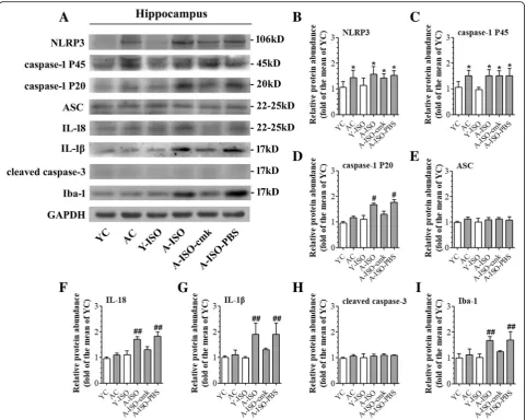
Compared to group YC, group AC had an increased expression of NLRP3 in the hippocampus. The levels of caspase-1 P20, cleaved IL-18, cleaved IL-1β, and Iba-1 were upregulated after isoflurane treatment in the aged mice, which was reversed by Ac-YVAD-cmk pretreatment (Fig.
2a–g, i). These results indicate that the NLRP3 inflamma-some activation may contribute to isoflurane-induced age-related hippocampal inflammation. The levels of cleaved caspase-3 were not upregulated in the hippocampus of aged mice no matter whether they were exposed to isoflurane (Fig.2a, h).
NLRP3 inflammasome priming was induced by LPS stimulation
To determine the characteristics of LPS-induced priming of NLRP3 inflammasome pathway, BV-2 cells and primary microglial cultures were incubated with various doses of LPS. LPS induced a dose-dependent increase of NLRP3 mRNA. Interestingly, this increase was induced by much lower doses of LPS in the primary microglial cultures than in the BV-2 cells. Nevertheless, most doses that induced NLRP3 mRNA expression did not increase the concentra-tion of IL-1βin the culture medium of the BV-2 cells and primary microglial cultures. The increase of IL-1β hap-pened only when a very high concentration (25μg/mL in BV-2 cells or 10 ng/mL in primary microglial cultures) of LPS was used (Fig. 3). These results suggest that those concentrations of LPS that increased NLRP3 mRNA but did not increase IL-1β induced a priming status of the microglial cells.
NLRP3 priming was necessary in isoflurane-induced NLRP3 inflammasome activation
LPS, 4% isoflurane treatment induced a significant IL-1β and IL-18 production (Figs.4a, band5a). Interestingly, 4% isoflurane alone could not increase IL-1βor IL-18 produc-tion (Fig. 4a, h). Similarly, isoflurane-induced caspase-1 activation only in NLRP3-primed in BV-2 cells, indicating that NLRP3 priming is necessary in isoflurane-induced NLRP3 inflammasome activation (Figs. 4a–g and 5d). To determine whether the findings from the immortalized BV-2 cell line were reproducible in the primary microglial cultures, we performed a similar experiment. In primary microglial cultures, the combination of LPS and isoflurane also increased the expression of NLRP3 (Figs.4h, iand5b). After NLRP3 priming by 5 ng/mL LPS, 2% isoflurane treat-ment significantly increased IL-18 and IL-1β production, while no significant caspase-1 activation or IL-1β produc-tion was induced by isoflurane alone (Figs.4h–nand 5e). We further investigated the effect of isoflurane on the viability of BV-2 cells and primary microglial cultures by using CCK-8 cell viability assay. No significant differences in the cell viability were observed between cells that were harvested immediate after LPS priming and primed cells
that were harvested 12 h after an exposure to isoflurane for 6 h (Fig.5c).
NLRP3 knock-down reduced isoflurane-induced NLRP3 inflammasome activation
To determine the role of NLRP3 in the isoflurane effects, NLRP3 protein was knocked-down in the BV-2 cells. This downregulation significantly reduced the increase of caspase-1 P20 and P45 caused by the priming and isoflur-ane exposure (Fig.6a–g). These results suggest that down-regulating NLRP3 protein inhibits caspase-1 activation, an event that indicates NLRP3 inflammasome activation.
NLRP3 inflammasome assembly might be enhanced by isoflurane
Isoflurane appeared to increase the amount of pro-caspase-1 and ASC associated with NLRP3 (Fig.6h). ASC is an adaptor protein for NLRP3 inflammasome [25]. Isoflurane did not change the input of ASC and pro-caspase-1 (Fig. 6h). These results suggest that isoflurane increases NLRP3 inflammasome assembly.
Fig. 2Contribution of NLRP3 inflammasome activation to isoflurane-induced age-related neuroinflammation. Hippocampus was harvested at 24 h after the anesthesia.aRepresentative western blot images of NLRP3, caspase-1 P45, caspase-1 P20, ASC, IL-18, IL-1β, cleaved caspase-3, and Iba-1.b–iGraphic presentation of abundance of each protein. All values are expressed as fold changes over the mean values of group YC and are presented as mean ± SD (n≥3). *P< 0.05 compared with the corresponding data of group YC. #P< 0.05 and ##P< 0.05 compared with the corresponding data of group AC
Discussion
Elderly patients have a higher incidence rate of POCD after surgery and anesthesia [29]. The precise mechanism for the phenomenon remains unknown. Compared to young mice, the priming status of NLRP3 inflammasome has been found in the microglia of aged brain. Canonical NLRP3 inflammasome activation is reported to be linked to inflammation-induced cognitive function decline in aging [20] and Alzheimer’s disease [21]. Our study was performed to determine the effect of isoflurane on NLRP3 inflammasome activation.
that isoflurane could induce NLRP3 inflammasome activation in both BV-2 cells and primary microglial cultures after they were primed. We further observed that isoflurane might induce NLRP3 inflammasome activation by increasing NLRP3 inflammasome assembly. These findings suggest a potential target for reducing isoflurane-induced, age-related cognitive dysfunction.
Isoflurane, a widely used inhalational anesthetic, has been reported to impair the cognitive function in rodents, especially in the aged rodents [13,15,16]. Since neuroin-flammation has been found to play an important role in cognitive dysfunction after surgery or anesthesia [4,13,30], anesthetic effects on neuroinflammation have been a focus of research. Isoflurane alone has been reported to have no or a small effect on cytokine expression in microglia under control condition [31]. In addition, treatment with 2% isoflurane alone on primary neuron cultures for 6 h did not
enhance the transcription binding activity of NF-kB [32] but increased the death and NF-kB transcriptional activities in SH-SY5Y neuroblastoma cells after oxygen-glucose deprivation [33]. One minimum alveolar concentration (the concentration at which 50% of animals have no motor response to painful stimuli) of isoflurane in 14-month-old mice is 1.53 +/−0.14% [34]. According to our previous study and reports from others, 1.5% isoflurane treatment for 2 h impaired the cognitive functions of rodents, includ-ing learninclud-ing and memory function and anxiety-related behaviors, while the blood gas analysis demonstrated no hypoxia or acidosis immediately after the isoflurane expos-ure [16, 35, 36]. Our previous study showed that IL-1β
proinflammatory cytokines was observed in the aged hippo-campus and the aged mice had learning and memory dys-function after isoflurane anesthesia. Isoflurane did not induce learning and memory impairment in young mice. These findings suggest that aged brain have an overactive inflammatory response after isoflurane exposure, which may be a mechanism for cognitive impairment.
Aging is known to impair microglial functions with increased susceptibility to pro-inflammatory activation, thereby promoting aging-related neurodegeneration [38]. Previous studies have demonstrated that a common start of neuroinflammation is microglial activation and the subsequent inflammatory cascade, which lead to micro-environment for central nervous system disorder and ultimately affect cognitive function [39, 40]. Microglia secrete pro-inflammatory cytokines, like IL-1βand
TNF-α, after being stimulated by ischemia or infection [39,41]. These pro-inflammatory cytokines, together with astro-cytes, neurons, and oligodendroastro-cytes, regulate neuroin-flammation. In the aging brain, it has been proposed that microglia may be in a primed status to be activated [11]. A consequence of microglial priming with age is a hyper-active response to an immune challenge with an exagger-ated pro-inflammatory response.
protected from spatial memory impairment and have a decreased Aβ plaque burden [43]. Known as a major sensor of age-related accumulation of DAMPs and an upstream regulator that controls caspase-1-mediated pro-inflammatory state in aging [38, 44], it is important to identify how NLRP3 inflammasome expression and activa-tion occurred in aged brain after isoflurane exposure. A novel finding of our study is that isoflurane may induce NLRP3 inflammasome activation of the primed microglial cells. This result suggests that NLRP3 priming might be a condition for isoflurane to induce cognitive dysfunction and may be one of the reasons why elderly patients are more prone to POCD than young patients.
We showed that isoflurane induced age-related hippo-campal neuroinflammation via NLRP3 inflammasome acti-vation. We further showed this effect in a microglial cell line and primary microglial cultures. Of note, it is reported that many aspects of microglial signature are not present in microglial cell lines [45]. However, studies have reported that the BV-2 cell line and neonatal primary microglial cultures react in a similar way on NLRP3 inflammasome activation [46, 47]. Our preliminary work also found that the NLRP3-caspase-1 pathway could be regulated by different doses of LPS. Thus, we chose the BV-2 cells and neonatal primary microglial cultures for mechanistic study. Our results suggest a critical role of NLRP3 primed status in inducing mature proinflammatory cytokine production. We also showed that isoflurane activated NLRP3 inflamma-some possibly by increasing the assembly of NLRP3 inflam-masome. It is reported that a small amount of NLRP3 inflammasome complex is assembled but remains in resting status under control condition. The activation process recruits additional members and activates caspase-1 [17]. The detailed mechanism for isoflurane to induce NLRP3 inflammasome activation needs to be investigated in the future studies.
We have shown that isoflurane induces learning and memory dysfunction in elderly rats and that this effect is IL-1β-mediated [13, 15]. In this current study, we showed that isoflurane-induced impairment of learning and mem-ory is age-dependent. This phenomenon may be due to the primed status of microglia in the aged brain. Specifically, we found that the NLRP3 inflammasome might contribute to isoflurane-induced neuroinflammation and cognitive impairment in the aged mice. These findings advance our understanding on how isoflurane induces neuroinflamma-tion and cognitive dysfuncneuroinflamma-tion and suggest that NLRP3 inflammasome may be a therapeutic target for POCD.
Our study has limitations. First, we did not perform any surgical stimulation during the isoflurane exposure. The surgical effects on NLRP3 inflammasome activation are not known. Second, we only used male mice in this study to reduce the potential influence of fluctuating estrogen and progesterone concentrations in learning and memory
in female mice. Thus, the role of NLRP3 activation in isoflurane effects on female animals is not known. Third, we did not study cell viability in depth after isoflurane exposure because it is not a focus of our current study. However, we showed that isoflurane did not change the viability of the BV-2 cells and primary microglial cultures and did not induce apoptosis in the hippocampus, indicating that isoflurane-induced, age-related cognitive impairment may be independent of changes in brain cell viability. Fourth, there are differences between adult microglia in the brain and BV-2 cells or neonatal primary microglial cultures [45]. The in vivo system is very complex, and it may be difficult to use in vivo system to dissect cell type-specific effects. In addition, it has been reported that BV-2 cells and neonatal primary microglia cultures react in a similar way on NLRP3 inflammasome activation to that under in vivo condition [47, 48]. Also, NLRP3 may mainly be expressed in the microglia [42,49]. Thus, our results suggest the role of microglial activation in the effects we observed here.
In summary, we have shown that aged mice had learn-ing and memory impairment after isoflurane anesthesia, and this isoflurane effect was not apparent in young mice. The impairment in the aged mice was reversed by the inhibition of NLRP3-caspase-1 pathway. Isoflurane also induced age-related hippocampal inflammation through NLRP3-caspase-1 pathway. NLRP3 inflammasome prim-ing is necessary in isoflurane-induced IL-1β production. These results suggest that isoflurane induces age-related cognitive dysfunction via NLPR3 activation.
Conclusions
NLRP3 priming status in aged mouse brain may be involved in isoflurane-induced hippocampal inflammation and cognitive impairment.
Additional file
Additional file 1:Supplementary material for NLRP3 priming induced by LPS stimulation. (DOCX 90 kb)
Abbreviations
AC:Aged mice not being exposed to isoflurane or any drugs; A-ISO: Aged mice exposed to isoflurane; A-ISO-cmk: Ac-YVAD-cmk administered before aged mice were exposed to isoflurane; A-ISO-PBS: Solvent administered before aged mice were exposed to isoflurane; ANOVA: Analysis of variance; ASC: Apoptosis associated speck-like protein containing a caspase recruit-ment domain; CCK-8: Cell Counting Kit-8; CNS: Central nervous system; DAMPs: Damage-associated molecular patterns; DMSO: Dimethylsulphoxide; ELISA: Enzyme-Linked Immunosorbent Assay; FBS: Fetal bovine serum; GAPDH: Glyceraldehyde-3-phosphate dehydrogenase; IL-1β: Interleukin -1β; IL-18: Interleukin−18; LPS: lipopolysaccharide; MWM: Morris water maze;
Acknowledgements
The authors would thank Research Center of Medicine, Sun Yat-Sen Memorial Hospital for providing necessary infrastructure support to accomplish this work.
Funding
This work was supported by grants from the National Natural Science Foundation of China (8157050894 to Z Wang and 81571041 to L Cao and 81641160 to Z Zuo), grants from Guangdong Science and Technology Department (2014A030313204 to S Peng and 2015A030313178 to Z Wang) a grant (201704020222 to Z Zuo) from Guangzhou Science, Technology and Innovation Commission, Guangzhou, China and a grant from External Cooperation Program of Guangdong Science and Technology Department (2013B051000023 to L Cao).
Availability of data and materials Please contact author for data requests.
Authors’contributions
ZW, SM, ZZ, and SP conceived and designed the study. SM and YC performed the research and analyzed the data. LC and SP contributed reagents/materials/analysis tools. SM wrote the first draft of the paper. ZW, LC, SP, and ZZ contributed to the writing of the paper. All authors read and approved the final manuscript.
Ethics approval
All animal experiments were performed in accordance with current Chinese regulations and standards regarding the use of laboratory animals, and approved by the animal ethics committee of Sun Yat-Sen University.
Consent for publication Not applicable.
Competing interests
The authors declare that they have no competing interests with the material presented in the paper.
Publisher’s Note
Springer Nature remains neutral with regard to jurisdictional claims in published maps and institutional affiliations.
Author details
1Department of Anesthesiology, Sun Yat-Sen Memorial Hospital, Sun Yat-Sen University, Guangzhou 510289, Guangdong, China.2Laboratory of RNA and Major Diseases of Brain and Heart, Sun Yat-Sen Memorial Hospital, Sun Yat-Sen University, Guangzhou 510120, China.3Department of Anesthesiology, University of Virginia, Charlottesville, USA.
Received: 11 December 2017 Accepted: 26 March 2018
References
1. Akbaraly TN, Hamer M, Ferrie JE, Lowe G, Batty GD, Hagger-Johnson G, Singh-Manoux A, Shipley MJ, Kivimaki M. Chronic inflammation as a determinant of future aging phenotypes. CMAJ. 2013;185:E763.
2. Bittner EA, Yue Y, Xie Z. Brief review: anesthetic neurotoxicity in the elderly, cognitive dysfunction and Alzheimer’s disease. Can J Anaesth. 2011;58:216. 3. Androsova G, Krause R, Winterer G, Schneider R. Biomarkers of postoperative
delirium and cognitive dysfunction. Front Aging Neurosci. 2015;7:112. 4. Terrando N, Eriksson LI, Kyu Ryu J, Yang T, Monaco C, Feldmann M, Jonsson
Fagerlund M, Charo IF, Akassoglou K, Maze M. Resolving postoperative neuroinflammation and cognitive decline. Ann Neurol. 2011;70:986. 5. Zuo Z. Postoperative cognitive effects in newborns: the role of
inflammatory processes. Anesthesiology. 2013;118:481.
6. Zhang Z, Li X, Li F, An L. Berberine alleviates postoperative cognitive dysfunction by suppressing neuroinflammation in aged mice. Int Immunopharmacol. 2016;38:426.
7. Hovens IB, van Leeuwen BL, Nyakas C, Heineman E, van der Zee EA, Schoemaker RG. Postoperative cognitive dysfunction and microglial activation in associated brain regions in old rats. Neurobiol Learn Mem. 2015;118:74.
8. Franceschi C, Bonafe M, Valensin S, Olivieri F, De Luca M, Ottaviani E, De Benedictis G. Inflamm-aging. An evolutionary perspective on immunosenescence. Ann N Y Acad Sci. 2000;908:244.
9. Goto M. Inflammaging (inflammation + aging): a driving force for human aging based on an evolutionarily antagonistic pleiotropy theory? Biosci Trends. 2008;2:218.
10. Koellhoffer EC, McCullough LD, Ritzel RM. Old maids: aging and its impact on microglia function. Int J Mol Sci. 2017;18:e769
11. Harry GJ. Microglia during development and aging. Pharmacol Ther. 2013;139:313.
12. Li W. Phagocyte dysfunction, tissue aging and degeneration. Ageing Res Rev. 2013;12:1005.
13. Cao L, Li L, Lin D, Zuo Z. Isoflurane induces learning impairment that is mediated by interleukin 1beta in rodents. PLoS One. 2012;7:e51431. 14. Ji M, Dong L, Jia M, Liu W, Zhang M, Ju L, Yang J, Xie Z, Yang J. Epigenetic
enhancement of brain-derived neurotrophic factor signaling pathway improves cognitive impairments induced by isoflurane exposure in aged rats. Mol Neurobiol. 2014;50:937.
15. Lin D, Cao L, Wang Z, Li J, Washington JM, Zuo Z. Lidocaine attenuates cognitive impairment after isoflurane anesthesia in old rats. Behav Brain Res. 2012;228:319.
16. Li X, Zhou M, Wang X, Ji M, Zhou Z, Yang J. Resveratrol pretreatment attenuates the isoflurane-induced cognitive impairment through its anti-inflammation and -apoptosis actions in aged mice. J Mol Neurosci. 2014;52:286.
17. Lamkanfi M, Dixit VM. Mechanisms and functions of inflammasomes. Cell. 2014;157:1013.
18. Heneka MT, Kummer MP, Latz E. Innate immune activation in neurodegenerative disease. Nat Rev Immunol. 2014;14:463.
19. Sutterwala FS, Haasken S, Cassel SL. Mechanism of NLRP3 inflammasome activation. Ann N Y Acad Sci. 2014;1319:82.
20. Youm Y, Grant RW, McCabe LR, Albarado DC, Nguyen KY, Ravussin A, Pistell P, Newman S, Carter R, Laque A, Münzberg H, Rosen CJ, Ingram DK, Salbaum JM, Dixit VD. Canonical Nlrp3 inflammasome links systemic low-grade inflammation to functional decline in aging. Cell Metab. 2013;18:519. 21. Heneka MT, Kummer MP, Stutz A, Delekate A, Schwartz S, Vieira-Saecker A,
Griep A, Axt D, Remus A, Tzeng T, Gelpi E, Halle A, Korte M, Latz E, Golenbock DT. NLRP3 is activated in Alzheimer’s disease and contributes to pathology in APP/PS1 mice. Nature. 2012;493:674.
22. Goldmann T, Tay TL, Prinz M. Love and death: microglia, NLRP3 and the Alzheimer's brain. Cell Res. 2013;23:595.
23. Zhuang J, Wen X, Zhang YQ, Shan Q, Zhang ZF, Zheng GH, Fan SH, Li MQ, Wu DM, Hu B, Lu J, Zheng YL. TDP-43 upregulation mediated by the NLRP3 inflammasome induces cognitive impairment in 2 2′,4,4′-tetrabromodiphenyl ether (BDE-47)-treated mice. Brain Behav Immun. 2017;65:99.
24. Latz E. The inflammasomes: mechanisms of activation and function. Curr Opin Immunol. 2010;22:28.
25. Guo H, Callaway JB, Ting JP. Inflammasomes: mechanism of action, role in disease, and therapeutics. Nat Med. 2015;21:677.
26. Abais JM, Xia M, Zhang Y, Boini KM, Li P. Redox regulation of NLRP3 inflammasomes: ROS as trigger or effector? Antioxid Redox Sign. 2015;22: 1111.
27. Zhang F, Wang L, Wang J, Luo P, Wang X, Xia Z. The caspase-1 inhibitor AC-YVAD-CMK attenuates acute gastric injury in mice: involvement of silencing NLRP3 inflammasome activities. Sci Rep. 2016;6:24166.
28. Pan K, Li X, Chen Y, Zhu D, Li Y, Tao G, Zuo Z. Deferoxamine pre-treatment protects against postoperative cognitive dysfunction of aged rats by depressing microglial activation via ameliorating iron accumulation in hippocampus. Neuropharmacology. 2016;111:180.
29. Johnson T, Monk T, Rasmussen LS, Abildstrom H, Houx P, Korttila K, Kuipers HM, Hanning CD, Siersma VD, Kristensen D, Canet J, Ibanaz MT, Moller JT. Postoperative cognitive dysfunction in middle-aged patients.
Anesthesiology. 2002;96:1351.
30. Cibelli M, Fidalgo AR, Terrando N, Ma D, Monaco C, Feldmann M, Takata M, Lever IJ, Nanchahal J, Fanselow MS, Maze M. Role of interleukin-1beta in postoperative cognitive dysfunction. Ann Neurol. 2010;68:360. 31. Ye X, Lian Q, Eckenhoff MF, Eckenhoff RG, Pan JZ. Differential general
anesthetic effects on microglial cytokine expression. PLoS One. 2013;8:e52887. 32. Zhang L, Zhang J, Yang L, Dong Y, Zhang Y, Xie Z. Isoflurane and
33. Schallner N, Ulbrich F, Engelstaedter H, Biermann J, Auwaerter V, Loop T, Goebel U. Isoflurane but not sevoflurane or desflurane aggravates injury to neurons in vitro and in vivo via p75NTR-NF-kB activation. Anesth Analg. 2014;119:1429.
34. Eckel B, Richtsfeld M, Starker L, Blobner M. Transgenic Alzheimer mice have a larger minimum alveolar anesthetic concentration of isoflurane than their nontransgenic littermates. Anesth Analg. 2010;110:438.
35. Wu J, Li H, Sun X, Zhang H, Hao S, Ji M, Yang J, Li K. A mitochondrion-targeted antioxidant ameliorates isoflurane-induced cognitive deficits in aging mice. PLoS One. 2015;10:e138256.
36. Tanino M, Kobayashi M, Sasaki T, Takata K, Takeda Y, Mizobuchi S, Morita K, Nagai T, Morimatsu H. Isoflurane induces transient impairment of retention of spatial working memory in rats. Acta Med Okayama. 2016;70:455. 37. Wu X, Lu Y, Dong Y, Zhang G, Zhang Y, Xu Z, Culley DJ, Crosby G,
Marcantonio ER, Tanzi RE, Xie Z. The inhalation anesthetic isoflurane increases levels of proinflammatory TNF-α, IL-6, and IL-1β. Neurobiol Aging. 2012;33:1364.
38. Fougere B, Boulanger E, Nourhashemi F, Guyonnet S, Cesari M. Chronic inflammation: accelerator of biological aging. J Gerontol A Biol Sci Med Sci. 2016;72:1218.
39. Fernandes A, Miller-Fleming L, Pais TF. Microglia and inflammation: conspiracy, controversy or control? Cell Mol Life Sci. 2014;71:3969. 40. Norden DM, Godbout JP. Review: microglia of the aged brain: primed to be
activated and resistant to regulation. Neuropath Appl Neuro. 2013;39:19. 41. Tang Y, Le W. Differential roles of M1 and M2 microglia in
neurodegenerative diseases. Mol Neurobiol. 2015;53:1181.
42. Gustin A, Kirchmeyer M, Koncina E, Felten P, Losciuto S, Heurtaux T, Tardivel A, Heuschling P, Dostert C. NLRP3 Inflammasome is expressed and functional in mouse brain microglia but not in astrocytes. PLoS One. 2015;10:e130624.
43. Heneka MT. Inflammasome activation and innate immunity in Alzheimer’s disease. Brain Pathol. 2017;27:220.
44. Trott DW, Henson GD, Ho MH, Allison SA, Lesniewski LA, Donato AJ. Age-related arterial immune cell infiltration in mice is attenuated by caloric restriction or voluntary exercise. Exp Gerontol. 2016;(16)30348-5. 45. Butovsky O, Jedrychowski MP, Moore CS, Cialic R, Lanser AJ, Gabriely G,
Koeglsperger T, Dake B, Wu PM, Doykan CE, Fanek Z, Liu L, Chen Z, Rothstein JD, Ransohoff RM, Gygi SP, Antel JP, Weiner HL. Identification of a unique TGF-β–dependent molecular and functional signature in microglia. Nat Neurosci. 2013;17:131.
46. Weber MD, Frank MG, Tracey KJ, Watkins LR, Maier SF. Stress induces the danger-associated molecular pattern HMGB-1 in the Hippocampus of male Sprague Dawley rats: a priming stimulus of microglia and the NLRP3 Inflammasome. J Neurosci. 2015;35:316.
47. You T, Cheng Y, Zhong J, Bi B, Zeng B, Zheng W, Wang H, Xu J. Roflupram, a phosphodiesterase 4 inhibitior, suppresses inflammasome activation through autophagy in microglial cells. ACS Chem Neurosci. 2017;8:2381. 48. Yang H, Chen Y, Yu L, Xu Y. Esculentoside a exerts anti-inflammatory activity
in microglial cells. Int Immunopharmacol. 2017;51:148.
49. Kaushal V, Dye R, Pakavathkumar P, Foveau B, Flores J, Hyman B, Ghetti B, Koller BH, LeBlanc AC. Neuronal NLRP1 inflammasome activation of Caspase-1 coordinately regulates inflammatory interleukin-1-beta production and axonal degeneration-associated Caspase-6 activation. Cell Death Differ. 2015;22:1676.
• We accept pre-submission inquiries
• Our selector tool helps you to find the most relevant journal
• We provide round the clock customer support
• Convenient online submission
• Thorough peer review
• Inclusion in PubMed and all major indexing services
• Maximum visibility for your research
Submit your manuscript at www.biomedcentral.com/submit