R E V I E W
Open Access
Endometrial stem cells in regenerative medicine
Javad Verdi
1,2, Aaron Tan
1,3, Alireza Shoae-Hassani
2and Alexander M Seifalian
1,4*Abstract
First described in 2004, endometrial stem cells (EnSCs) are adult stem cells isolated from the endometrial tissue. EnSCs comprise of a population of epithelial stem cells, mesenchymal stem cells, and side population stem cells. When secreted in the menstrual blood, they are termed menstrual stem cells or endometrial regenerative cells. Mounting evidence suggests that EnSCs can be utilized in regenerative medicine. EnSCs can be used as immuno-modulatory agents to attenuate inflammation, are implicated in angiogenesis and vascularization during tissue regeneration, and can also be reprogrammed into induced pluripotent stem cells. Furthermore, EnSCs can be used in tissue engineering applications and there are several clinical trials currently in place to ascertain the therapeutic potential of EnSCs. This review highlights the progress made in EnSC research, describing their mesodermal, ectodermal, and endodermal potentials bothin vitroandin vivo.
Keywords:Endometrial stem cells, Menstrual blood stem cells, Endometrial regenerative cells, Endometrium, Regenerative medicine
Introduction
Cells in the earliest developmental stages in the embryo can generate embryonic and extra-embryonic tissues [1]. The ability to generate other cell types is known as po-tency. In the inner cell mass of the blastocyst in the em-bryo, the cells pluripotent, meaning they can give rise to the ectoderm, mesoderm, and endoderm lineages (entire organism). Pluripotent stem cells include embryonic stem cells (ESCs) from the inner cell mass of blastocysts, epiblast derived stem cells from embryos after implant-ation, embryonic germ cells (EGCs) from primordial germ cells, embryonic carcinoma cells (ECCs) derived from germ cells tumors, and germ line stem cells from testicu-lar tissue [2-4]. As the reproductive cycle progresses, cell potency decreases and only a small fraction of cells retain their potency, namely adult stem and progenitor cells. These are limited to tissue generation within specific line-ages. Adult stem cells (ASCs) are found in numerous hu-man tissues including: intestines [5], muscles [6], skin [7], blood [8], nervous system [9-11], endometrium [12], heart, liver [13,14], dental pulp, adipose tissue, synovial membrane, umbilical cord blood, amniotic fluid [15,16],
and the endometrium [17]. In comparison to pluripotent stem cells, ASCs are considered safer for therapeutic pur-poses and several are currently used in clinical trials. The concept of using ASCs over embryonic stem (ES) cells in regenerative medicine has recently gained traction. The rationale for this trend include: (1) The complicated con-trol of the culture conditions of ESCs; (2) the existence of several intermediate stages before reaching terminal differ-entiation of ESCs; (3) Teratoma formation is a major obs-tacle for clinical development of ESCs; (4) immunological rejection of cells derived from ES cells to the recipient and (5) ethical scrutiny involved in ESCs application.
Endometrium and endometrial stem cells (EnSCs)
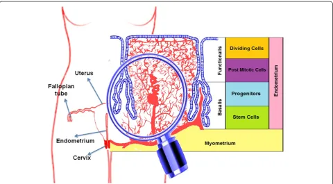
The human endometrium is an extraordinary model of controlled tissue remodeling, unparalleled in other or-gans, which grows about 7 mm within one week in every menstrual cycle [18]. At this rate, there is a very rapid rate of angiogenesis for approximately 500 cycles within a tightly controlled manner in a woman’s lifetime. The human endometrium, derived from the mucosal lining of the fused paramesonephric tubes during embryogen-esis, is a dynamic tissue (Figure 1). It is comprised of two major zones: (1) the functionalis, a transient layer containing glands extending from the surface epithelium as well as the supportive stroma, and (2) the basalis, comprised of the basal region of the glands, stroma, * Correspondence:a.seifalian@ucl.ac.uk
1
Centre for Nanotechnology and Regenerative Medicine, UCL Division of Surgery & Interventional Science, University College London (UCL), London NW3 2QG, UK
4Royal Free London NHS Foundation Trust Hospital, London, UK
Full list of author information is available at the end of the article
© 2014 Verdi et al.; licensee BioMed Central Ltd. This is an Open Access article distributed under the terms of the Creative Commons Attribution License (http://creativecommons.org/licenses/by/4.0), which permits unrestricted use, distribution, and reproduction in any medium, provided the original work is properly credited. The Creative Commons Public Domain Dedication waiver (http://creativecommons.org/publicdomain/zero/1.0/) applies to the data made available in this article, unless otherwise stated.
supporting vasculature, and lymphoid aggregates. Pro-genitor cells are located in the basal layer (Figure 1) of human endometrium [19]. These rapidly proliferating cells (transient cells) then move to the functional layer [20] and actively participate in the regeneration and re-modeling of the endometrium. Endometrial mesenchy-mal stem cells (MSCs) have been identified in the endometrium (lining of the uterus). Indeed, cloning of human endometrial cells confirmed the existence of endometrial epithelial progenitor cells for the first time in 2004 [21]. There are three distinct endometrial stem cells including: epithelial progenitor cells, MSCs and endothelial progenitor cells [22]. Recent studies reported the isolation of multipotent EnSCs from menstrual blood [23,24] or endometrial biopsies [25]. These cells are called endometrial regenerative cells (ERCs), and are heterogenic and have morphological differences among their different isolation sites. Stromal cells have been cultured from menstrual blood [24,26], suggesting that endometrial MSCs are shed during menstruation [22] but epithelial cells have not been detected [27], suggest-ing that epithelial progenitors may not be shed dursuggest-ing menstruation and more likely reside in the basalis. Dur-ing the proliferative menstrual phase, the endometrium prepares for recruiting new EnSCs to cover the new blood vessels. The variation in the numbers of endothe-lial progenitor cells and stem cell derived factor-1 is a pivotal factor for release and homing of stem cells [28].
These EnSCs have properties similar to bone marrow or adipose tissue stem cells. They proliferate and differentiate throughout the estrous cycle and during pregnancy under the control of the ovarian hormones. Clonogenic MSCs are responsible for regenerating the endometrium, and they exist in peri-menopausal women, postmenopausal women, and women on oral contraceptives [29]. Irregular function of these SCs may contribute to the pathogenesis of endometriosis and the growth of tissue outside the uterine cavity, causing dysmenorrhoea, subfertility and endometrial carcinoma [30]. Stem cells that are ectopically distributed through lymphovascular metastasis [31] may also contribute to the pathogenetic process, because their high proliferation promotes rapid clonal expansion [32].
The progenitor cells in the endometrium has a high proliferative potential that can generate 6 × 10¹¹ cells from a single cell, with the ability to differentiate into large cytokeratin-expressing structures when cultured in Matrigel [17], comprising laminin, collagen IV and hepa-ran sulfate proteoglycan. It was used instead of mouse embryonic fibroblast feeder layers for ES cells prolifera-tion. Our data shows that mesenchymal stem cells from endometrium can be grown extensively bothin vivoand
in vitro. These cells are not tumorigenic in nude mice and could be passaged approximately 40 timesin vitro.
The fact that about 600,000 hysterectomies are per-formed yearly in the United States creates a potential source of endometrial cells [33]. Studies have indicated
that EnSCs reside in the superficial layers accessible by endometrial biopsy [34,35]. Indeed, endometrial biopsies may represent a viable technique in harvesting EnSCs, as they are routinely performed for gynecologic pur-poses, and does not impair endometrial function. Thus, endometrial tissue specimens from women who are not receiving hormonal therapy are suitable for isolation of stem cells (Figure 2).
Endometrial stem cells may be derived from fetal stem cells or bone marrow stem cells, including haemopoietic SCs, MSCs and endothelial progenitor cells [36-38]. The human endometrium shows expression of the pluripo-tency factors Sox-2, Oct-4 and Nanog. Sox-2 co-localized with telomerase, contribute to the immortality of embry-onic stem cells [39]. Reprogramming of differentiated cells to an induced pluripotent stem (iPS) cell is through induc-tion or increasing in the expression of Oct-4, Sox-2, Klf-4, and c-Myc [40] that easily could be achieved from EnSCs that already express these factors. As a result, iPS cells from EnSCs have been established as early as 12 days after transduction rather than the usually reported 4 weeks for other cell types [41]. As patient- or disease-specific biomedical research using iPS cells becomes more widespread, the endometrium may play a key role as re-programmable cells into donor- or disease-specific pluri-potent cell lines for the female population.
Markers of human EnSCs
Flow cytometric analysis of important endometrial stem cell markers are indicated in Figure 3. Cultured EnSCs
express typical markers used for bone marrow stem cells; CD9, CD13, CD14, CD29, CD31, CD44, CD73, CD90, CD105, CD117, CD133, CD146 [42], but not STRO-1, CD31 (endothelial) and CD34 (haemopoietic stem cell and endothelial) [43]. The EnSCs could be purified on the basis of their co-expression of two perivascular markers CD140b and CD146 [44]. Cells with a hematopoietic stem cell phenotype (CD34 + CD45+) co-expressing CD7 and CD56 have been identified in human endometrial cell sus-pensions and may be lymphoid progenitors [45]. Musashi-1, an RNA-binding protein in neural stem cells and an epithelial progenitor cell marker that regulates self-renewal signaling pathways, has been recognized in hu-man endometrium. NAC1 is a transcription repressor that is involved in self-renewal and the maintenance of pluri-potency in embryonic stem cells [46]. Ishikawa et al. dem-onstrated that NAC1 was over-expressed in the normal cyclic endometrium in the early and mid proliferative phases [47]. MSI1 and NOTCH1, which maintain SCs in an undifferentiated state, are expressed in EnSCs. Tissue non-specific alkaline phosphatase, also immune-localizes to a perivascular location in human endometrium and may be useful for the prospective isolation of EnSCs [48]. Leucine rich repeat containing G protein-coupled receptor-5 (Lgr-5) is also expressed in human endometrial epithelial progenitor cells and endometrial [49] stromal cells [50]. W5C5 is a single marker for purifying EnSC, which differentiates into adipogenic, osteogenic, chondro-genic, and myogenic cell lineages [51]. One-third of these EnSCs expressedα-smooth muscle actin (αSMA) but not
Figure 2The endometrial stem cell isolation.A diagram that shows the stepwise isolation of endometrial stem cells by collagenase digestion, then serial filtration through 70μm and 40μm meshes, ficoll separation and selection the colony forming cells.
CD31, suggesting that they are vascular smooth muscle cells and that they occupy a peri-vascular niche.
Immunogenicity of EnSCs
Utilizing ES cells in regenerative therapy is associated with many hazards. Without adequate ES cells screening procedures, patients would face tissue rejection similar as rejection of organ transplants. A solution to this problem is to create patient specific stem cells. Unlike ES cells, adult stem cells are not rejected by the immune system [52]. The human endometrium has unique im-munological requirements. In pregnancy, the endomet-rium must tolerate the invading embryo, which expresses both paternal and maternal antigens. In 2012, Peron et al. demonstrated that endometrial MSCs were able to sup-press neuro-inflammation in a mouse model of multiple sclerosis. This suppression is dependent on the secretion of anti-inflammatory cytokines such as IL-10 and IL-27, and also the expression of indoleamine-2,3-dioxigenase
[53]. Menstrual blood SCs were tested for allogeneic re-sponse in culture with peripheral blood mononuclear cells (PBMN). The SCs were analyzed at a 1:2 stimulator (mito-mycin C treated PBMN) to responder cell (menstrual blood SCs) ratio for 6 days. The menstrual blood SCs demonstrated a moderately weak stimulatory response in a mixed lymphocyte reaction (MLR). An emerging bank of data allows for the classification of the mesenchymal stem cell from bone marrow as immunosuppressive cell source derived from studies with human cells [54,55]. Some studies support that SC administration is a potential way to suppress tumor growth. It has been demonstrated that human skin-derived progenitor cells have selective tropism for malignant tissues. Also more interestingly, these cells have the ability to inhibit tumor growth [56]. MSCs could selectively integrate into gliomas after intra-vascular or local delivery [57-59]. ERC administration inhibits C6 tumor growth and its administration associ-ated with reduced neovascularization [60]. Despite the
angiogenic potential of ERC in the hindlimb ischemia model, these data support a paradoxical tumor inhibi-tory activity of ERC. Further studies are needed to de-termine the qualitative differences between physiological angiogenesis, which seems to be supported by ERC and tumor angiogenesis which appeared to be inhibited. Other studies have demonstrated that MSCs directly se-crete tumor inhibitory factors [61,62].
Potency and differentiation of EnSCs
The main features of mesenchymal stem cells are their ability for self-renewal, colony-forming ability, and dif-ferentiation into mesoderm-derived lineages including adipogenic, chondrogenic and osteogenic differentiation [63-65]. Endometrial SCs that are persistent in the uter-ine endometrium from the fetal to the postmenopausal period [66] were found to differentiate into adipocytes, osteoblasts and chondrocytes as well [67,68] (Figure 4). The modulation of p38 and c-jun might play an import-ant role for the differentiation and proliferation of nor-mal human endometrial cells [69]. These differentiations can identify by positive staining, with Oil Red O (for lipid droplets), alizarin red (for osteogenesis), von Kossa
(for calcified extracellular matrix), and Alcian blue (for sulfated proteoglycans), respectively. Clonogenicity of EnSCs does not vary from the proliferative to secretory stage of the menstrual cycle, or between active, cycling and inactive endometrium for both epithelial and stro-mal cells [29].
Identification and isolation of differentiated cells
Research pertaining to stem cell biology in the female re-productive tract is still in its infancy, and although surface markers of prospective isolation of human endometrial stromal colony-forming cells (putative endometrial stro-mal/progenitor cells) have recently identified [17,44], there remains a need for definitive markers of both myometrial and endometrial stem cells for more selective isolation and enrichment. Complete characterization of uterine stem/ progenitor cells will improve our understanding of the mechanisms supporting physiological regeneration of the female reproductive tract. In addition, such studies will enhance our understanding of uterine cancer, hyper-plasia, endometriosis, leiomyomas and adenomyosis. In-deed, our data and the published observations from other laboratories have enabled us to propose a novel
Figure 4Multipotent differentiation of endometrial stem cells.Endometrial stem cells were cultured under appropriate differentiation media as described in box1-15 and assessed for differentiation using the indicated staining methods.A)Adipocytic differentiation, the red color indicates lipid vacuoles stained by Oil-Red.B)Osteocytic differentiation, red indicates calcium stained by Alizarin Red andC)Von-Kossa staining of osteoclasts differentiated from EnSCs.D)Chondrocytic differentiation, the blue color resulted from Alcian blue staining.E)Neuronal differentiation, the red conjugated mAb indicates pre-neural marker Nestin stain.F)Angiogenesis of EnSCs, the green indicated CD31 stain.G)Oligodendrocytic differentiation, the red conjugated mAb indicates receptor interacting protein (RIP).H)Hepatocytic differentiation, green indicates albumin stain.I)Myocytic
differentiation, red indicatesα-actinin stain.
hypothetical model for eutopic and ectopic endometrial regeneration. Finally, availability of these stem cells sug-gests new approaches to reconstruction of the human uterus and perhaps other organs as well.
The final step for processing stem cells before trans-plantation would be the identification and isolation of the differentiated cells. Recognition and selection of desired cells can be done via reverse transcriptase polymerase chain reaction, flow cytometry or fluorescence-activated cell sorting (FACS). Unfortunately, these methods would destroy many of the differentiated cells, thereby rendering them unusable. Also, flow cytometry and FACS is de-pendent on the presence of specific markers on the cell surface, which are not always displayed on every type of differentiated cells [70]. Thus, a more viable technique for identification of differentiated cells can be made with the transient transfection of a reporter gene into a stem cell source. The reporter gene can encode a fluorescent (green fluorescent protein) or a drug selectable protein. The transi-ent transfection would be favorable for human cell therapy because the reporter is only meant for selection purposes.
Comparison between EnSCs, MSCs, and umbilical cord blood stem cells
Genetic profiling of EnSCs and bone marrow MSCs re-vealed that they had similar (although not identical) cytokine production, miRNAs, and gene expression [71]. It was reported that EnSCs express higher levels of ICAM-1 and IL-8, while bone marrow MSCs express higher levels of TGFβ1, TGFβ2, IL-6, and vascular endo-thelial growth factor (VEGF). This observation suggests that EnSCs may play a role in acute inflammation, and may be more suitable for tissue engineering applications. It was also reported that, compared to MSCs, EnSCs had a 27-fold higher level of PDGF-BB, and 14-fold higher level of angiopoietin. This indicates that EnSCs may activate alternative angiogenesis pathways. However, more studies are required to ascertain if EnSCs stimulate higher levels of angiogenesis than bone marrow MSCs
in vivo. It was shown that EnSCs produced collagen type II and proteoglycans when cultured on nanofibrous polycaprolactone material, suggesting their cartilaginous tissue differentiation potential [72]. It was also observed that proliferation of EnSCs was faster than bone marrow MSCs, and EnSCs express the pluripotency marker OCT-4, but lack the MSC marker STRO1 [72]. Umbilical cord blood was first used in bone marrow transplant in a patient with Fanconi’s anaemia in 1988 [73]. Consider-ing that a large proportion of haematopoietic stem cells are found in the fetal circulation, there is also a corres-pondingly large proportion of haematopoietic stem cells in the umbilical cord. Bone marrow MSCs are distinct from haematopoietic stem cells, as they are CD45−. Ma-ture bone marrow MSCs are a heterogeneous population
of cells that are able to support haematopoiesis, and cap-able of differentiating into different lineages such as neural cells [74,75], muscle [76-78], liver cells [79,80], among many others. Various studies have reported that a single mesenchymal progenitor cell is responsible for all lineages are produced from bone marrow MSC
in vitro. It has been shown that bone marrow MSCs
contain more mesenchymal progenitor cells when com-pared to umbilical cord blood stem cells. Indeed, the proportion of MSCs within umbilical cord blood is ex-tremely low and difficult to detect [81]. Nevertheless, more studies comparing EnSCs, MSCs and umbilical cord blood stem cells are required to accurately ascertain their similarities and differences.
EnSCs in regenerative therapeutics
To date, EnSCs have been used in several pre-clinical and small animal studies. The regenerative potential of EnSCs was first seen in an experimental model of Du-chenne muscular dystrophy in immunodeficient mdx mice, whereby EnSCs that were transplanted into atro-phied skeletal muscle fibres contributed to muscle repair [26]. Although the exact mechanism of repair has not been elucidated, it was postulated that cell fusion andin
situdifferentiation might have been involved, in part due to the observation that the transplanted EnSCs homed to peri-muscle fibre regions and promoted angiogenesis. The concept of angiogenesis was also supported by an-other study, where EnSCs were improved critical limb is-chaemia induced by femoral artery ligation [82].
The use of EnSCs to treat myocardial infarction in a murine model was also seen. In this study, EGFP-labelled EnSCs were grafted into the infarct area of nude rat hearts, which subsequently differentiated into α-actinin+, troponin+striated cardiac muscle cells [83]. Furthermore, it was observed that a significantly larger reduction in in-farct area was seen in animals treated with EnSCs, com-pared to control bone marrow MSCs.
Gargett et al., the first group that reported the existence of EnSCs in 2004 [21], are currently developing an autolo-gous tissue engineered scaffold using artificial meshes and EnSCs for the treatment of pelvic organ prolapse, and was tested in vivo, on an animal skin wound repair model [84,85]. Results indicated that EnSCs promoted neovascu-larization, increased tissue integration, reduced chronic inflammation, increased deposition of collagen fibres and distensibility of the mesh at 3 months post-implantation. They also found that EnSCs downregulated foreign body reactions and enhanced mesh integration, indicating they have a role to play in modulating tissue response with regards to implanted foreign materials.
seeded with EnSCs for the reconstruction of urinary bladder wall for women [86]. We had also shown that EnSCs were capable of differentiating into smooth muscle cells [86], potentially functioning as an autolo-gous cell source for bladder tissue engineering.
Intraperitoneal delivery of EnSCs were also studied in a murine model of encephalomyelitis [53]. It was founds that EnSCs exerted a an anti-inflammatory effect, as evi-denced by a lower number of infiltrating mononuclear cells in the lesions, upregulation of IL-10 and IL-27 in the spleen, and reduced recruitment of Th1 and Th17 cells in the central nervous system.
EnSCs were also used to demonstrate neural regenera-tive capabilities, whereby reduced neuronal cell death was seen when oxygen-deprived primary neuronal cell cultures were exposed to EnSCs [87]. It was also ob-served that EnSCs released neuroprotective trophic factors such as neurotrophin-3 (NT-3), brain-derived neurotrophic factor (BDNF), and VEGF. Thein vivopart of the study was done in a murine model of ischaemic stroke, whereby injection of EnSCs resulted in signifi-cantly lower histological and behavioural impairments. It was reported that EnSCs exerted a trophic effect, releas-ing factors that promoted survival of neural cells.
The use of EnSCs to treat glioma was observed in a murine model. In this study, EnSCs were administered intravenously in a murine model of intracranial glioma. Results revealed a reduction of tumour size of almost 50%, possibly due to its anti-angiogenic effects [60].
The applications of EnSCs have also been reported in several human studies. The first reported use of EnSCs was demonstrated by Zhong et al. [88]. Clinical-grade menstrual blood-derived EnSCs have been used in a small Phase I clinical trial of 4 patients suffering from multiple sclerosis, whereby EnSCs were delivered via intravenous and intrathecal routes. Results showed no immunological reactions or adverse side effects after 1 year [88].
Another human study involved a patient suffering from Duchenne muscular dystrophy that was given intramuscular injections of EnSCs. Follow-up observa-tions reported no adverse effects even after 3 years, and increased muscle strength and decreased respiratory in-fections was also reported [89].
The third reported use of EnSCs in human was a pa-tient with congestive heart failure, who was given intra-venous administration of EnSCs. Results revealved that ejection fraction of the patient increased from 30% to 40%, decreased basic natriuretic peptide values (Pro-BNP), and decreased Minnesota Living with Heart Fail-ure Questionnaire score at 1-year follow up [90].
The promise and limitations of EnSCs
EnSCs are an attractive source of stem cells for regen-erative therapeutics as they are easily obtainable and
easily expandable in culture, as has been demonstrated to be safe for clinical use. Protocols and methods for extraction and isolation of EnSCs are well established, as purified EnSCs can be obtained using magnetic bead sorting using the W5C5/SUSD2 marker. In addition, clinical-grade good manufacturing practice (cGMP) are currently being developed for culture expansion of EnSCs, and have been tested in animals. However, there is a lack of published information on the exact cGMP protocols in place for the production of EnSCs. This is compounded by the fact that there is no general scien-tific consensus regarding specific MSC markers to detect EnSCs; rather, researchers rely on the ability of MSCs to adhere to plastic. Hence, the purity of EnSCs is not guaranteed as the cultures could potentially contain fibroblasts.
EnSCs can be obtained from menstrual blood; hence no invasive procedures are needed to harvest these cells. A menstrual cup is used to collect menstrual blood over several hours on days 2 to 3 of the menstrual period. Al-though there is a potential risk of infection via vaginal contact, there have been no reports of any complications after antibiotic use.
Although the ability of EnSCs to re-integrate into tis-suein vivo has been demonstrated, there is a theoretical risk that endometriosis could develop from using EnSCs. However, none of the animal model studies have reported this. Nevertheless, it is an aspect of EnSC application that warrants attention. Indeed, transdifferentiation (some-times referred to as adult stem cell plasticity) is a contro-versial topic in the field of stem cells. It is highly probable that nuclear reprogramming and altered transcriptional activity of important developmental genes are in part re-sponsible for transdifferentiation. Transdifferentiation can be seen as a form of metaplasia due to alterations in the extracellular environment, and often happens during tis-sue damage [91]. Hence, one probable cause of endomet-riosis is thought to be due to metaplasia of the peritoneal lining. However, there is also evidence to suggest that circulating stem cells can be a source of metaplastic trans-differentiation when it gains access into the pelvic cavity. Genetically-tagged bone marrow MSCs were tracked in a murine transplant model, and it was established that a small population of it integrated into endometriosis le-sions and transdifferentiated into stromal cells (0.1%) and epithelial cells (<0.04%) [92]. It was found that these bone marrow MSCs contributed to the progression of endomet-riosis rather than initiating it [93]. Changes in cellular phenotype may involve processes that are associated with both embryogenesis and carcinogenesis, such as chymal epithelial transition (MET) and epithelial mesen-chymal transition (EMT) [94]. Correspondingly, the invasiveness of endometriotic cells may be attributed to the changes in cellular phenotype in endometriotic lesions.
For instance, well differentiated CK+E+-Cadherin cells, CK− E-Cadherin- stromal cells, and an invasive CK+ E-Cad-herin−N-Cadherin+ epithelial cells (similar to carcinoma micrometastasis) are found in endometriotic lesions [95]. In line with carcinoma characteristics, a regression of endometriotic lesion is seen in estrogen depletion therapy, but recur when therapy stops. This indicates that a quies-cent stem cell population is present within the lesion, which reactivates on exposure to estrogen. Thus it may also follow that EnSCs within lesions may also contribute to subsequent lesions.
In terms of regenerative medicine applications, it is envisioned that bone marrow MSCs are more suited for osteogenic differentiation and therefore used for bone tissue engineering, while EnSCs are more suited for soft tissue engineering, such as bladder reconstruction. Due to the intense interest generated with using EnSCs for regenerative medicine, we believe that EnSCs were intro-duced too quickly into human tests without adequately assessing its impact on large animal models. Further-more, the exact mechanism of how EnSCs exert is re-generative potential is not clearly understood. Hence more robust large animal studies are needed to fill this information gap that currently exists.
Concluding remarks and future perspectives
Although EnSCs would form a non-invasive and steady supply of autologous stem cells for women, we must not forget that this means that 50% of the population (i.e. the male population) is excluded. Furthermore, non-invasive harvesting of EnSCs would not be possible from post-menopausal women (in this case, endometrial bi-opsy might be possible, but data regarding the potency of EnSCs in post-menopausal women has not been pub-lished). In addition, the issue of EnSCs storage could also develop into the same debates we are facing regard-ing the storage of cord blood; is there enough scientific evidence to justify storage of EnSCs for future use?
Nevertheless, given the significant progress made in the application of EnSCs in regenerative medicine, it is envi-sioned that human clinical trials would be conducted in the near future. Indeed, the RECOVER-ERC trial was re-cently launched by Medistem Inc (a start-up company based in San Diego, California), which aims to evaluate the potency of EnSCs in 60 heart failure patients in a double-blind placebo controlled Phase II clinical trial [96]. Furthermore, a search in theclinicaltrials.govdatabase re-veals that several clinical trials regarding EnSCs are under-way, with applications ranging from treating critical limb ischaemia (NCT 01558908), liver cirrhosis (NCT01483248), type I diabetes (NCT01496339), and in vitro fertilization (NCT01649752). Hence, it can be envisioned that data from human patients would be available in due course to evaluate the clinical impact of EnSCs.
In conclusion, the discovery of EnSCs represents a paradigm shift for the use of adult stem cells by offering an “off the shelf” therapeutic application regenerative medicine. The EnSCs niche and in situ role highlight these cells as being important for cell and tissue regener-ation. This work highlights crucial features of an inter-esting population of mesenchymal progenitors isolated form endometrial tissue. Cells grownin vitrowere char-acterized by a high clonogenic potential and a long-term survival. Advantages in comparison with MSC from other sources include a greater ease of supply and the protracted availability during a woman’s lifetime with the additional benefit of deriving stem cells from a waste tissue, thus avoiding critical ethical issues.
In the light of their capacity to differentiate into mes-enchymal tissues and their propensity to undergo gen-etic manipulation, EnSCs hold considerable promise for novel therapeutic approaches for diseases in the wider field of regenerative medicine.
Competing interests
The authors declare that no competing interests exist.
Authors’contributions
JV, AT, AS, AMS conceived and planned the structure of the manuscript. JV, AT, AS, AMS collated and analyzed the data. JV, AT, AS, AMS wrote the manuscript. All authors read and approved the final manuscript.
Author details 1
Centre for Nanotechnology and Regenerative Medicine, UCL Division of Surgery & Interventional Science, University College London (UCL), London NW3 2QG, UK.2Applied Cell Sciences Department, School of Advanced Technologies in Medicine, Tehran University of Medical Sciences, Tehran, Iran.3UCL Medical School, University College London (UCL), London, UK.
4Royal Free London NHS Foundation Trust Hospital, London, UK.
Received: 5 April 2014 Accepted: 30 June 2014 Published: 1 August 2014
References
1. Kelly SJ:Studies of the developmental potential of 4‐and 8‐cell stage mouse blastomeres.J Exp Zool1977,200(3):365–376.
2. Conrad S, Renninger M, Hennenlotter J, Wiesner T, Just L, Bonin M, Aicher W, Bühring H-J, Mattheus U, Mack A:Generation of pluripotent stem cells from adult human testis.Nature2008,456(7220):344–349.
3. Ko K, Araúzo-Bravo MJ, Tapia N, Kim J, Lin Q, Bernemann C, Han DW, Gentile L, Reinhardt P, Greber B:Human adult germline stem cells in question.Nature2010,465(7301):E1–E1.
4. Stadtfeld M, Hochedlinger K:Induced pluripotency: history, mechanisms, and applications.Genes Dev2010,24(20):2239–2263.
5. Bjerknes M, Cheng H:Clonal analysis of mouse intestinal epithelial progenitors.Gastroenterology1999,116(1):7–14.
6. Jankowski R, Deasy B, Huard J:Muscle-derived stem cells.Gene Ther2002,
9(10):642–647.
7. Alonso L, Fuchs E:Stem cells of the skin epithelium.Proc Natl Acad Sci U S A
2003,100(Suppl 1):11830–11835.
8. Spangrude GJ, Smith L, Uchida N, Ikuta K, Heimfeld S, Friedman J, Weissman I:Mouse hematopoietic stem cells.Blood1991,78(6):1395–1402. 9. Morrison SJ, White PM, Zock C, Anderson DJ:Prospective identification,
isolation by flow cytometry, and in vivo self-renewal of multipotent mammalian neural crest stem cells.Cell1999,96(5):737–749. 10. Uchida N, Buck DW, He D, Reitsma MJ, Masek M, Phan TV, Tsukamoto AS,
11. Rietze RL, Valcanis H, Brooker GF, Thomas T, Voss AK, Bartlett PF:
Purification of a pluripotent neural stem cell from the adult mouse brain.Nature2001,412(6848):736–739.
12. Chan RW, Gargett CE:Identification of Label‐Retaining Cells in Mouse Endometrium.Stem Cells2006,24(6):1529–1538.
13. Beltrami AP, Cesselli D, Bergamin N, Marcon P, Rigo S, Puppato E, D’Aurizio F, Verardo R, Piazza S, Pignatelli A:Multipotent cells can be generated in vitro from several adult human organs (heart, liver, and bone marrow).Blood2007,110(9):3438–3446.
14. Crisan M, Yap S, Casteilla L, Chen C-W, Corselli M, Park TS, Andriolo G, Sun B, Zheng B, Zhang L:A perivascular origin for mesenchymal stem cells in multiple human organs.Cell Stem Cell2008,3(3):301–313.
15. da Silva ML, Chagastelles PC, Nardi NB:Mesenchymal stem cells reside in virtually all post-natal organs and tissues.J Cell Sci2006,
119(11):2204–2213.
16. Kalervo Väänänen H:Mesenchymal stem cells.Ann Med2005,37(7):469–479. 17. Gargett CE, Schwab KE, Zillwood RM, Nguyen HP, Wu D:Isolation and
culture of epithelial progenitors and mesenchymal stem cells from human endometrium.Biol Reprod2009,80(6):1136–1145.
18. McLennan CE, Rydell AH:Extent of endometrial shedding during normal menstruation.Obstet Gynecol1965,26(5):605–621.
19. Padykula HA:Regeneration in the primate uterus: the role of stem cells.
Ann NY Acad Sci1991,622:47–56.
20. Okulicz WC, Ace CI, Scarrell R:Zonal changes in proliferation in the rhesus endometrium during the late secretory phase and menses.Exp Biol Med
1997,214(2):132–138.
21. Chan RW, Schwab KE, Gargett CE:Clonogenicity of human endometrial epithelial and stromal cells.Biol Reprod2004,70(6):1738–1750. 22. Gargett CE, Masuda H:Adult stem cells in the endometrium.Mol Hum
Reprod2010,16(11):818–834.
23. Meng X, Ichim TE, Zhong J, Rogers A, Yin Z, Jackson J, Wang H, Ge W, Bogin V, Chan KW:Endometrial regenerative cells: a novel stem cell population.J Transl Med2007,5(1):57.
24. Patel AN, Park E, Kuzman M, Benetti F, Silva FJ, Allickson JG:Multipotent menstrual blood stromal stem cells: isolation, characterization, and differentiation.Cell Transplant2008,17(3):303–311.
25. Schüring AN, Schulte N, Kelsch R, Röpke A, Kiesel L, Götte M:
Characterization of endometrial mesenchymal stem-like cells obtained by endometrial biopsy during routine diagnostics.Fertil Steril2011,
95(1):423–426.
26. Cui CH, Uyama T, Miyado K, Terai M, Kyo S, Kiyono T, Umezawa A:
Menstrual blood-derived cells confer human dystrophin expression in the murine model of Duchenne muscular dystrophy via cell fusion and myogenic transdifferentiation.Mol Biol Cell2007,18(5):1586–1594. 27. Musina R, Belyavski A, Tarusova O, Solovyova E, Sukhikh G:Endometrial
mesenchymal stem cells isolated from the menstrual blood.Bull Exp Biol Med2008,145(4):539–543.
28. Elsheikh E, Sylven C, Ericzon BG, Palmblad J, Mints M:Cyclic variability of stromal cell-derived factor-1 and endothelial progenitor cells during the menstrual cycle.Int J Mol Med2011,27(2):221–226.
29. Schwab KE, Chan RWS, Gargett CE:Putative stem cell activity of human endometrial epithelial and stromal cells during the menstrual cycle.Fertil Steril2005,84:1124–1130.
30. Gargett C:Uterine stem cells: what is the evidence?Hum Reprod Update
2007,13(1):87–101.
31. Sasson IE, Taylor HS:Stem cells and the pathogenesis of endometriosis.
Ann NY Acad Sci2008,1127(1):106–115.
32. Kim C, Oh Y, Cho S, Chung D, Hwang J, Park K, Cho D, Choi Y, Lee B:
Increased telomerase activity and human telomerase reverse transcriptase mRNA expression in the endometrium of patients with endometriosis.Hum Reprod2007,22(3):843–849.
33. Wu JM, Wechter ME, Geller EJ, Nguyen TV, Visco AG:Hysterectomy rates in the United States, 2003.Obstet Gynecol2007,110(5):1091–1095. 34. Götte M, Wolf M, Staebler A, Buchweitz O, Kelsch R, Schüring A, Kiesel L:
Increased expression of the adult stem cell marker Musashi‐1 in endometriosis and endometrial carcinoma.J Pathol2008,215(3):317–329. 35. Shoae Hassani A, Mortazavi Tabatabaei SA, Sharif S, Seifalian AM, Azimi A,
Samadikuchaksaraei A, Verdi J:Differentiation of human endometrial stem cells into urothelial cells on a three‐dimensional nanofibrous silk– collagen scaffold: an autologous cell resource for reconstruction of the urinary bladder wall.J Tissue Eng Regen Med2013, [In press].
36. Mayani H, Alvarado-Moreno JA, Flores-Guzmán P:Biology of human hematopoietic stem and progenitor cells present in circulation.Arch Med Res2003,34(6):476–488.
37. He Q, Wan C, Li G:Concise review: multipotent mesenchymal stromal cells in blood.Stem Cells2007,25(1):69–77.
38. Robb A, Mills N, Smith I, Short A, Tura-Ceide O, Barclay G, Blomberg A, Critchley H, Newby D, Denison F:Influence of menstrual cycle on circulating endothelial progenitor cells.Hum Reprod2009,24(3):619–625.
39. Kyo S, Masutomi K, Maida Y, Kanaya T, Yatabe N, Nakamura M, Tanaka M, Takarada M, Sugawara I, Murakami S:Significance of immunological detection of human telomerase reverse transcriptase: re-evaluation of expression and localization of human telomerase reverse transcriptase.
Am J Pathol2003,163(3):859–867.
40. Takahashi K, Tanabe K, Ohnuki M, Narita M, Ichisaka T, Tomoda K, Yamanaka S:Induction of pluripotent stem cells from adult human fibroblasts by defined factors.Cell2007,131(5):861–872.
41. Park JH, Daheron L, Kantarci S, Lee BS, Teixeira JM:Human endometrial cells express elevated levels of pluripotent factors and are more amenable to reprogramming into induced pluripotent stem cells.
Endocrinology2011,152(3):1080–1089.
42. Dominici M, Le Blanc K, Mueller I, Slaper-Cortenbach I, Marini F, Krause D, Deans R, Keating A, Prockop D, Horwitz E:Minimal criteria for defining multipotent mesenchymal stromal cells. The International Society for Cellular Therapy position statement.Cytotherapy2006,8(4):315–317. 43. Dimitrov R, Timeva T, Kyurkchiev D, Stamenova M, Shterev A, Kostova P,
Zlatkov V, Kehayov I, Kyurkchiev S:Characterization of clonogenic stromal cells isolated from human endometrium.Reproduction2008,135(4):551–558. 44. Schwab K, Gargett C:Co-expression of two perivascular cell markers
isolates mesenchymal stem-like cells from human endometrium.Hum Reprod2007,22(11):2903–2911.
45. Lynch L, Golden-Mason L, Eogan M, O’Herlihy C, O’Farrelly C:Cells with haematopoietic stem cell phenotype in adult human endometrium: relevance to infertility?Human Reprod2007,22(4):919–926. 46. Wang J, Rao S, Chu J, Shen X, Levasseur DN, Theunissen TW, Orkin SH:
A protein interaction network for pluripotency of embryonic stem cells.
Nature2006,444(7117):364–368.
47. Ishikawa M, Nakayama K, Yeasmin S, Katagiri A, Iida K, Nakayama N, Miyazaki K:NAC1, a potential stem cell pluripotency factor expression in normal endometrium, endometrial hyperplasia and endometrial carcinoma.Int J Oncol2010,36(5):1097–1103.
48. Sobiesiak M, Sivasubramaniyan K, Hermann C, Tan C, Örgel M, Treml S, Cerabona F, de Zwart P, Ochs U, Müller CA:The mesenchymal stem cell antigen MSCA-1 is identical to tissue non-specific alkaline phosphatase.
Stem Cells Dev2009,19(5):669–677.
49. Krusche CA, Kroll T, Beier HM, Classen-Linke I:Expression of leucine-rich repeat-containing G-protein-coupled receptors in the human cyclic endometrium.Fertil Steril2007,87(6):1428–1437.
50. Gil Sanchis C, Cervelló I, Mas A, Faus A, Pellicer A, Simón C:Leucine-rich repeat-containing G-protein-coupled receptor 5 (Lgr5) as a putative human endometrial stem cell marker.Mol Hum Reprod2013,19(7):407–414. 51. Masuda H, Anwar SS, Bühring H-J, Rao JR, Gargett CE:A novel marker of
human endometrial mesenchymal stem-like cells.Cell Transplant2012,
21(10):2201–2214.
52. Awong G, Herer E, Surh CD, Dick JE, La Motte-Mohs RN, Zúñiga-Pflücker JC:
Characterization in vitro and engraftment potential in vivo of human progenitor T cells generated from hematopoietic stem cells.Blood2009,
114(5):972–982.
53. Peron J, Jazedje T, Brandao W, Perin P, Maluf M, Evangelista L, Halpern S, Nisenbaum M, Czeresnia C, Zatz M:Human endometrial-derived mesenchymal stem cells suppress inflammation in the central nervous system of EAE mice.Stem Cell Rev2012,8(3):940–952.
54. Tse WT, Pendleton JD, Beyer WM, Egalka MC, Guinan EC:Suppression of allogeneic T-cell proliferation by human marrow stromal cells: implications in transplantation.Transplantation2003,75(3):389–397. 55. Kiss J, Urban V, Dudics V, Vas V:Uher F: [Mesenchymal stem cells and the
immune system–immunosuppression without drugs?].Orv Hetil2008,
149(8):339–346.
56. Pisati F, Belicchi M, Acerbi F, Marchesi C, Giussani C, Gavina M, Javerzat S, Hagedorn M, Carrabba G, Lucini V:Effect of human skin-derived stem cells on vessel architecture, tumor growth, and tumor invasion in brain tumor animal models.Cancer Res2007,67(7):3054–3063.
57. Ehtesham M, Kabos P, Kabosova A, Neuman T, Black KL, John SY:The use of interleukin 12-secreting neural stem cells for the treatment of intracranial glioma.Cancer Res2002,62(20):5657–5663.
58. Ehtesham M, Kabos P, Gutierrez MA, Chung NH, Griffith TS, Black KL, John SY:Induction of glioblastoma apoptosis using neural stem cell-mediated delivery of tumor necrosis factor-related apoptosis-inducing ligand.
Cancer Res2002,62(24):7170–7174.
59. Nakamizo A, Marini F, Amano T, Khan A, Studeny M, Gumin J, Chen J, Hentschel S, Vecil G, Dembinski J:Human bone marrow–derived mesenchymal stem cells in the treatment of gliomas.Cancer Res2005,
65(8):3307–3318.
60. Han X, Meng X, Yin Z, Rogers A, Zhong J, Rillema P, Jackson JA, Ichim TE, Minev B, Carrier E:Inhibition of intracranial glioma growth by endometrial regenerative cells.Cell Cycle2009,8(4):606–610.
61. Qiao L, Xu ZL, Zhao TJ, Ye LH, Zhang XD:Dkk-1 secreted by mesenchymal stem cells inhibits growth of breast cancer cells < i > via</i > depression of Wnt signalling.Cancer Lett2008,269(1):67–77.
62. Qiao L, Zhao TJ, Wang FZ, Shan CL, Ye LH, Zhang XD:NF-kappaB downregulation may be involved the depression of tumor cell proliferation mediated by human mesenchymal stem cells.Acta Pharmacol Sin2008,29(3):333–340.
63. Pittenger MF, Mackay AM, Beck SC, Jaiswal RK, Douglas R, Mosca JD, Moorman MA, Simonetti DW, Craig S, Marshak DR:Multilineage potential of adult human mesenchymal stem cells.Science1999,
284(5411):143–147.
64. Colter DC, Class R, DiGirolamo CM, Prockop DJ:Rapid expansion of recycling stem cells in cultures of plastic-adherent cells from human bone marrow.Proc Natl Acad Sci U S A2000,97(7):3213–3218. 65. Zuk PA, Zhu M, Mizuno H, Huang J, Futrell JW, Katz AJ, Benhaim P, Lorenz
HP, Hedrick MH:Multilineage cells from human adipose tissue: implications for cell-based therapies.Tissue Eng2001,7(2):211–228. 66. Cho NH, Park YK, Kim YT, Yang H, Kim SK:Lifetime expression of stem cell
markers in the uterine endometrium.Fertil and Steril2004,81(2):403–407. 67. Gargett CE, Chan RW, Schwab KE:Endometrial stem cells.Curr Opin Obstet
Gynecol2007,19(4):377–383.
68. Kao AP, Wang KH, Chang CC, Lee JN, Long CY, Chen HS, Tsai CF, Hsieh TH, Tsai EM:Comparative study of human eutopic and ectopic endometrial mesenchymal stem cells and the development of an in vivo endometriotic invasion model.Fertil Steril2011,95(4):1308–1315. e1301. 69. Hong I-S, Kim S-H, Koong MK, Jun JH, Kim S-H, Lee Y-S, Kang K-S:Roles of
p38 and c-jun in the differentiation, proliferation and immortalization of normal human endometrial cells.Hum Reprod2004,19(10):2192–2199. 70. Giudice A, Trounson A:Genetic modification of human embryonic stem
cells for derivation of target cells.Cell Stem Cell2008,2(5):422–433. 71. Wang H, Jin P, Sabatino M, Ren J, Civini S, Bogin V, Ichim TE, Stroncek DF:
Comparison of endometrial regenerative cells and bone marrow stromal cells.J Transl Med2012,10(1):207.
72. Kazemnejad S, Akhondi M-M, Soleimani M, Zarnani AH, Khanmohammadi M, Darzi S, Alimoghadam K:Characterization and chondrogenic differentiation of menstrual blood-derived stem cells on a nanofibrous scaffold.Int J Artif Organs2012,35(1):55–66.
73. Gluckman E, Broxmeyer H, Auerbach AD, Friedman HS, Douglas GW, Devergie A, Esperou H, Thierry D, Socie G, Lehn P:Hematopoietic reconstitution in a patient with Fanconi’s anemia by means of umbilical-cord blood from an HLA-identical sibling.N Engl J Med1989,
321(17):1174–1178.
74. Zhao LR, Duan WM, Reyes M, Keene CD, Verfaillie CM, Low WC:Human bone marrow stem cells exhibit neural phenotypes and ameliorate neurological deficits after grafting into the ischemic brain of rats.
Exp Neurol2002,174(1):11–20.
75. Meletis K, Frisen J:Have the bloody cells gone to our heads?J Cell Biol
2001,155(5):699–702.
76. Hakuno D, Fukuda K, Makino S, Konishi F, Tomita Y, Manabe T, Suzuki Y, Umezawa A, Ogawa S:Bone marrow–derived regenerated
cardiomyocytes (CMG cells) express functional adrenergic and muscarinic receptors.Circulation2002,105(3):380–386.
77. Wakitani S, Saito T, Caplan AI:Myogenic cells derived from rat bone marrow mesenchymal stem cells exposed to 5‐azacytidine.Muscle Nerve
1995,18(12):1417–1426.
78. Prockop DJ:Marrow stromal cells as stem cells for nonhematopoietic tissues.Science1997,276(5309):71–74.
79. Theise ND, Badve S, Saxena R, Henegariu O, Sell S, Crawford JM, Krause DS:
Derivation of hepatocytes from bone marrow cells in mice after radiation‐induced myeloablation.Hepatology2000,31(1):235–240. 80. Theise ND, Nimmakayalu M, Gardner R, Illei PB, Morgan G, Teperman L,
Henegariu O, Krause DS:Liver from bone marrow in humans.Hepatol
2000,32(1):11–16.
81. Wexler SA, Donaldson C, Denning Kendall P, Rice C, Bradley B, Hows JM:
Adult bone marrow is a rich source of human mesenchymal‘stem’cells but umbilical cord and mobilized adult blood are not.Br J Haematol
2003,121(2):368–374.
82. Murphy MP, Wang H, Patel AN, Kambhampati S, Angle N, Chan K, Marleau AM, Pyszniak A, Carrier E, Ichim TE, Riordan NH:Allogeneic endometrial regenerative cells: an“Off the shelf solution”for critical limb ischemia?
J Transl Med2008,6:45.
83. Hida N, Nishiyama N, Miyoshi S, Kira S, Segawa K, Uyama T, Mori T, Miyado K, Ikegami Y, Cui C:Novel Cardiac Precursor‐Like Cells from Human Menstrual Blood‐Derived Mesenchymal Cells.Stem Cells2008,
26(7):1695–1704.
84. Ulrich D, Edwards SL, White JF, Supit T, Ramshaw JA, Lo C, Rosamilia A, Werkmeister JA, Gargett CE:A preclinical evaluation of alternative synthetic biomaterials for fascial defect repair using a rat abdominal hernia model.PLoS One2012,7(11):e50044.
85. Edwards SL, Werkmeister JA, Rosamilia A, Ramshaw JA, White JF, Gargett CE:
Characterisation of clinical and newly fabricated meshes for pelvic organ prolapse repair.J Mech Behav Biomed Mater2013,23:53–61.
86. Shoae-Hassani A, Sharif S, Seifalian AM, Mortazavi-Tabatabaei SA, Rezaie S, Verdi J:Endometrial stem cell differentiation into smooth muscle cell: a novel approach for bladder tissue engineering in women.BJU Int2013,
112(6):854–863.
87. Borlongan CV, Kaneko Y, Maki M, Yu S-J, Ali M, Allickson JG, Sanberg CD, Kuzmin-Nichols N, Sanberg PR:Menstrual blood cells display stem cell-like phenotypic markers and exert neuroprotection following transplantation in experimental stroke.Stem Cells Dev2010,19(4):439–452.
88. Zhong Z, Patel AN, Ichim TE, Riordan NH, Wang H, Min W-P, Woods EJ, Reid M, Mansilla E, Marin GH:Feasibility investigation of allogeneic endometrial regenerative cells.J Transl Med2009,7(15):29–37.
89. Ichim TE, Alexandrescu DT, Solano F, Lara F, Campion RDN, Paris E, Woods EJ, Murphy MP, Dasanu CA, Patel AN:Mesenchymal stem cells as anti-inflammatories: implications for treatment of Duchenne muscular dystrophy.Cell Immunol2010,260(2):75–82.
90. Ichim TE, Solano F, Lara F, Rodriguez JP, Cristea O, Minev B, Ramos F, Woods EJ, Murphy MP, Alexandrescu DT, Patel AN, Riordan NH:
Combination stem cell therapy for heart failure.Int Arch Med2010,3(1):5. 91. Tosh D, Slack JM:How cells change their phenotype.Nat Rev Mol Cell Biol
2002,3(3):187–194.
92. Du H, Taylor HS:Contribution of Bone Marrow‐Derived Stem Cells to Endometrium and Endometriosis.Stem Cells2007,25(8):2082–2086. 93. Du H, Taylor HS:Reviews: stem cells and female reproduction.Reprod Sci
2009,16(2):126–139.
94. Polyak K, Weinberg RA:Transitions between epithelial and mesenchymal states: acquisition of malignant and stem cell traits.Nature Rev Cancer
2009,9(4):265–273.
95. Starzinski-Powitz A, Zeitvogel A, Schreiner A, Baumann R:In search of pathogenic mechanisms in endometriosis: the challenge for molecular cell biology.Curr Mol Med2001,1(6):655–664.
96. Bockeria L, Bogin V, Bockeria O, Le T, Alekyan B, Woods EJ, Brown AA, Ichim TE, Patel AN:Endometrial regenerative cells for treatment of heart failure: a new stem cell enters the clinic.J Transl Med2013,11:56.
doi:10.1186/1754-1611-8-20