Open Access
R E S E A R C H
© 2010 Caenepeel et al; licensee BioMed Central Ltd. This is an Open Access article distributed under the terms of the Creative Com-mons Attribution License (http://creativecomCom-mons.org/licenses/by/2.0), which permits unrestricted use, distribution, and reproduc-tion in any medium, provided the original work is properly cited.
Research
Motesanib inhibits Kit mutations associated with
gastrointestinal stromal tumors
Sean Caenepeel
1, Lisa Renshaw-Gegg
2, Angelo Baher
1, Tammy L Bush
1, Will Baron
2, Todd Juan
2, Raffi Manoukian
3,
Andrew S Tasker
4, Anthony Polverino
5and Paul E Hughes*
1Abstract
Background: Activating mutations in Kit receptor tyrosine kinase or the related platelet-derived growth factor receptor (PDGFR) play an important role in the pathogenesis of gastrointestinal stromal tumors (GIST).
Methods: This study investigated the activity of motesanib, an inhibitor of vascular endothelial growth factor receptors (VEGFR) 1, 2, and 3; PDGFR; and Kit, against primary activating Kit mutants and mutants associated with secondary resistance to imatinib. Single- and double-mutant isoforms of Kit were evaluated for their sensitivity to motesanib or imatinib in autophosphorylation assays and in Ba/F3 cell proliferation assays.
Results: Motesanib inhibited Kit autophosphorylation in CHO cell lines expressing primary activating mutations in exon 9 (AYins503-504, IC50 = 18 nM) and exon 11 (V560 D, IC50 = 5 nM; Δ552-559, IC50 = 1 nM). Motesanib also demonstrated activity against kinase domain mutations conferring imatinib resistance (V560D/V654A, IC50 = 77 nM; V560D/T670I, IC50 = 277 nM; Y823 D, IC50 = 64 nM) but failed to inhibit the imatinib-resistant D816V mutant (IC50 > 3000 nM). Motesanib suppressed the proliferation of Ba/F3 cells expressing Kit mutants with IC50 values in good agreement with those observed in the autophosphorylation assays.
Conclusions: In conclusion, our data suggest that motesanib possesses inhibitory activity against primary Kit mutations and some imatinib-resistant secondary mutations.
Background
Approximately 85% to 90% of all cases of gastrointestinal stromal tumors (GIST) are associated with gain-of-func-tion mutagain-of-func-tions in the gene KIT [1-4]. A further 5% to 10% of cases of GIST are associated with activating mutations in the platelet-derived growth factor receptor alpha (PDGFRα) gene [1,4,5]. Activating Kit mutations in GIST occur principally in the extracellular domain, the jux-tamembrane domain (which regulates receptor dimeriza-tion), kinase domain I, and kinase domain II (or activation loop) [1]. Imatinib, a small-molecule inhibitor of Kit and PDGFRα, represents an effective first-line ther-apy option for patients with advanced GIST [6]. Imatinib is a potent inhibitor of wild-type Kit and juxtamembrane domain Kit mutants, while Kit activation loop mutants are resistant [1,7]. Secondary imatinib resistance is most
commonly associated with the acquisition of a secondary mutation in Kit (either in the kinase domain I or the acti-vation loop) or in PDGFRα [8].
Motesanib is an orally administered small-molecule antagonist of vascular endothelial growth factor recep-tors (VEGFR) 1, 2, and 3; PDGFR and Kit [9,10]. In clini-cal studies, motesanib has shown encouraging efficacy in the treatment of patients with advanced solid tumors [10-13]. In biochemical assays, motesanib potently inhibits the activity of both Kit (50% inhibitory concentration [IC50] = 8 nM) and PDGFR (IC50 = 84 nM) [9], suggesting that it may have direct antitumor activity in GIST [14,15]. The aim of this study was to characterize the ability of motesanib to inhibit the activity of wild-type Kit in vitro and in vivo, and to investigate differences in the potency of motesanib and imatinib against clinically important primary activating Kit mutants and mutants associated with secondary imatinib resistance. The results suggest that motesanib has inhibitory activity against primary Kit muta-tions and some imatinib-resistant secondary mutamuta-tions. * Correspondence: phughes@amgen.com
1 Department of Oncology Research, Amgen Inc., One Amgen Center Drive,
Thousand Oaks, CA, 91320-1799, USA
Methods
Reagents
Unless specified otherwise all reagents were purchased from Sigma Aldrich; all cell culture reagents were pur-chased from Invitrogen (Carlsbad, CA).
In Vivo Hair Depigmentation Assay
Female C57B6 mice (6 to 8 weeks old; 20 to 30 g; Charles River Laboratories, Wilmington, MA) were anesthetized, and an area of skin 2 × 2 cm on the right flank was depil-ated. Oral administration of either 75 mg/kg motesanib (Amgen Inc., Thousand Oaks, CA) or vehicle (water, pH 2.5) was initiated on the same day as depilation and con-tinued for 21 days. On day 21, photographs were taken for assessment of hair depigmentation. The same patch of skin was depilated again on day 28, and photographs for assessment of depigmentation were taken on day 35. All animal experimental procedures were conducted in accordance with the guidelines of the Amgen Animal Care and Use Committee and the Association for Assess-ment and Accreditation of Laboratory Animal Care stan-dards.
Preparation of Wild-Type and Mutant KIT Constructs
KIT mutants (Table 1) were identified from published reports [8] and generated using PCR-based site-directed mutagenesis. PCR products were cloned into the pcDNA3.1+ hygro vector or the pDSRα22 vector (Amgen Inc), gel purified, and then ligated with a common 5' frag-ment of human wild-type KIT to yield full-length, mutant constructs in pcDNA3.1+ hygro or pDSRα22 expression vectors.
Stable Transfection of CHO and Ba/F3 Cells With Wild-Type and Mutant KIT
AM-1/D Chinese Hamster Ovary (CHO) cells (Amgen Inc.) were maintained under standard conditions. Cells were transfected with wild-type or mutant KIT using Lipofectamine2000 and Opti-MEM (Invitrogen)
follow-ing the manufacturer's instructions. Four days after trans-fection, cells were transferred into selection medium: Gibco DMEM High Glucose with 10% FBS plus 300 μg/ mL hygromycin (Roche Applied Sciences, Indianapolis, IN) for cells transfected with pcDNA3.1+ hygro; DMEM High Glucose with 10% dialyzed FBS for cells transfected with pDSRα22. Stably transfected CHO cells were selected 2 weeks later and maintained as described above. Interleukin 3 (IL-3)-dependent Ba/F3 cells were main-tained under standard conditions including 3 ng/mL murine IL-3 (Cat # PMC0035; Invitrogen/BioSource). Cells were transfected with wild-type or mutant KIT in the pDSRa22 expression vector along with linearized pcDNA Neo using the Nucleofector Kit V and a Nucleo-porator (Lonza; Cologne, Germany) following the manu-facturer's instructions. Two to 3 days post transfection, cells were transferred into selection medium (supple-mented RPMI medium plus 750 μg/mL G418). Stably transfected Ba/F3 cells were maintained in supplemented RPMI medium plus 3 ng/mL murine IL-3.
Fluorescence activated cell sorting (FACS) was utilized to isolate pools of CHO and Ba/F3 cells stably expressing wild-type and mutant KIT variants. FACS was performed on a FACS Aria cell sorter (BD Biosciences San Jose, CA), under sterile conditions using 488 nm laser excitation.
KIT transfected cells were labeled with the anti-Kit monoclonal antibody SR1 (prepared at Amgen Inc.; data on file) followed by incubation with FITC-labeled sec-ondary anti-mouse IgG antibody (SouthernBiotech, Bir-mingham, AL). Cells were then resuspended in Dulbecco's phosphate-buffered saline with 0.5% bovine serum albumin at a final concentration of 1 × 10⁶ cells per mL to ensure a constant and viable sorting rate of 5000 cells/sec. Cells transfected with vector control were used to adjust the baseline instrument settings. Forward and side scatter gating enabled the exclusion of dead cells and debris. The top 10% to 15% of Kit-positive cells within the overall transfected cell population were then isolated to ensure collection of high-expressing cells. Cells were
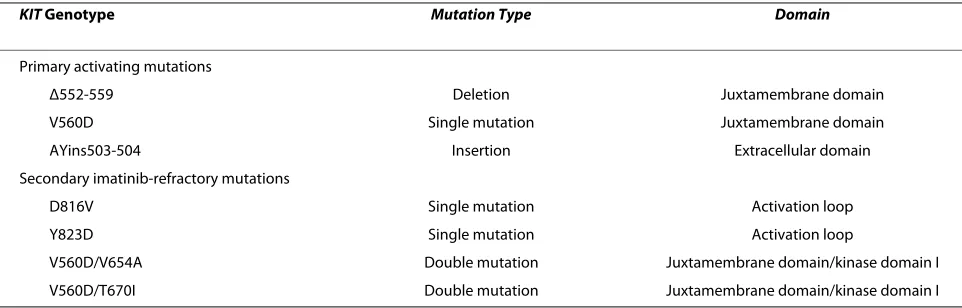
Table 1: Clinically Relevant KIT Mutations
KIT Genotype Mutation Type Domain
Primary activating mutations
Δ552-559 Deletion Juxtamembrane domain
V560D Single mutation Juxtamembrane domain
AYins503-504 Insertion Extracellular domain
Secondary imatinib-refractory mutations
D816V Single mutation Activation loop
Y823D Single mutation Activation loop
V560D/V654A Double mutation Juxtamembrane domain/kinase domain I
sorted directly into 15 mL conical tubes containing the appropriate growth media. Cell pools were then cultured and maintained under the respective selection condi-tions, and were reanalyzed for Kit expression prior to characterization of Kit autophosphorylation.
Cell-Based Kit Autophosphorylation Assay
CHO cells stably transfected with wild-type or mutant isoforms of KIT were seeded in a 96-well tissue culture plate at a density of 2 × 10⁴ cells per well. For stem cell factor (SCF) characterization experiments, cells were stimulated with serial dilutions of SCF for varying times. To determine IC50 values, the cells were treated for 2 hours with single 10-fold serial dilutions of motesanib or imatinib starting at 3 μM. Cell lines transfected with wild-type KIT were stimulated for 10 minutes with 100 ng/mL SCF following treatment with motesanib or ima-tinib. Cell lines transfected with activating KIT mutants were not stimulated with SCF in IC50 experiments. Cells were washed with phosphate-buffered saline and lysed in RIPA buffer (50 mM Tris, pH 7; 150 mM NaCl, 1% Igepal, 0.5% sodium deoxycholate, 0.1% SDS, 300 μM activated sodium vanadate, 1× protease inhibitor cocktail) for 30 minutes at 4°C with shaking. Cell lysates were added to a 96-well DELFIA microplate (PerkinElmer Inc.) coated with anti-Kit antibody (1 μg per well; AF332, R&D Sys-tems, Inc.; Minneapolis, MN) and incubated for 2 hours. Lysates were then removed and the plate was washed 3 times with DELFIA wash buffer (PerkinElmer Inc.). Recombinant anti-phosphotyrosine antibody 4G10 (Cat. # 05-777; Upstate/Millipore, Billerica, MA) was added to each well (0.1 μg per well) and incubated at room temper-ature for 1 hour. The plate was then washed 3 times with DELFIA wash buffer before 0.01 μg of Eu-N1-labeled anti-mouse antibody (Cat. # AD0124, PerkinElmer Inc.) was added to each well. The plate was again incubated at room temperature for 1 hour and then washed 3 times with DELFIA wash buffer before the signal was detected by adding DELFIA enhancement buffer (PerkinElmer Inc.) to each well. Luminescence was measured using a Victor Model 1420 multilabel counter (PerkinElmer Inc.). Kit autophosphorylation at each motesanib or imatinib concentration was expressed as a percentage of the vehi-cle control (0.2% DMSO).
Ba/F3 Functional Viability Assay
The ability of Kit mutants to act as survival factors was assessed in Kit-dependent Ba/F3 cells. Ba/F3 cells stably transfected with various KIT mutants were seeded in a 96-well tissue culture plate at a density of 5 × 10³ cells per well. To determine IC50 values, cells were treated for 24 hours with single 10-fold serial dilutions of motesanib or imatinib starting at 3 μM (0.1 μM for motesanib-treated V560 D and Δ552-559 Kit mutants). Cell viability was
assessed by measuring the level of adenosine triphos-phate using ATPlite assays (PerkinElmer Life Sciences, Boston, MA). Reconstituted ATPLite 1-step solution was added to each well followed by incubation with shaking for 2 minutes. The plate was read on a Victor Model 1420 multilabel counter (PerkinElmer Inc.) under the lumines-cence setting. Viability at each motesanib or imatinib concentration was expressed as a percentage of the vehi-cle control (0.2% DMSO).
Results
In Vitro Inhibition of Wild-Type Kit by Motesanib
Motesanib potently inhibited SCF-induced autophospho-rylation of Kit in CHO cells stably transfected with the wild-type KIT gene (IC50 = 36 nM). In comparison, ima-tinib inhibited wild-type Kit with an IC50 of 165 nM.
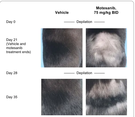
Inhibition of Wild-Type Kit Activity in Mice by Motesanib Hair depigmentation was used as a surrogate marker to assess the ability of motesanib to inhibit Kit activity in vivo [16]. Following depilation, female C57B6 mice were administered either 75 mg/kg motesanib (n = 8) or vehi-cle (n = 8) twice daily for 21 days. In mice receiving mote-sanib, hair regrowth was markedly depigmented compared with mice receiving vehicle (Figure 1). This effect was reversible. Following the cessation of mote-sanib treatment on day 21, the mice were depilated again
Figure 1 Effect of treatment with motesanib or vehicle on hair de-pigmentation, a surrogate marker of Kit activity [16], in female C57B6 mice. Anesthetized animals were depilated and immediately treated with either vehicle (water; left panels) or motesanib 75 mg/kg BID (right panels) for 21 days. On day 21, hair depigmentation was as-sessed. Depilation was repeated on day 28 and hair depigmentation was again assessed on day 35. Representative images from each treat-ment group for the day-21 and day-35 time points are shown. BID = twice daily.
Day 28 Day 21 (Vehicle and motesanib treatment ends)
Motesanib, 75 mg/kg BID Vehicle
Day 0
Day 35
——— Depilation ———
on day 28. There was no apparent depigmentation of regrown hair on day 35. Similar results were obtained in male mice (data not shown).
Characterization of Kit Mutants
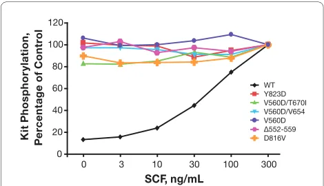
Figure 2 summarizes the results from the autophosphory-lation experiments using CHO cells stably transfected with the wild-type KIT gene or various KIT mutant genes. Tyrosine phosphorylation of wild-type Kit was dose-dependent, with the greatest intensity of autophos-phorylation occurring after a 30 minute incubation of the cells with 300 ng/mL of SCF. In contrast, tyrosine phos-phorylation of activated Kit mutants occurred in the absence of SCF with no further phosphorylation induced by treatment with SCF.
Activity of Motesanib against Primary Activating Kit Mutants
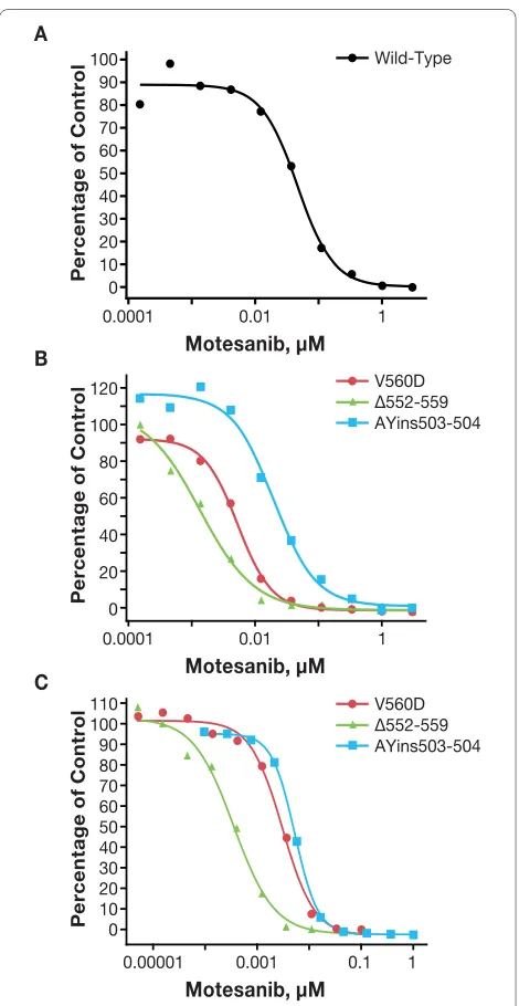
In CHO cells, motesanib inhibited the autophosphoryla-tion of the primary activating Kit mutants V560 D, Δ552-559, and AYins503-504 (Table 2; Figure 3B). In each instance, motesanib was a more potent inhibitor of Kit autophosphorylation than imatinib. For example, mote-sanib inhibited the AYins503-504 mutant with an IC50 of 18 nM, whereas imatinib inhibited this mutant with an IC50 of 84 nM. Interestingly, the IC50 values for inhibition of these Kit mutants were lower than the IC50 for inhibi-tion of wild-type Kit by motesanib. Consistent results were obtained in a functional viability assay utilizing IL-3-independent growth of Ba/F3 cells (Figure 3C). For example, when testing the AYins503-504 mutant, the IC50 for motesanib was 11 nM versus 47 nM for imatinib.
Activity of Motesanib against Imatinib-Resistant Kit Mutants
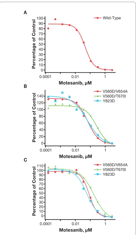
Motesanib inhibited the activity of Kit mutants associated with secondary imatinib resistance. In Kit autophospho-rylation assays, motesanib inhibited tyrosine phosphory-lation of the juxtamembrane domain/kinase domain I double mutants V560D/V654A and V560D/T670I with IC50 values of 77 nM and 277 nM, respectively. Imatinib had limited activity against the V560D/V654A mutant and no activity against the V560D/T670I mutant at con-centrations of up to 3000 nM (Table 3; Figure 4B). Consis-tent results were obtained in the Ba/F3 cells expressing the V560D/V654A and V560D/T670I mutants with motesanib IC50 values of 91 nM and 180 nM, respectively. Again, motesanib was a more potent inhibitor of these mutants than imatinib (Table 3; Figure 4C).
Similarly, motesanib inhibited autophosphorylation of the imatinib-resistant activation loop mutant Y823 D (IC50 = 64 nM) more potently than imatinib (IC50 > 3000 nM) (Table 3: Figure 4B). However, neither motesanib nor imatinib inhibited autophosphorylation of the D816V mutant (Table 3). Consistent with these results, mote-sanib inhibited the growth of Ba/F3 cells transfected with the V560D/V654A, V560D/T670I, or Y823 D mutant more potently than imatinib. Of note, the IC50 of ima-tinib against the Y823 D mutant when established in the functional viability assay was at least 10-fold lower than the IC50 measured in the autophosphorylation assay. IL-3-independent Ba/F3 cells expressing the D816V Kit mutant could not be established.
Discussion
In this study, motesanib was found to be a potent inhibi-tor of wild-type Kit, both in vitro and in vivo. In a surro-gate marker assay, we observed reversible hair Figure 2 Effect of stem cell factor (SCF) treatment on tyrosine
phosphorylation of wild-type Kit and mutant Kit isoforms stably expressed in Chinese hamster ovary cells. Chinese hamster ovary
cells stably transfected with wild-type (WT) or mutant KIT isoforms
were stimulated with single serial dilutions of stem cell factor, and Kit phosphorylation was assessed. For mutant Kit isoforms, data are ex-pressed as the percentage of vehicle control. For wild-type Kit, data are expressed as the percentage of phosphorylation observed following stimulation with 300 ng/mL SCF. The results of a single experiment are shown.
Table 2: Inhibition of the Activity of Wild-Type Kit and Primary Activating Kit Mutants by Motesanib and Imatinib*
IC50 of Kit
Autophosphorylation, nM
IC50 of Stably Transfected Ba/F3 Cell Survival, nM
KIT Genotype Motesanib Imatinib Motesanib Imatinib
Wild-type 36 165 -
-V560D 5 18 3 7
Δ552-559 1 5 0.4 1
AYins503-504 18 84 11 47
depigmentation in mice treated with motesanib 75 mg/kg twice daily. This dose is comparable to the doses used in xenograft studies demonstrating antitumor and antian-giogenic properties of motesanib [9,17]. Kit signaling plays an important role in the regulation of hair follicle
melanocytes, likely through control of tyrosinase and tyrosinase-related protein 1 (TRP1) expression [16]. Depigmentation has previously been observed in mice treated with anti-Kit antibodies [16,18] or with sunitinib [18]. Importantly, motesanib had inhibitory activity against Kit mutants associated with GIST and inhibited these mutants more potently than imatinib and generally with an IC50 that was less than or similar to the 24-hour trough concentration of motesanib at therapeutic doses in humans [10].
Motesanib was a more potent inhibitor of the primary activating juxtamembrane domain and extracellular domain Kit mutants V560 D, Δ552-559, and AYins503-504, compared with imatinib. Importantly, motesanib also inhibited the activity of an activation loop mutant (Y823D) associated with imatinib resistance. Imatinib did not inhibit this mutant at concentrations of up to 3000 nM, suggesting that there are marked differences in how the two inhibitors interact with Kit. We previously solved the structure of motesanib bound to the VEGFR2 kinase domain at 2.2 Å resolution (PDB Accession Code 3EFL) [19]. This structure superimposes favorably with that of Kit co-crystallized with imatinib (PDB Accession Code 1T46) [20]. Both inhibitors bind the inactive, auto-inhib-ited form of the kinases with the backbone of the protein reorganized into the so-called "DFG-out" conformation. Based on the structural similarities and the similar poten-cies of motesanib against VEGFR2 and Kit, we reasoned that motesanib binds these target kinases in exactly the same fashion.
Modeling studies suggest that motesanib engages Kit via three polar interactions and a multitude of van der Waals contacts (Figure 5). In the context of this study, the most important of these interactions are those with thre-onine 670 via a non-classical CH-O pseudo hydrogen bond and interactions with valine 654 through hydropho-Figure 3 Inhibition of the activity of wild-type Kit and primary
ac-tivating Kit mutants by motesanib. Autophosphorylation (ex-pressed as a percentage of vehicle control) of wild-type Kit (panel A) and primary activating Kit mutants (panel B) was assessed in stably transfected Chinese hamster ovary cells treated for 2 hours with single 10-fold serial dilutions of motesanib. Representative data from 1 of 2 experiments are shown. Viability (expressed as the percentage of vehi-cle control) of Ba/F3 cells expressing the same primary activating Kit mutants treated for 24 hours with single 10-fold serial dilutions of motesanib was also assessed (panel C). Viability experiments were per-formed once (representative curves are shown).
Motesanib, μM A
B
C
P
ercent
a
ge
o
f C
o
ntrol
0.0001
V560D ∆552-559 AYins503-504
V560D ∆552-559 AYins503-504 0.01 1
100 90 80 70 60 50 40 30 20 10 0
Motesanib, μM
P
ercent
a
ge
o
f C
o
ntrol
0.0001 0.01 1 100
120
80
60
40
20
0
Motesanib, μM
P
ercent
a
ge
o
f C
o
ntrol
0.00001 0.001 100
110
80
60
40
20
0 90
70
50
30
10
Wild-Type
1 0.1
Table 3: Inhibition of the Activity of Kit Mutants Associated With Imatinib Resistance by Motesanib and Imatinib*
IC50 of Kit
Autophosphorylation, nM
IC50 of Stably
Transfected Ba/F3 Cell Survival, nM
KIT Genotype Motesanib Imatinib Motesanib Imatinib
V560D/V654A 77 319 91 145
V560D/T670I 277 >3000 180 >3000
Y823D 64 >3000 62 330
D816V >3000 >3000 -
bic contacts. The fifteen-fold loss of motesanib activity (5 nM versus 77 nM) noted with the V560D/V654A double mutant, compared with V560 D alone, is rationalized by the loss of two van der Waals contacts with alanine 654 in a similar fashion to that described for imatinib [21,22].
Motesanib and imatinib have much diminished activity against the activation loop mutant (D816V). The D816V
mutant destabilizes the inactivated form of Kit, in a way that the ability of the protein to adopt the "DFG out" (inactive) conformation is much reduced or even elimi-nated; thus, the mutation prevents both motesanib and imatinib from binding to the ATP pocket [23,24]. The failure to potently inhibit the D816V mutation is a feature of Kit inhibitors in the clinic, with the exception for dasa-tinib [23,25,26], which binds the "DFG in", or activated form, of the kinase [27]. However, the ability of motesanib to inhibit the Y823 D mutant suggests that its activity may not be entirely restricted to an inactive protein con-formation, or alternatively it may reflect that in contrast to the D816V mutation, the conformational equilibrium of the Y823 D mutant is not shifted permanently to the active conformation.
The data from the present study are of translational rel-evance, supporting evidence indicating that targeted therapy molecules with different binding sites and/or mode of action may be required in the treatment of can-cers for which mutations are the primary oncogenic event. A recent study has demonstrated that differences in the conformational structure of Kit mutants influences the ability of sunitinib [28], and imatinib to bind and inhibit receptor autophosphorylation, thus providing a unique mechanism of drug resistance for each mutant that is unlikely to be overcome using a single treatment [23].
Conclusions
In summary, the results of this study demonstrate that different Kit mutations respond differently to motesanib or imatinib. This likely reflects differences in the mole-cules' mode of action. The data also show that motesanib is active against Kit mutations associated with resistance, Figure 5 A model of motesanib bound to the active site of Kit ki-nase derived from a 2.2 Ångstrom resolution crystal structure of motesanib bound to the active site of VEGFR2 kinase (PDB code 2EFL).
Figure 4 Inhibition of the activity of Kit mutants associated with secondary imatinib resistance by motesanib. Autophosphorylation (expressed as a percentage of vehicle control) of wild-type Kit (panel A) and Kit mutants associated with secondary imatinib resistance (panel B) was assessed in stably transfected Chinese hamster ovary cells treat-ed for 2 hours with single 10-fold serial dilutions of motesanib. Repre-sentative data from 1 of 2 experiments are shown. Viability (expressed as the percentage of vehicle control) of Ba/F3 cells expressing the same Kit mutants treated for 24 hours with single 10-fold serial dilu-tions of motesanib was also assessed (panel C; not shown: D816V,
which had a motesanib IC50 > 3 μM). Viability experiments were
per-formed once and representative curves are shown (D816V was not evaluated because Ba/F3 cells expressing this mutant could not be es-tablished).
suggesting that it may have clinical utility in the treat-ment of patients with primary and secondary imatinib-resistant GIST.
Competing interests
All authors are employees of and shareholders in Amgen Inc.
Authors' contributions
SC designed the cell viability and Kit autophosphorylation assays. LRG contrib-uted to the generation of cell lines expressing wild-type and mutant Kit. AB performed the depilation experiments. TLB performed the depilation
experi-ments. WB designed and generated wild-type and mutant KIT gene expression
vectors. TJ designed and generated wild-type and mutant KIT gene expression
vectors. RM contributed to the generation of cell lines expressing wild-type and mutant Kit. AST contributed the molecular modelling and assisted with the writing of the manuscript. AP was responsible for the overall experimental design and contributed to the writing of the manuscript. PEH was responsible for individual experimental designs and contributed to the writing of the man-sucript. All authors have read and approved the final manuscript.
Acknowledgements
The authors wish to acknowledge Douglas Whittington and Joseph Kim (Amgen Inc., Cambridge, MA) for generating the model of motesanib bound to Kit. Additionally, the authors would like to thank Ali Hassan, PhD (Complete Healthcare Communications, Inc.), whose work was funded by Amgen Inc., and Beate Quednau, PhD (Amgen Inc.), for their assistance in the preparation of this manuscript.
Author Details
1Department of Oncology Research, Amgen Inc., One Amgen Center Drive,
Thousand Oaks, CA, 91320-1799, USA, 2Department of Protein Science, Amgen
Inc., One Amgen Center Drive, Thousand Oaks, CA, 91320-1799, USA,
3Department of Molecular Sciences, Amgen Inc., One Amgen Center Drive,
Thousand Oaks, CA, 91320-1799, USA, 4Department of Medicinal Chemistry,
Amgen Inc., One Amgen Center Drive, Thousand Oaks, CA, 91320-1799, USA
and 5Department of Oncology, Amgen, Inc., 1201 Amgen Court West, Seattle,
WA, 98119-3105, USA
References
1. Heinrich MC, Corless CL, Demetri GD, Blanke CD, von Mehren M, Joensuu
H, McGreevey LS, Chen CJ, Van den Abbeele AD, Druker BJ, Kiese B, Eisenberg B, Roberts PJ, Singer S, Fletcher CD, Silberman S, Dimitrijevic S,
Fletcher JA: Kinase mutations and imatinib response in patients with
metastatic gastrointestinal stromal tumor. J Clin Oncol 2003,
21:4342-4349.
2. Hirota S, Isozaki K, Moriyama Y, Hashimoto K, Nishida T, Ishiguro S, Kawano
K, Hanada M, Kurata A, Takeda M, Muhammad Tunio G, Matsuzawa Y,
Kanakura Y, Shinomura Y, Kitamura Y: Gain-of-function mutations of
c-kit in human gastrointestinal stromal tumors. Science 1998,
279:577-580.
3. Corless CL, McGreevey L, Haley A, Town A, Heinrich MC: KIT mutations are
common in incidental gastrointestinal stromal tumors one centimeter or less in size. Am J Pathol 2002, 160:1567-1572.
4. Corless CL, Fletcher JA, Heinrich MC: Biology of gastrointestinal stromal
tumors. J Clin Oncol 2004, 22:3813-3825.
5. Heinrich MC, Corless CL, Duensing A, McGreevey L, Chen CJ, Joseph N,
Singer S, Griffith DJ, Haley A, Town A, Demetri GD, Fletcher CD, Fletcher JA:
PDGFRA activating mutations in gastrointestinal stromal tumors.
Science 2003, 299:708-710.
6. Demetri GD, von Mehren M, Blanke CD, Van den Abbeele AD, Eisenberg B,
Roberts PJ, Heinrich MC, Tuveson DA, Singer S, Janicek M, Fletcher JA, Silverman SG, Silberman SL, Capdeville R, Kiese B, Peng B, Dimitrijevic S,
Druker BJ, Corless C, Fletcher CD, Joensuu H: Efficacy and safety of
imatinib mesylate in advanced gastrointestinal stromal tumors. N Engl J Med 2002, 347:472-480.
7. Frost MJ, Ferrao PT, Hughes TP, Ashman LK: Juxtamembrane mutant
V560GKit is more sensitive to Imatinib (STI571) compared with
wild-type c-kit whereas the kinase domain mutant D816VKit is resistant.
Mol Cancer Ther 2002, 1:1115-1124.
8. Heinrich MC, Corless CL, Blanke CD, Demetri GD, Joensuu H, Roberts PJ,
Eisenberg BL, von Mehren M, Fletcher CD, Sandau K, McDougall K, Ou WB,
Chen CJ, Fletcher JA: Molecular correlates of imatinib resistance in
gastrointestinal stromal tumors. J Clin Oncol 2006, 24:4764-4774.
9. Polverino A, Coxon A, Starnes C, Diaz Z, DeMelfi T, Wang L, Bready J,
Estrada J, Cattley R, Kaufman S, Chen D, Gan Y, Kumar G, Meyer J, Neervannan S, Alva G, Talvenheimo J, Montestruque S, Tasker A, Patel V,
Radinsky R, Kendall R: AMG 706, an oral, multikinase inhibitor that
selectively targets vascular endothelial growth factor, platelet-derived growth factor, and kit receptors, potently inhibits angiogenesis and induces regression in tumor xenografts. Cancer Res 2006, 66:8715-8721. 10. Rosen LS, Kurzrock R, Mulay M, Van Vugt A, Purdom M, Ng C, Silverman J,
Koutsoukos A, Sun YN, Bass MB, Xu RY, Polverino A, Wiezorek JS, Chang
DD, Benjamin R, Herbst RS: Safety, pharmacokinetics, and efficacy of
AMG 706, an oral multikinase inhibitor, in patients with advanced solid tumors. J Clin Oncol 2007, 25:2369-2376.
11. Price TJ, Lipton L, McGreivy J, McCoy S, Sun YN, Rosenthal MA: Safety and
pharmacokinetics of motesanib in combination with gemcitabine for the treatment of patients with solid tumours. Br J Cancer 2008,
99:1387-1394.
12. Schlumberger MJ, Elisei R, Bastholt L, Wirth LJ, Martins RG, Locati LD, Jarzab B, Pacini F, Daumerie C, Droz JP, Eschenberg MJ, Sun YN, Juan T,
Stepan DE, Sherman SI: Phase II study of safety and efficacy of
motesanib in patients with progressive or symptomatic, advanced or metastatic medullary thyroid cancer. J Clin Oncol 2009, 27:3794-3801. 13. Sherman SI, Wirth LJ, Droz JP, Hofmann M, Bastholt L, Martins RG, Licitra L,
Eschenberg MJ, Sun YN, Juan T, Stepan DE, Schlumberger MJ: Motesanib
diphosphate in progressive differentiated thyroid cancer. N Engl J Med
2008, 359:31-42.
14. Benjamin R, Schöffski P, Hartmann JT, Bui BN, Duyster J, Schuetze S, Blay J,
Reichard P, Rosen L, Skubitz K, Eschenberg M, Stepan D, Baker L: Initial
results of a multicenter single arm phase 2 study of AMG 706, an oral multi-kinase inhibitor, for the treatment of advanced imatinib-resistant gastrointestinal stromal tumors (GIST) [abstract 641]. Connective Tissue Oncology Society 12th Annual Meeting 2006. Venice, Italy. Year
15. Sawaki A, Yamada Y, Komatsu Y, Kanda T, Doi T, Koseki M, Baba H, Sun YN,
Murakami K, Nishida T: Phase II study of motesanib in Japanese patients
with advanced gastrointestinal stromal tumors with prior exposure to imatinib mesylate. Cancer Chemother Pharmacol 2009, 65:961-967. 16. Botchkareva NV, Khlgatian M, Longley BJ, Botchkarev VA, Gilchrest BA:
SCF/c-kit signaling is required for cyclic regeneration of the hair pigmentation unit. FASEB J 2001, 15:645-658.
17. Coxon A, Bush T, Saffran D, Kaufman S, Belmontes B, Rex K, Hughes P, Caenepeel S, Rottman JB, Tasker A, Patel V, Kendall R, Radinsky R, Polverino A: Broad antitumor activity in breast cancer xenografts by motesanib, a highly selective, oral inhibitor of vascular endothelial growth factor, platelet-derived growth factor, and Kit receptors. Clin Cancer Res 2009,
15:110-118.
18. Moss KG, Toner GC, Cherrington JM, Mendel DB, Laird AD: Hair
depigmentation is a biological readout for pharmacological inhibition of KIT in mice and humans. J Pharmacol Exp Ther 2003, 307:476-480.
19. Tasker AS, Patel VF: Discovery of motesanib. In Kinase Inhibitor Drugs
Edited by: Li R, Stafford JA. Hoboken, NJ: John Wiley & Sons, Inc.; 2009:113-130.
20. Mol CD, Dougan DR, Schneider TR, Skene RJ, Kraus ML, Scheibe DN, Snell
GP, Zou H, Sang BC, Wilson KP: Structural basis for the autoinhibition
and STI-571 inhibition of c-Kit tyrosine kinase. J Biol Chem 2004,
279:31655-31663.
21. McLean SR, Gana-Weisz M, Hartzoulakis B, Frow R, Whelan J, Selwood D,
Boshoff C: Imatinib binding and cKIT inhibition is abrogated by the cKIT
kinase domain I missense mutation Val654Ala. Mol Cancer Ther 2005,
4:2008-2015.
22. Roberts KG, Odell AF, Byrnes EM, Baleato RM, Griffith R, Lyons AB, Ashman LK: Resistance to c-KIT kinase inhibitors conferred by V654A mutation.
Mol Cancer Ther 2007, 6:1159-1166.
23. Gajiwala KS, Wu JC, Christensen J, Deshmukh GD, Diehl W, Dinitto JP, English JM, Greig MJ, He YA, Jacques SL, Lunney EA, McTigue M, Molina D, Quenzer T, Wells PA, Yu X, Zhang Y, Zou A, Emmett MR, Marshall AG,
Zhang HM, Demetri GD: KIT kinase mutants show unique mechanisms
Received: 30 March 2010 Accepted: 15 July 2010 Published: 15 July 2010
This article is available from: http://www.jeccr.com/content/29/1/96 © 2010 Caenepeel et al; licensee BioMed Central Ltd.
This is an Open Access article distributed under the terms of the Creative Commons Attribution License (http://creativecommons.org/licenses/by/2.0), which permits unrestricted use, distribution, and reproduction in any medium, provided the original work is properly cited.
of drug resistance to imatinib and sunitinib in gastrointestinal stromal tumor patients. Proc Natl Acad Sci USA 2009, 106:1542-1547.
24. Foster R, Griffith R, Ferrao P, Ashman L: Molecular basis of the
constitutive activity and STI571 resistance of Asp816Val mutant KIT receptor tyrosine kinase. J Mol Graph Model 2004, 23:139-152. 25. Schittenhelm MM, Shiraga S, Schroeder A, Corbin AS, Griffith D, Lee FY,
Bokemeyer C, Deininger MW, Druker BJ, Heinrich MC: Dasatinib
(BMS-354825), a dual SRC/ABL kinase inhibitor, inhibits the kinase activity of wild-type, juxtamembrane, and activation loop mutant KIT isoforms associated with human malignancies. Cancer Res 2006, 66:473-481.
26. Shah NP, Lee FY, Luo R, Jiang Y, Donker M, Akin C: Dasatinib
(BMS-354825) inhibits KITD816V, an imatinib-resistant activating mutation that triggers neoplastic growth in most patients with systemic mastocytosis. Blood 2006, 108:286-291.
27. Tokarski JS, Newitt JA, Chang CY, Cheng JD, Wittekind M, Kiefer SE, Kish K,
Lee FY, Borzillerri R, Lombardo LJ, Xie D, Zhang Y, Klei HE: The structure of
Dasatinib (BMS-354825) bound to activated ABL kinase domain elucidates its inhibitory activity against imatinib-resistant ABL mutants. Cancer Res 2006, 66:5790-5797.
28. Demetri GD, van Oosterom AT, Garrett CR, Blackstein ME, Shah MH, Verweij J, McArthur G, Judson IR, Heinrich MC, Morgan JA, Desai J, Fletcher
CD, George S, Bello CL, Huang X, Baum CM, Casali PG: Efficacy and safety
of sunitinib in patients with advanced gastrointestinal stromal tumour after failure of imatinib: a randomised controlled trial. Lancet 2006,
368:1329-1338.
doi: 10.1186/1756-9966-29-96