R E S E A R C H
Open Access
Mesenchymal stem cells exert anti-proliferative
effect on lipopolysaccharide-stimulated BV2
microglia by reducing tumour necrosis factor-
α
levels
Shinsmon Jose
1,2,3, Shi Wei Tan
1, Yin Yin Ooi
4, Rajesh Ramasamy
2,5and Sharmili Vidyadaran
1,2*Abstract
Background:Progression of neurodegenerative diseases occurs when microglia, upon persistent activation, perpetuate a cycle of damage in the central nervous system. Use of mesenchymal stem cells (MSC) has been suggested as an approach to manage microglia activation based on their immunomodulatory functions. In the present study, we describe the mechanism through which bone marrow-derived MSC modulate the proliferative responses of lipopolysaccharide-stimulated BV2 microglia.
Methods:BV2 microglia were cultured with MSC and stimulated with 1μg/ml lipopolysaccharide. Using an inducible nitric oxide synthase inhibitor, tritiated thymidine (3H-TdR) incorporation assay was performed to
determine the role of nitric oxide in the anti-proliferative effect of MSC. We also studied apoptosis and the cell cycle of both cell types using flow cytometry and explored their cytokine profile using protein and cytometric arrays. Moreover, the role of IL-6 and TNF-αin immunomodulation was deduced using specific blocking antibodies and recombinant proteins.
Results:MSC reduces microglia proliferation upon lipopolysaccharide stimulation by 21 to 28% and modulates the levels of nitric oxide, IL-6 and TNF-α. The role of nitric oxide in conferring the anti-proliferative effect of MSC was ruled out. Furthermore, we found that MSC exert their anti-proliferative effect by restoring the percentage of BV2 cells at S and G2/M phase to levels similar to unstimulated cells. MSC undergo a G0/G1 arrest while exerting this effect. We have also identified that MSC-mediated modulation of microglia is independent of IL-6, whilst reduction of TNF-αin co-culture is critical for inhibition of microglia proliferation.
Conclusions:Our study demonstrates that MSC inhibit microglia proliferation independent of nitric oxide and IL-6, although reduction of TNF-αis critical for this effect. The inhibition of proliferation is through cell cycle modulation. These findings shed light on the mechanisms of microglial immunomodulation by MSC.
Keywords:Microglia, Mesenchymal stem cells, Nitric oxide, Cell cycle, Interleukin-6, Tumour necrosis factor-α
* Correspondence:sharmili@upm.edu.my
1Neuroinflammation Group, Immunology Laboratory, Department of
Pathology, Faculty of Medicine and Health Sciences, Universiti Putra Malaysia, 43400 Serdang, Selangor, Malaysia
2
Genetic Medicine Research Centre, Faculty of Medicine and Health Sciences, Universiti Putra Malaysia, 43400 Serdang, Selangor, Malaysia
Full list of author information is available at the end of the article
Background
Mesenchymal stem cells (MSC) are a group of clono-genic, non-haematopoietic, plastic adherent multi-potent stromal cells that possess potential to differentiate into cells of mesodermal lineage [1,2]. MSC have been shown to ameliorate disease in numerous clinical trials and animal models. Approximately 400 clinical trials using MSC are currently underway according to the clinical trials registry of the US National Institute of Health (http://clinicaltrials. gov). It is noteworthy that a good proportion of these studies are exploring the immunomodulatory properties of MSC. This aspect of MSC is widely explored in managing graft-versus-host disease [3] and in a variety of experimen-tal disease models, including autoimmune encephalomyeli-tis [4], stroke [5], amyotrophic lateral sclerosis [6], spinal cord injury [7], diabetes [8] and myocardial infarction [9]. These advances at the therapeutic level are a result of de-tailed descriptions on the potential of MSC to modulate a range of immune cells including T cells, B cells, dendritic cells, monocytes and macrophages [10-15].
Microglia are resident macrophages of the central ner-vous system (CNS), derived from primitive myeloid pro-genitors that migrate from the embryonic yolk sac to the brain rudiment [16]. In adults, microglia survey the entire brain every couple of hours through concerted movements [17] for damaged neurons, endogenous disease proteins, and infection [18]. Microglia assume functions beyond being immune sentinels by maintaining a healthy CNS environment through synaptic pruning [19-21] and secretion of neurotrophic factors [22,23]. In response to disturbances in CNS homeostasis microglia undergo morphological, phenotypic and functional changes. These changes include an increase in numbers through prolifera-tion, a shift from ramified to amoeboid morphology, secretion of inflammatory mediators such as cytokines, chemokines and reactive oxygen and nitrogen species, and an increase in phagocytic activity. Experiments have shown that these changes which microglia undergo upon activation can cause deleterious effects within the CNS microenvironment and play a key role in patho-genesis of neurodegenerative disease [24-27].
Modulating microglia responses is being considered as an effective approach to manage progression of neu-rodegenerative diseases. Recently, with the rise in re-ports on immunomodulatory prospects of MSC, we and others have explored and described the potential of MSC to dampen inflammatory responses of microglia [28-32]. However, the mechanisms underlying these effects are poorly understood. The present study deter-mined the mechanisms through which MSC confer an anti-proliferative effect on microglia by examining apop-tosis and cell cycle. We also determined the role of nitric oxide, IL-6 and TNF-α in conferring the modulatory effects of MSC. It was also demonstrated that MSC
experience growth arrest in co-culture while modulating microglial responses.
Materials and methods
Mouse bone marrow mesenchymal stem cell culture MSC previously isolated from ICR mouse bone marrow were obtained from the culture collection of the Im-munology Laboratory, Faculty of Medicine and Health Sciences, Universiti Putra Malaysia. Frozen vials stored in liquid nitrogen were thawed at 37°C and cultured in MSC complete medium (high glucose DMEM (GIBCO Invitrogen, CA, USA), supplemented with 15% (v/v) MesenCult® Mesenchymal Stem Cell Stimulatory Sup-plements (Mouse) (Stemcell™ Technologies, Canada), 1% penicillin and streptomycin (iDNA), 250 μg/ml fun-gizone (GIBCO Invitrogen, CA, USA), 2.0 mM Gluta-MaX and 1.5 g sodium bicarbonate) at 37°C, 5% CO2in
a humidified incubator. Cells were routinely sub-cultured using 0.25% Trypsin-EDTA (GIBCO Invitrogen, CA, USA) before reaching 90% confluency and immunophenotyped using a panel of markers comprising CD106, CD45, CD44, CD11b, Sca-1, MHC-I and MHC-II (all from Becton Dickinson, BD, San Jose, CA, USA) [1]. MSC phenotype was confirmed by positivity to CD106, CD44, Sca-1 and MHC-I and negativity to CD45, CD11b and MHC-II.
BV2 microglia culture
BV2 cells were a generous gift from Dr Thameem Dheen of the National University of Singapore. BV2 is a murine microglial cell line immortalised with v-raf/v-myc genes carrying retrovirus J2 [33]. Cells were cultured in DMEM with 5% heat-inactivated foetal bovine serum, 100 U/ml penicillin, 100μg/ml streptomycin, 10 g/ml genta-micin (all Invitrogen), 1 × non-essential amino acids and 6.25 μg/ml insulin (Sigma-Aldrich, St. Louis, MO, USA). Cells were either sub-cultured or used for downstream assays before reaching 90% confluency by harvesting with 0.25% trypsin containing 1 mM EDTA for 5 mi-nutes at 37°C.
MSC/BV2 co-cultures
BV2 and MSC were seeded simultaneously at a ratio of 1:0.2 and incubated overnight at 37°C in a 5% CO2
3
H-TdR incorporation assay
BV2 cell proliferation was determined by assessing triti-ated thymidine (3H-TdR; Perkin Elmer, Boston, USA) in-corporation. In 96-well plates, 1 × 10³MSC were seeded in triplicate and allowed to adhere overnight. The follow-ing day, MSC were treated with 10 μg/ml mitomycin-C (Sigma) for 2 hours to halt their proliferation. Plates were washed thoroughly with DMEM to remove any traces of the mitotic inhibitor and BV2 cells were then seeded at 5 × 10³cells/well. Co-cultures were activated with 1μg/ml LPS for 48 hours and3H-TdR (0.037 MBq/well (0.5μCi/ well)) was added to wells at the final 6 hours of incuba-tion. Plates were exposed to a freeze/thaw cycle at -20°C to ease cell harvesting. Cells were harvested onto a filter mat by using an automated cell harvester (Harvester Mach III M, TOMTEC, CT, USA Thymidine incorpor-ation was measured by liquid scintillincorpor-ation spectroscopy on a beta counter (MicroBetaTriLux, Perkin Elmer Boston, USA) after the addition of scintillation fluid (OptiPhaseSu-perMix Cocktail; Perkin Elmer Boston, USA) and readouts were in counts per minute (cpm).
Griess assay
Nitric oxide (NO) was detected in the supernatant of cultures using the Griess assay. For this, 50 μl culture supernatant from each sample was transferred to a 96-well plate in triplicate and an equal volume of Griess reagent added (1% sulphanilamide/0.1% N-1-napthylethylene-diamine dihydrochloride/2.5% phosphoric acid; all from Sigma). Absorbance was read at 530 nm (MRX II micro-plate reader, Dynex, VA, USA) after 10 minutes incuba-tion. Nitrite concentration was calculated with reference to a standard curve of freshly prepared sodium nitrite (0 to 100μM). The results are displayed as concentration of NO2-inμM.
Apoptosis assay
Apoptosis of cells in co-culture was determined by flow cytometry after double staining with FITC-Annexin-V and propidium iodide (PI). BV2 cells and MSC were co-cultured overnight at a 1:0.2 ratio, stimulated with 1μg/ ml LPS the following day, and left in culture for 48 hours. Cells were then harvested using 0.25% trypsin-EDTA. Cells were washed twice in ice-cold PBS and suspended in 100μl of 1X binding buffer at a concentration of 1 × 10⁶cells/ml. Cells were stained for CD45 by incubating with 0.5μl antibody (Rat anti-mouse CD45, BioLegend®, San Diego, CA, USA ) at 4°C for 15 minutes followed by 15 minutes incubation with secondary antibody (DyLight™ 649 Goat anti-rat IgG, BioLegend®). CD45 staining was performed to distinguish BV2 microglia from the MSC population during flow cytometry analysis. Five microlitres each of FITC-conjugated Annexin-V and PI was added to each tube and incubated for 15 minutes at room
temperature. Four hundred microlitres of 1X binding buffer was added to each tube before acquisition and analysis using a flow cytometer.
Cell cycle analysis
Distribution of cells through the three distinct phases of cell cycle (G0/G1, S and G2/M phase) was analysed using PI staining. Co-cultures were harvested using 0.25% trypsin-EDTA and washed once in PBS by centrifugation at 1,000 rpm for 7 minutes. Cells were then suspended in 1 ml of ice-cold PBS at a density of 0.5 × 10⁶cells per tube. The cell suspension was subsequently added drop wise to 3 ml ice-cold 95% ethanol for fixation. The tubes were in-cubated at -20°C for at least 2 hours. The tubes were re-moved from -20°C and spun at 1,200 rpm for 10 minutes and washed once using ice-cold PBS to remove traces of ethanol. The cells were then suspended in 500μl of 50μg/ ml PI staining solution and incubated at 37°C for 15 mi-nutes. Stained cells were washed once with PBS to remove excess PI and suspended in 400μl PBS. The cell cycle data for individual samples were acquired using the BD LSR Fortessa™ flow cytometer equipped with BD FACSDiva™ software (BD) and analysed using ModFit LT™ software (Verity Software House, ME, USA).
Cytokine bead array/protein array for TNF-αand IL-6 detection
Co-culture supernatants were assayed at 24 hours using a custom RayBio® mouse cytokine array kit (RayBiotech, Inc., GA, USA ), according to manufacturer’s instructions. The results are expressed as relative protein levels com-pared with LPS-stimulated BV2 cells. Absolute quantity of IL-6 and TNF-αin culture supernatants were then deter-mined at 24 hours by using a multiplex bead array kit (BD Cytometric Bead Array mouse inflammation kit; BD Biosciences, San Jose, CA, USA), according to the manu-facturer’s instructions. Samples were assayed on a FACS Fortessa flow cytometer (BD Biosciences) and analysed with FCAP array software (BD Biosciences). Concentra-tion of cytokines in samples was calculated using individ-ual standard curves and expressed as pg/ml.
Cytokine blocking studies
after overnight incubation. Equal volumes of diluted anti-body were added to cultures to obtain a final concentration of 10.0, 1.0 and 0.1μg/ml. Culture supernatants were col-lected at 24 and 48 hours and assayed for NO production using Griess assay. Proliferation was analysed at 48 hours by pulsing the cultures with3H-TdR for the final 6 hours of incubation.
Statistical analysis
Mean values and standard deviations (SD) were calcu-lated from three independent experiments and signifi-cance was assessed using one-way analysis of variance followed by the Tukey post hoc test or student’s t test using GraphPad Prism version 6 (GraphPad software, San Diego, CA, USA).
Results
Mesenchymal stem cells inhibit BV2 microglia proliferation independent of nitric oxide
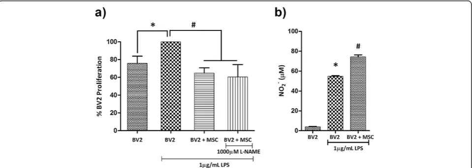
An in vitro model of microglia proliferation was estab-lished by examining BV2 microglia proliferation in re-sponse to LPS stimulation at 6, 24 and 48 hours. The proliferation rate of BV2 microglia appeared unaffected by LPS at 6 and 24 hours (Additional file 1: Figure S1a,b). At 48 hours, LPS-stimulated microglia displayed an approxi-mate 30% increase in proliferation (Figure 1a, P< 0.01; Additional file 1: Figure S1c). This increase in microglia proliferation was used as a model to test the anti-proliferative effect of MSC.
To study the mechanisms through which MSC exert their anti-proliferative effect on microglia, the BV2:MSC
ratio of 0.2 was selected based on previous reports from our laboratory [28,34] (see Additional file 2: Figure S2 for images of BV2/MSC co-culture). Co-culturing with MSC at a 0.2 ratio decreased LPS-induced proliferation of BV2 microglia by 28.77 ± 7.90% at 48 hours (Figure 1a, P< 0.01), similar to proliferation rates of unstimulated BV2 microglia. MSC were mitotically arrested by mitomycin-C (10μg/ml) treatment (Additional file 1: Figure S1d,e) to fa-cilitate accurate measurement of microglia proliferation in co-culture.
A previous report from our laboratory demonstrated that MSC generate NO upon exposure to soluble factors from microglia, while BV2/MSC co-culture induces a surge in NO levels beyond that of LPS-stimulated microglia [34]. When tested in this study, co-culture of BV2:MSC at a 1:0.2 ratio induced a 25% surge in NO levels from 56.94 ± 2.65 μM to 76.59 ± 3.08 μM at 48 hours (Figure 1b; P< 0.01). Within an immunomodulatory paradigm, NO has been shown to play a vital role in MSC-mediated suppres-sion of activated T cell proliferation [35,36]. These reports, with the use of MSC derived from inducible nitric oxide synthase (iNOS)-/- mice, have pinpointed that NO gen-erated by MSC in the co-culture paradigm plays a vital role in reducing T cell proliferation. We used N-nitro-L-arginine methyl ester (L-NAME), a specific inhibitor of NOS, to address the role of NO in conferring the anti-proliferative effect of MSC on BV2 microglia. We tested a series of L-NAME concentrations (50, 125, 250, 500, 750 and 1000μM) on LPS-stimulated BV2 microglia (Additional file 3: Figure S3) and demonstrated that even reducing the NO levels in co-culture to levels comparable
to unstimulated BV2 microglia (using 1000μM L-NAME) does not alter the anti-proliferative effect conferred by MSC (Figure 1a).
Co-culture does not induce apoptosis
Data from the3H-TdR assays indicate that the reduction of microglial proliferation results from a decreased num-ber of actively proliferating cells. Such reductions could also occur due to induced death of the responder cells as reported for MSC–T cell interactions [37], apart from the inhibition of proliferation postulated here. Thus, we sought to determine whether co-culture induces cell death in microglia. When BV2 cells were co-cultured with MSC, the proportion of apoptotic cells as examined by Annexin V and PI staining (Figure 2a) of the CD45+ population (Additional file 4: Figure S4a,c) was not altered. The aver-age percentaver-age viability (Annexin-V-/PI-) of BV2 cells remained higher than 94.0 ± 1.7% (Additional file 5: Table S1) across all samples tested, ruling out the process of apoptosis as a compounding factor in inhibition of BV2 proliferation by co-culture. Similarly, no significant cell death was noted for MSC (CD45- population; Additional file 4: Figure S4b,c) in co-culture (Figure 2b and Additional file 5: Table S1).
Mesenchymal stem cells inhibit BV2 microglia proliferation through contact-dependent cell cycle modulation
MSC in co-culture are shown to induce cell cycle arrest of target cells (splenocytes, dendritic cells and in tumour cells) at G0/G1 phase [12,14,38]. Experiments were per-formed in order to test whether MSC induce similar ef-fects on microglia. At 48 hours, LPS stimulation induced a surge of BV2 microglia in the S phase of the cell cycle from 40.0 ± 0.4% to 57.4 ± 1.2% (P< 0.01) and direct co-culture with MSC reduced the percentage of BV2 micro-glia in S phase to 42.7 ± 1.1 (P< 0.01), levels comparable to resting BV2 cultures (Figure 3a). Experiments were also performed with MSC separated from BV2 cells in culture wells by transwell inserts of 1 μm pore size to determine the importance of cell-to-cell contact in cell cycle modulation. The transwell system physically sepa-rates BV2 microglia and MSC but permits the transfer of soluble factors and thus enables the demarcation of the role of soluble factors and cell-to-cell contact. MSC in transwell (in the absence of cell-to-cell contact) did not induce a significant change in the percentage popu-lation of BV2 cells in S phase (53.4 ± 2.4) compared with LPS-stimulated BV2 cells, revealing that cell-to-cell con-tact is critical for MSC-mediated modulation of BV2 microglia cell cycling.
In order to test whether the slowdown of LPS-stimulated BV2 proliferation in co-culture is indeed an arrest, their cell cycle was further analysed at 24, 48, 72 and 96 hours (Table 1) in separate experiments. MSC did not induce cell cycle arrest in BV2 cells at any of the time points tested but continued to restore the cell cycle of LPS-stimulated BV2 microglia to that of resting BV2 cells (BV2 microglia cells alone).
Interestingly, co-culture exerted a G0/G1 arrest in MSC cell cycle by 48 hours. Presence of microglia in the culture induced an approximate 20% increase of MSC at the G0/G1 phase (Figure 3b; P <0.001). The surge was more prominent when cultures were stimulated with LPS; LPS-activated microglia retained most of the MSC at G0/G1 phase (86.32 ± 2.05%;P< 0.001). Neither MSC cell cycle (Figure 3b) nor proliferation (Additional file 1: Figure S1f ) was affected by exposure to LPS alone. Cell cycle arrest of MSC in co-culture may also be induced by contact inhibition and/or media deprivation. In order to negate these possibilities, the cell seeding density of both MSC and BV2 were reduced by 10 times in co-culture and MSC cell cycle was analysed. Even at such low seeding density, where probabilities of contact inhibition
or media deprivation was minimal, MSC cell cycle was arrested as early as 24 hours when in co-culture with BV2 microglia (Additional file 6: Figure S5).
Mesenchymal stem cells modulate the cytokine profile of co-cultures
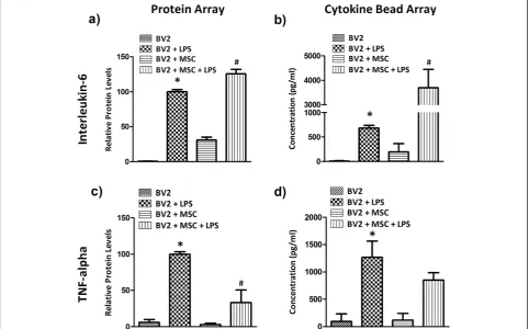
Culture supernatants were examined using a glass slide-based protein array to identify soluble proteins involved in MSC-mediated immunomodulation of microglia. From this array it was observed that IL-6 and TNF-αwere sig-nificantly modulated by co-culture. BV2 microglia in cul-ture produced negligible amounts of IL-6 and TNF-α (Figure 4a,c). LPS stimulation increased the expression of both cytokines at 24 hours. Co-culture with MSC in the presence of LPS further induced a 25.5 ± 6.5% surge in IL-6 (Figure 4a,P< 0.001), whilst TNF-αlevels were reduced to 33.2 ± 17.3% (Figure 4c, P< 0.001). These cytokines were then quantified using a cytometric bead array. Con-sistent with the results from protein array, IL-6 produc-tion was negligible in BV2 cultures (~9 pg/ml; Figure 4b). LPS stimulation increased IL-6 levels to 683.1 ± 55.4 pg/ ml by 24 hours and the presence of MSC increased IL-6 levels to 3,693.8 ± 751.1 pg/ml in LPS-treated BV2
Figure 3Mesenchymal stem cells inhibit microglia proliferation through contact-dependent cell cycle modulation.BV2 and Mesenchymal stem cells (MSC) were cultured at a 1:0.2 ratio and stimulated with 1μg/ml lipopolysaccharide (LPS) after overnight incubation. Cells were harvested 48 hours post-LPS stimulation and cell cycle was analysed by propidium iodide staining. Separation of BV2 and MSC populations on flow cytometry was enabled by CD45 staining (see Additional file 3: Figure S3). Histograms show distribution of(a)BV2 cells and
(b)MSC from direct co-culture across G0/G1, S and G2/M phases of cell cycle at 48 hours after LPS stimulation and/or co-culture. Values are expressed as mean ± standard deviation of percentage of cells from three independent experiments.(a)*P< 0.01, versus BV2 cells; #P< 0.01, versus BV2 + LPS.(b)*P< 0.001, versus MSC; #P< 0.001, versus MSC + LPS.
Table 1 Co-culture with Mesenchymal stem cells continues to modulate BV2 cell cycle until 96 hours but does not induce a cell cycle arrest
Cell cycle distribution (BV2 Microglia: MSC direct coculture)
BV2 BV2 + LPS BV2 + MSC + LPS
G0/G1 S G2/M G0/G1 S G2/M G0/G1 S G2/M
24 hrs 42.8 ± 1.5 49.3 ± 3.7 7.0 ± 2.5 34.2 ± 0.6* 62.0 ± 3.8# 3.8 ± 3.3 44.3 ± 1.8† 50.1 ± 1.0‡ 5.6 ± 4.9
48 hrs 50.7 ± 1.9 44.2 ± 1.1 4.7 ± 1.2 38.2 ± 1.2* 59.0 ± 2.7# 1.8 ± 2.2 43.9 ± 1.9† 48.6 ± 1.2‡ 6.3 ± 3.7
72 hrs 77.6 ± 0.9 19.3 ± 0.9 3.1 ± 0.1 48.2 ± 1.5* 51.6 ± 1.8# 0.3 ± 0.3 52.4 ± 5.6 41.3 ± 2.4‡ 1.9 ± 3.0
96 hrs 83.8 ± 4.0 14.1 ± 3.7 2.1 ± 2.3 63.2 ± 1.3* 35.7 ± 21.1# 1.0 ± 1.1 75.6 ± 1.7† 18.4 ± 0.9‡ 6.0 ± 1.1€
BV2 cells and Mesenchymal stem cells (MSC) were seeded at a 1:0.2 ratio in 6-well plates and left overnight prior to lipopolysaccharide (LPS) stimulation (1μg/ml). Cell cycle analysis of BV2 cells (CD45 + ve population in co-culture) was performed using propidium iodide (PI) staining at the time points indicated. Data show mean percentage population of BV2 cells ± standard deviation from three independent experiments. *P< 0.05,#P
< 0.05, versus respective BV2 alone controls.
Figure 4Mesenchymal stem cells modulate the expression of IL-6 and TNF-αin microglia co-culture.Microglia (BV2 cells or primary microglia) and Mesenchymal stem cells (MSC) were seeded simultaneously into 24-well plates at a 1:0.2 ratio. Cells were left overnight in culture and stimulated with 1μg/ml lipopolysaccharide (LPS). Culture supernatants were collected 24 hours later and expression of(a,b)interleukin (IL)-6 and(c,d)tumour necrosis factor (TNF)-αwere analysed using the RayBio antibody array (a,c) or BD Cytokine Bead Array (b,d). Protein array results are expressed as mean relative protein level ± standard deviation from two independent experiments and cytokine bead array results are expressed in mean concentration (pg/ml) ± standard deviation from three independent experiments. *P< 0.001, versus BV2 cells; #P< 0.001, versus BV2 + LPS.
microglia cultures (Figure 4b). Conversely the presence of MSC reduced TNF-α levels to 852.1 ± 134.9 pg/ml compared with 1,271.2 ± 294.2 pg/ml (Figure 4d) in LPS-stimulated BV2 cells alone.
Mesenchymal stem cell-mediated inhibition of microglial proliferation is independent of IL-6
LPS stimulation induced proliferation of BV2 microglia by 32.24 ± 4.5% and co-culture of BV2 microglia and MSC at the 0.2 ratio reduced LPS-stimulated proliferation of BV2 microglia by 21.50 ± 3.32% (Figure 5a,P< 0.05). In order to identify whether the surge in IL-6 in BV2/MSC co-culture is responsible for the anti-proliferative effect of MSC, IL-6 from cultures was neutralised using anti-mouse IL-6 blocking antibody. An isotype IgG was used as a con-trol to negate any non-specific effects. Addition of isotype IgG did not induce any changes (data not shown). Neutra-lising IL-6 from cultures did not influence the proliferation of LPS-stimulated microglia nor the anti-proliferative effect
of MSC (Figure 5a). It was also demonstrated that IL-6 did not influence the NO production within the MSC-mediated immunomodulatory paradigm (Figure 5b).
Mesenchymal stem cells inhibit microglial proliferation by reducing TNF-α
We sought to determine the role of TNF-α in MSC-mediated immunomodulation of microglial responses and first addressed whether application of blocking anti-bodies could affect the NO profile. Similar to that demonstrated previously, co-culture induced a surge in LPS-induced NO production (Figure 6a). Neutralising TNF-αdid not affect NO levels in LPS-stimulated BV2 cultures, but 10 μg/ml TNF-α blocking antibody signifi-cantly reduced NO levels in co-culture (Figure 6a, P< 0.05). Next, we applied recombinant TNF-αto co-cultures and its effect on NO expression was studied. TNF-αlevels detected in LPS-stimulated BV2 cultures by the cytokine bead array were approximately 1.5 ng/ml. Addition of
Figure 6Nitric oxide surge in co-culture and reduction in microglial proliferation are mediated by TNF-αmodulation.BV2 cells and mesenchymal stem cells (MSC) were seeded at a 0.2 ratio into 96 well plates. After overnight incubation, 1μg/ml lipopolysaccharide (LPS) and
recombinant TNF-α at low concentrations (0.5 and 5.0 ng/ml) did not affect the NO profile of any of the culture conditions tested (Figure 6b). However, at 50 ng/ml, TNF-α induced NO levels in co-culture by approximately two-fold (Figure 6b, P< 0.001 compared to activated co-culture without recombinant TNF-α).
Next we sought to address the effect of TNF-α deprivation on microglial proliferation. LPS stimulation induced the proliferation of BV2 microglia by approxi-mately 30% and in co-culture MSC reduced the prolifer-ation of LPS-stimulated BV2 microglia by 26.13 ± 7.24% (Figure 6c,P< 0.05). Neutralising TNF-αin LPS-stimulated BV2 microglia cultures (without MSC) reduced their prolif-eration in a dose-dependent manner; at 10μg/ml, prolifera-tion was reduced to levels similar to unstimulated BV2 microglia. As MSC reduced TNF-αlevels (Figure 4c,d) in LPS-stimulated BV2 microglia cultures, we presume that a reduction of TNF-α by MSC may be the mechanism through which MSC exert their anti-proliferative effect on microglia. Further reduction in TNF-αlevels did not influence the anti-proliferative effect of MSC; percent-age proliferation of BV2 remained between 70.57 ± 5.6% and 72.70 ± 3.5% for all three antibody concentrations tested (Figure 6c). Addition of recombinant TNF-αto co-cultures abolished the anti-proliferative effect of MSC at concentrations as low as 0.5 ng/ml, with higher concentra-tions inducing a dose-dependent increase in microglial proliferation (Figure 6d). At 50 ng/ml TNF-α, microglia proliferation was induced in co-culture to 119.75 ± 5.56% (Figure 6d, P< 0.05) compared to 73.87 ± 7.24% in the control co-culture.
On the contrary, TNF-α exerted an anti-proliferative effect on resting BV2 microglia (Figure 6d). Addition of TNF-αto unstimulated BV2 culture at 50 ng/ml reduced their proliferation to 41.26 ± 4.62% (Figure 6d, P< 0.05) compared to 66.94 ± 6.58% in the control, whereas addition of TNF-α did not alter the proliferation of LPS-induced BV2 microglia.
Discussion
Here, for the first time, we demonstrate the role of TNF-α in MSC-mediated immunomodulation of microglia. Treat-ment of BV2 microglia cultures with TNF-αblocking anti-bodies reduced LPS-induced proliferation while addition of recombinant TNF-αto co-cultures abolished the anti-proliferative effect of MSC on microglia. Microgliosis is a common response towards injury in the CNS, including Alzheimer’s disease [39], Parkinson’s disease [40], multiple sclerosis [41] and stroke [42], and the findings here may prove to be beneficial for these conditions.
In addition, the present study negated the role of apop-tosis and soluble factors such as NO and IL-6 in conferring this effect. We also provide the first evidence that MSC confer the inhibitory effect on microglia proliferation
through modulation of microglia cell cycle and MSC them-selves undergo a G0/G1 arrest while exerting this effect.
Following co-culture with MSC, microglia prolifera-tion in response to LPS stimulaprolifera-tion was dampened and NO levels were increased. Surge in NO levels in MSC co-cultures have been previously identified as the key mechanism conferring anti-proliferative effects on T cells [35,36]. To examine similar prospects, NO production in coculture was abolished using a specific iNOS inhibitor -L-NAME - and microglia proliferation was analysed. Con-verse to the reports in MSC/T cell co-cultures, abolishing NO did not restore microglia proliferation in our study. This is contradictory to a recent report which suggested the role of NO in MSC-mediated inhibition of microglia proliferation [43]. In the mentioned report, conditioned media from MSC cultures treated with an iNOS inhibitor was used to study the anti-proliferative effect, and thus the role of microglial cues that may be required by MSC to exert such effects was not negated. Our laboratory has previously reported that MSC requires microglial cue to induce a NO surge [34]. The current approach to elimin-ate NO directly from co-culture by addition of L-NAME efficiently negates such doubts and rules out the role of NO in inhibition of microglial proliferation by MSC.
The inhibition of microglial proliferation reported here could also be contributed by cell death. MSC are shown to induce indoleamine 2,3-dioxygenase-dependent apop-tosis of activated T cells [37]. Through examination using Annexin-V/PI staining, we demonstrate that co-culture with MSC does not induce apoptosis in microglia. Such differences in interaction of MSC with T cells and micro-glia emphasises diversity in the modulatory functions that MSC exert on different target cell types.
a cell cycle arrest is presumably a beneficial one. Tumouri-genic transformation of MSC has been implicated in sev-eral experimental transplantations including osteogenic sarcomas [44], myocardial infarction and diabetic neur-opathy [45]. Therefore, careful evaluation and under-standing of proliferative potential of MSC within the inflamed milieu is necessary for the success of clinical interventions.
We have also demonstrated here that MSC modulate the expression of TNF-αand IL-6. Co-culture with MSC significantly reduced the TNF-α which was upregulated upon LPS stimulation whereas co-culture induced a surge in IL-6. Similar modulation of cytokines was also reported previously by us [34] and others [29,43]. The impact of differential modulation of IL-6 and TNF-αwas further de-duced using blocking antibodies and recombinant TNF-α. Through addition of blocking antibodies, we have identi-fied that the IL-6 surge in co-culture does not influence the anti-proliferative effect of MSC or NO modulation. Distinct from IL-6, TNF-α seems to play a context-dependent role on modulation of NO and microglial pro-liferation in co-culture. Reducing TNF-α level in culture using blocking antibodies did not influence the NO pro-file of activated microglia. However, addition of recom-binant TNF-α induced a dose-dependent NO surge in co-cultures. It has been previously described by our group that soluble factors from activated co-culture in-duce NO production in MSC and these soluble factors include TNF-α[34]. It is possible that, in co-culture, the additional TNF-α added to the co-culture acts in tan-gent with microglial signals to induce a surge in NO, or TNF-αitself acts as the signal to induce NO production by MSC. MSC are known to respond to TNF-αsignals and elicit downstream responses including NFκB trans-location which is required for iNOS transcription [46]. However, such postulations need to be further validated. We also showed distinct responses in microglia prolifera-tion upon altering TNF-α levels in culture. TNF-α has been previously identified as vital for beta-amyloid-induced proliferation of microglia; abolishing TNF signals through addition of anti-TNF-αantibody or a soluble TNF receptor inhibitor both prevented beta-amyloid-induced prolifera-tion of microglia [47]. Similarly in an MPTP mouse model of Parkinson’s disease, knocking out TNF-α significantly reduced microglial activation as measured by cell number and morphology compared to wild-type controls [48], sug-gesting the important role played by TNF-α in microglial activation. Managing TNF-αlevels in the inflammatory mi-lieu is thus considered an efficient way to modulate inflam-mation [48,49]. At present, there is no evidence to suggest that modulation of TNF-α is the mechanism through which MSC confer an anti-proliferative effect on microglia. Likewise, there is no report on how MSC reduce microglial TNF-α. However, earlier reports from our laboratory [34]
and the present study confirms the potential of MSC to downregulate TNF-α production in an LPS-stimulated microglia co-culture paradigm. Similarly, intravenous transplantation of MSC into rat traumatic brain injury models significantly decreased TNF-α levels in the in-jured cortex and decreased the number of glial cells at the site of injury [50]. Thus, we hypothesised that modula-tion of TNF-α by MSC may be vital in conferring their anti-proliferative effect and monitored the effects of neu-tralising TNF-αand adding recombinant TNF-αon prolif-eration of LPS-stimulated BV2 microglia in co-culture. Abolishing TNF-α from co-culture did not enhance the anti-proliferative effect of MSC, indicating that a further decrease of TNF-αfrom that seen in co-cultures does not have an additive effect on MSC anti-proliferation of BV2. Also, the basal proliferation of BV2 cells may not require TNF-α, as addition of anti-TNF onto unstimulated BV2 did not affect their proliferation. However, the prolifera-tion of LPS-stimulated microglia was reduced in a dose-dependent manner upon addition of TNF-α neutralising antibodies, thus confirming the role of TNF-αin inducing proliferation of microglia. It is noteworthy that addition of recombinant TNF-α to LPS-stimulated BV2 culture did not increase their proliferation. This indicates that a critical concentration of TNF-α may be required for microglial activation above which the cells may become non-responsive. Also, other cues may be necessary for a proliferative response. Considering the requirement of cell-to-cell contact in modulating the cell cycle of activated microglia, we suggest the involvement of one or few cell surface molecules that may work in tandem with TNF-α modulation to achieve the inhibition of microglia prolifera-tion by MSC. Recent studies have strongly suggested the role of cell surface molecules such as CD200 [51], HLA-G [52] and PD-1 [53] expressed by MSC on their immuno-modulatory potential. It is possible that these cell surface ligands could be involved in the MSC-mediated modula-tion of BV2 microglia proliferamodula-tion.
Next, in order to confirm the relation of MSC-mediated reduction of TNF-αand inhibition of microglial prolifera-tion in co-culture, we used recombinant TNF-α. At con-centrations as low as 0.5 ng/ml, TNF-α abolished the inhibition of microglia proliferation in co-culture. More-over, higher concentrations of recombinant TNF-α in-duced the proliferation of microglia in co-culture to levels beyond that of LPS-stimulated culture. This confirms downregulation of TNF-α in co-culture by MSC as the key mechanism leading to the inhibition of microglial proliferation.
Conclusions
soluble factors NO and IL-6 in conferring MSC-mediated inhibition of microglia proliferation. We also provide evi-dence that MSC confer the inhibitory effect on microglia proliferation through modulation of microglia cell cycle and MSC themselves undergo a G0/G1 arrest while exerting this effect. These findings suggest the potential use of MSC in modulating microglial responses in in-flammatory conditions.
Additional files
Additional file 1: Figure S1.Effect of LPS on BV2 and MSC cell proliferation and mitomycin-C on MSC. MSCs and BV2 microglia were co-cultured at a 1:0.2 ratio and proliferation was analysed with3H-TdR incorporation at(a)6 hours,(b)24 hours and(c)48 hours after stimulation with 1μg/ml LPS. MSC were cultured independently at the same seeding density and treated with 10μg/ml mit-C for 2 hours after overnight culture.
(d)Proliferation and (e) cell cycle was analysed 48 hours post mit-C treatment with3H-TdR incorporation and PI staining respectively.(f) MSC culture was stimulated with 1μg/ml LPS and proliferation was analysed with3H-TdR incorporation at 48 hours post-LPS stimulation. Values are expressed as mean ± SD from three independent experiments. *P< 0.05, versus respective controls.3H-TdR, tritiated thymidine; LPS, lipopolysaccharide; mit-C, mitomycin C; MSC, mesenchymal stem cells; PI, propidium iodide; SD, standard deviation.
Additional file 2: Figure S2.Morphology of BV2 cells and MSC in culture. Phase contrast images showing morphology of BV2 cells and MSC seeded at a 1:0.2 ratio in a 6-well plate with 1μg/ml LPS at 48 hours. LPS, lipopolysaccharide; MSC, mesenchymal stem cells.
Additional file 3: Figure S3.L-NAME inhibits NO in a dose dependent manner. NO2-concentration in culture supernatant was determined using the Griess assay at 24 and 48 hours. BV2 cells were stimulated with 1μg/ ml LPS in the presence of different concentrations of L-NAME as indicated below the graph and NO was assayed. Values are expressed as mean ± SD of three independent experiments. *P< 0.01, versus BV2 cells; #P< 0.01, versus BV2 + LPS. LPS, lipopolysaccharide; L-NAME, N-nitro-L-ar-ginine methyl ester; NO, nitric oxide; SD, standard deviation.
Additional file 4: Figure S4.BV2 microglia and MSC show distinct CD45 expression. Scatter plots show CD45 expression of(a)BV2 microglia,(b)MSC and(c)co-culture after 48 hours in culture.(d)
Distribution of viable (lower left), early apoptotic (lower right), late apoptotic (upper right) and necrotic (upper left) population in DMSO treated BV2 cells (positive control for apoptosis assay) as determined by Annexin-V/PI staining. Numbers within each quadrant of plot indicate percentage of cells. Results are from a representative of three independent experiments. MSC, mesenchymal stem cells; PI, propidium iodide.
Additional file 5: Table S1.Co-culture does not induce apoptosis in BV2 cells and MSC. Table shows apoptosis assay results of BV2 microglia and MSC at 48 hours after co-culture. Results are expressed as mean ± SD from three independent experiments. BV2, BV2 microglia; LPS, lipopolysaccharide; MSC, mesenchymal stem cells; SD, standard deviation.
Additional file 6: Figure S5.Microglia-induced cell cycle arrest of MSC is not because of contact inhibition or nutrient deprivation. BV2 and MSC were co-cultured at a 1:0.2 ratio at a seeding density 10-fold lower than usual and were stimulated with 1μg/ml LPS after overnight incubation. Cells were harvested 48 hours post-LPS stimulation and cell cycle was analysed by propidium iodide staining. Separation of BV2 and MSC populations on flow cytometry was enabled by CD45 staining. LPS, lipopolysaccharide; MSC, mesenchymal stem cells.
Abbreviations
CNS:central nervous system; DMEM: Dulbecco's modified Eagle's medium; 3H-TdR: tritiated thymidine; Ig: immunoglobulin; IL: interleukin;
iNOS: inducible nitric oxide synthase; L-NAME: N-nitro-L-arginine methyl ester; LPS: lipopolysaccharide; MSC: mesenchymal stem cells; NO: nitric oxide;
PBS: phosphate-buffered saline; PI: propidium iodide; TNF: tumour necrosis factor.
Competing interests
The authors declare that they have no competing interests.
Authors’contributions
SJ designed and conducted the experiments, analysed the data and wrote the manuscript. SWT performed the Griess assays. YYO performed the cytometric bead array. RR assessed the cell cycle data. SV conceived and designed the study, obtained funding for the work, and analysed the data. All authors read and approved the final.
Acknowledgements
We thank Tong Chih Kong for his assistance with the apoptosis assay, Catherine Chieng Cheng Yun for discussions and critical comments on the manuscript, and Marsitah Abdul Jalil for maintaining the flow cytometer facility. This study was funded by the ScienceFund (Ministry of Science, Technology and Innovation, Malaysia) [02-01-04-SF1538] and Exploratory Research Grant Scheme (Ministry of Higher Education, Malaysia) [ERGS/1/2012/5527106].
Author details
1Neuroinflammation Group, Immunology Laboratory, Department of
Pathology, Faculty of Medicine and Health Sciences, Universiti Putra Malaysia, 43400 Serdang, Selangor, Malaysia.2Genetic Medicine Research Centre, Faculty of Medicine and Health Sciences, Universiti Putra Malaysia, 43400 Serdang, Selangor, Malaysia.3Current address: Tissue Engineering Centre, Universiti Kebangsaan Malaysia Medical Centre, 56000 Kuala Lumpur, Malaysia.4Department of Anatomy, Yong Loo Lin School of Medicine, NUS, MD10, 4 Medical Drive, Singapore 117597, Singapore.5Stem Cells & Immunity Group, Immunology Laboratory, Department of Pathology, Faculty of Medicine and Health Sciences, Universiti Putra Malaysia, 43400 Serdang, Selangor, Malaysia.
Received: 19 May 2014 Accepted: 9 August 2014 Published: 3 September 2014
References
1. Ooi YY, Rahmat Z, Jose S, Ramasamy R, Vidyadaran S:Immunophenotype and differentiation capacity of bone marrow-derived mesenchymal stem cells from CBA/Ca, ICR and Balb/c mice.World J Stem Cells2013,5:34–42. 2. Uccelli A, Moretta L, Pistoia V:Mesenchymal stem cells in health and
disease.Nat Rev Immunol2008,8:726–736.
3. Le Blanc K, Frassoni F, Ball L, Locatelli F, Roelofs H, Lewis I, Lanino E, Sundberg B, Bernardo ME, Remberger M, Dini G, Egeler RM, Bacigalupo A, Fibbe W, Ringden O:Mesenchymal stem cells for treatment of steroid-resistant, severe, acute graft-versus-host disease: a phase II study.Lancet2008,371:1579–1586.
4. Fisher-Shoval Y, Barhum Y, Sadan O, Yust-Katz S, Ben-Zur T, Lev N, Benkler C, Hod M, Melamed E, Offen D:Transplantation of placenta-derived mesenchymal stem cells in the EAE mouse model of MS.J Mol Neurosci
2012,48:176–184.
5. Gutierrez-Fernandez M, Rodriguez-Frutos B, Ramos-Cejudo J, Teresa Vallejo-Cremades M, Fuentes B, Cerdan S, Diez-Tejedor E:Effects of intravenous administration of allogenic bone marrow- and adipose tissue-derived mesenchymal stem cells on functional recovery and brain repair markers in experimental ischemic stroke.Stem Cell Res Ther2013,4:11.
6. Boucherie C, Schafer S, Lavand’homme P, Maloteaux JM, Hermans E:
Chimerization of astroglial population in the lumbar spinal cord after mesenchymal stem cell transplantation prolongs survival in a rat model of amyotrophic lateral sclerosis.J Neurosci Res2009,87:2034–2046. 7. Abrams MB, Dominguez C, Pernold K, Reger R, Wiesenfeld-Hallin Z, Olson L,
Prockop D:Multipotent mesenchymal stromal cells attenuate chronic inflammation and injury-induced sensitivity to mechanical stimuli in experimental spinal cord injury.Restor Neurol Neurosci2009,27:307–321. 8. Pileggi A:Mesenchymal stem cells for the treatment of diabetes.Diabetes
2012,61:1355–1356.
10. Krampera M, Glennie S, Dyson J, Scott D, Laylor R, Simpson E, Dazzi F:Bone marrow mesenchymal stem cells inhibit the response of naive and memory antigen-specific T cells to their cognate peptide.Blood2003,
101:3722–3729.
11. Corcione A, Benvenuto F, Ferretti E, Giunti D, Cappiello V, Cazzanti F, Risso M, Gualandi F, Mancardi GL, Pistoia V, Uccelli A:Human mesenchymal stem cells modulate B-cell functions.Blood2006,107:367–372.
12. Glennie S, Soeiro I, Dyson PJ, Lam EW, Dazzi F:Bone marrow mesenchymal stem cells induce division arrest anergy of activated T cells.Blood2005,
105:2821–2827.
13. Jiang XX, Zhang Y, Liu B, Zhang SX, Wu Y, Yu XD, Mao N:Human mesenchymal stem cells inhibit differentiation and function of monocyte-derived dendritic cells.Blood2005,105:4120–4126. 14. Ramasamy R, Fazekasova H, Lam EW, Soeiro I, Lombardi G, Dazzi F:
Mesenchymal stem cells inhibit dendritic cell differentiation and function by preventing entry into the cell cycle.Transplantation2007,
83:71–76.
15. English K, Barry FP, Mahon BP:Murine mesenchymal stem cells suppress dendritic cell migration, maturation and antigen presentation.Immunol Lett2008,115:50–58.
16. Ginhoux F, Greter M, Leboeuf M, Nandi S, See P, Gokhan S, Mehler MF, Conway SJ, Ng LG, Stanley ER, Samokhvalov IM, Merad M:Fate mapping analysis reveals that adult microglia derive from primitive macrophages.
Science2010,330:841–845.
17. Hughes V:Microglia: the constant gardeners.Nature2012,485:570–572. 18. Hanisch UK, Kettenmann H:Microglia: active sensor and versatile
effector cells in the normal and pathologic brain.Nat Neurosci2007,
10:1387–1394.
19. Schafer DP, Lehrman EK, Kautzman AG, Koyama R, Mardinly AR, Yamasaki R, Ransohoff RM, Greenberg ME, Barres BA, Stevens B:Microglia sculpt postnatal neural circuits in an activity and complement-dependent manner.Neuron2012,74:691–705.
20. Tremblay ME, Lowery RL, Majewska AK:Microglial interactions with synapses are modulated by visual experience.PLoS Biol2010,8:e1000527. 21. Nimmerjahn A, Kirchhoff F, Helmchen F:Resting microglial cells are highly
dynamic surveillants of brain parenchyma in vivo.Science2005,
308:1314–1318.
22. Aguzzi A, Barres BA, Bennett ML:Microglia: scapegoat, saboteur, or something else?Science2013,339:156–161.
23. Gomes C, Ferreira R, George J, Sanches R, Rodrigues DI, Goncalves N, Cunha RA:Activation of microglial cells triggers a release of brain-derived neurotrophic factor (BDNF) inducing their proliferation in an adenosine A2A receptor-dependent manner: A2A receptor blockade prevents BDNF release and proliferation of microglia.J Neuroinflammation2013,10:16. 24. Burguillos MA, Deierborg T, Kavanagh E, Persson A, Hajji N, Garcia-Quintanilla A,
Cano J, Brundin P, Englund E, Venero JL, Joseph B:Caspase signalling controls microglia activation and neurotoxicity.Nature2011,
472:319–324.
25. Ghoshal A, Das S, Ghosh S, Mishra MK, Sharma V, Koli P, Sen E, Basu A:
Proinflammatory mediators released by activated microglia induces neuronal death in Japanese encephalitis.Glia2007,55:483–496. 26. Wu DC, Jackson-Lewis V, Vila M, Tieu K, Teismann P, Vadseth C, Choi DK,
Ischiropoulos H, Przedborski S:Blockade of microglial activation is neuroprotective in the 1-methyl-4-phenyl-1,2,3,6-tetrahydropyridine mouse model of Parkinson disease.J Neurosci2002,22:1763–1771. 27. Saijo K, Crotti A, Glass CK:Regulation of microglia activation and
deactivation by nuclear receptors.Glia2013,61:104–111. 28. Ooi YY, Ramasamy R, Rahmat Z, Subramaiam H, Tan SW, Abdullah M,
Israf DA, Vidyadaran S:Bone marrow-derived mesenchymal stem cells modulate BV2 microglia responses to lipopolysaccharide.
Int Immunopharmacol2010,10:1532–1540.
29. Zhou C, Zhang C, Chi S, Xu Y, Teng J, Wang H, Song Y, Zhao R:Effects of human marrow stromal cells on activation of microglial cells and production of inflammatory factors induced by lipopolysaccharide.
Brain Res2009,1269:23–30.
30. Lanza C, Morando S, Voci A, Canesi L, Principato MC, Serpero LD, Mancardi G, Uccelli A, Vergani L:Neuroprotective mesenchymal stem cells are endowed with a potent antioxidant effect in vivo.J Neurochem2009,
110:1674–1684.
31. Lee JK, Jin HK, Bae JS:Bone marrow-derived mesenchymal stem cells reduce brain amyloid-beta deposition and accelerate the activation of
microglia in an acutely induced Alzheimer’s disease mouse model.
Neurosci Lett2009,450:136–141.
32. Kim JY, Kim DH, Kim JH, Lee D, Jeon HB, Kwon SJ, Kim SM, Yoo YJ, Lee EH, Choi SJ, Seo SW, Lee JI, Na DL, Yang YS, Oh W, Chang JW:Soluble intracellular adhesion molecule-1 secreted by human umbilical cord blood-derived mesenchymal stem cell reduces amyloid-beta plaques.
Cell Death Differ2012,19:680–691.
33. Blasi E, Barluzzi R, Bocchini V, Mazzolla R, Bistoni F:Immortalization of murine microglial cells by a v-raf/v-myc carrying retrovirus.
J Neuroimmunol1990,27:229–237.
34. Rahmat Z, Jose S, Ramasamy R, Vidyadaran S:Reciprocal interactions of mouse bone marrow-derived mesenchymal stem cells and BV2 microglia following lipopolysaccharide stimulation.Stem Cell Res Ther2013,4:12. 35. Ren G, Zhang L, Zhao X, Xu G, Zhang Y, Roberts AI, Zhao RC, Shi Y:
Mesenchymal stem cell-mediated immunosuppression occurs via concerted action of chemokines and nitric oxide.Cell Stem Cell2008,2:141–150. 36. Sato K, Ozaki K, Oh I, Meguro A, Hatanaka K, Nagai T, Muroi K, Ozawa K:
Nitric oxide plays a critical role in suppression of T-cell proliferation by mesenchymal stem cells.Blood2007,109:228–234.
37. Plumas J, Chaperot L, Richard MJ, Molens JP, Bensa JC, Favrot MC:
Mesenchymal stem cells induce apoptosis of activated T cells.Leukemia
2005,19:1597–1604.
38. Ramasamy R, Lam EW, Soeiro I, Tisato V, Bonnet D, Dazzi F:Mesenchymal stem cells inhibit proliferation and apoptosis of tumor cells: impact on in vivo tumor growth.Leukemia2007,21:304–310.
39. Block ML, Zecca L, Hong JS:Microglia-mediated neurotoxicity: uncovering the molecular mechanisms.Nat Rev Neurosci2007,8:57–69.
40. Vroon A, Drukarch B, Bol JG, Cras P, Breve JJ, Allan SM, Relton JK, Hoogland PV, Van Dam AM:Neuroinflammation in Parkinson’s patients and MPTP-treated mice is not restricted to the nigrostriatal system: microgliosis and differential expression of interleukin-1 receptors in the olfactory bulb.
Exp Gerontol2007,42:762–771.
41. Groebe A, Clarner T, Baumgartner W, Dang J, Beyer C, Kipp M:Cuprizone treatment induces distinct demyelination, astrocytosis, and microglia cell invasion or proliferation in the mouse cerebellum.Cerebellum2009,
8:163–174.
42. Li T, Pang S, Yu Y, Wu X, Guo J, Zhang S:Proliferation of parenchymal microglia is the main source of microgliosis after ischaemic stroke.
Brain2013,136:3578–3588.
43. Yan K, Zhang R, Sun C, Chen L, Li P, Liu Y, Peng L, Sun H, Qin K, Chen F, Huang W, Chen Y, Lv B, Du M, Zou Y, Cai Y, Qin L, Tang Y, Jiang X:Bone marrow-derived mesenchymal stem cells maintain the resting phenotype of microglia and inhibit microglial activation.PLoS One2013,
8:e84116.
44. Tolar J, Nauta AJ, Osborn MJ, Panoskaltsis Mortari A, McElmurry RT, Bell S, Xia L, Zhou N, Riddle M, Schroeder TM, Westendorf JJ, McIvor RS, Hogendoorn PC, Szuhai K, Oseth L, Hirsch B, Yant SR, Kay MA, Peister A, Prockop DJ, Fibbe WE, Blazar BR:Sarcoma derived from cultured mesenchymal stem cells.Stem Cells2007,25:371–379.
45. Jeong JO, Han JW, Kim JM, Cho HJ, Park C, Lee N, Kim DW, Yoon YS:
Malignant tumor formation after transplantation of short-term cultured bone marrow mesenchymal stem cells in experimental myocardial infarction and diabetic neuropathy.Circ Res2011,108:1340–1347. 46. van den Berk LC, Jansen BJ, Siebers-Vermeulen KG, Roelofs H, Figdor CG,
Adema GJ, Torensma R:Mesenchymal stem cells respond to TNF but do not produce TNF.J Leukoc Biol2010,87:283–289.
47. Jekabsone A, Mander PK, Tickler A, Sharpe M, Brown GC:Fibrillar beta-amyloid peptide Abeta1-40 activates microglial proliferation via stimulating TNF-alpha release and H2O2 derived from NADPH oxidase: a cell culture study.J Neuroinflammation2006,3:24.
48. Barcia C, Ros CM, Annese V, Gomez A, Ros-Bernal F, Aguado-Yera D, Martinez-Pagan ME, de Pablos V, Fernandez-Villalba E, Herrero MT:IFN-gamma signaling, with the synergistic contribution of TNF-alpha, mediates cell specific microglial and astroglial activation in experimental models of Parkinson’s disease.Cell Death Dis2011,2:e142.
49. Ferger B, Leng A, Mura A, Hengerer B, Feldon J:Genetic ablation of tumor necrosis factor-alpha (TNF-alpha) and pharmacological inhibition of TNF-synthesis attenuates MPTP toxicity in mouse striatum.J Neurochem
2004,89:822–833.
50. Zhang R, Liu Y, Yan K, Chen L, Chen XR, Li P, Chen FF, Jiang XD:
mesenchymal stem cell transplantation in experimental traumatic brain injury.J Neuroinflammation2013,10:106.
51. Najar M, Raicevic G, Jebbawi F, De Bruyn C, Meuleman N, Bron D, Toungouz M, Lagneaux L:Characterization and functionality of the CD200-CD200R system during mesenchymal stromal cell interactions with T-lymphocytes.
Immunol Lett2012,146:50–56.
52. Nasef A, Mathieu N, Chapel A, Frick J, Francois S, Mazurier C, Boutarfa A, Bouchet S, Gorin NC, Thierry D, Fouillard L:Immunosuppressive effects of mesenchymal stem cells: involvement of HLA-G.Transplantation2007,
84:231–237.
53. Luz-Crawford P, Noel D, Fernandez X, Khoury M, Figueroa F, Carrion F, Jorgensen C, Djouad F:Mesenchymal stem cells repress Th17 molecular program through the PD-1 pathway.PLoS One2012,7:e45272. doi:10.1186/s12974-014-0149-8
Cite this article as:Joseet al.:Mesenchymal stem cells exert anti-proliferative effect on lipopolysaccharide-stimulated BV2 microglia by reducing tumour necrosis factor-αlevels.Journal of Neuroinflammation 201411:149.
Submit your next manuscript to BioMed Central and take full advantage of:
• Convenient online submission
• Thorough peer review
• No space constraints or color figure charges
• Immediate publication on acceptance
• Inclusion in PubMed, CAS, Scopus and Google Scholar
• Research which is freely available for redistribution