1. Introduction
Parkinson’s disease (PD) is a progressive, disabling neurodegene rative movement disorder. Medical treatments that treat many of the motor symptoms of the disease are available.[1] Unfortunately, after
prolonged exposure to oral levodopa, motor complications may arise with patients rapidly changing from periods of limited movement (akinesia/‘off’) to periods of excessive movements that may range from mild to severe and disabling (dyskinesias). Although there are several medicationbased strategies to reduce these levodopa induced motor complications, ultimately the majority of these fail or become excessively burdensome to the patient.[2]
The history of deep brain stimulation (DBS) began in the 1940s, when because there was limited medication available for the treatment of PD, emphasis came to be placed on the use of surgically induced lesions of the thalamus and internal segment of the globus pallidus (GPi). With the advent and widespread use of dopamine in the 1960s, surgery fell out of favour until the reintroduction of pallidotomy by Laitinen.[3] It was subsequently shown that highfrequency DBS
was able to simulate the effects of ablation surgery of basal ganglia targets in a reversible and adjustable manner. With the renaissance of functional neurosurgery in this field, DBS is now considered an important treatment for PD, dystonia and tremor. DBS has been used for the treatment of movement disorders in South Africa (SA) since Dr Roger Melvill did the first procedure in 2000, and DBS procedures have been performed at a wide range of hospitals in both the private and the state sectors.
2. Stakeholder involvement
DBS guidelines were discussed at a meeting held on 4 July 2013 in Johannesburg, SA. At the time of writing, DBS has been provided at five academic centres in the state sector: Inkosi Albert Luthuli Central Hospital (KwaZuluNatal), Universitas Hospital (Free State) and Red Cross Children’s, Groote Schuur and Tygerberg hospitals (Western Cape), although financial constraints for obtaining DBS equipment
are significant in the state sector. DBS has also been carried out at a number of private hospitals (where the bulk of procedures have been performed). Potentially, all patients in SA with PD, severe tremor and dystonia who meet appropriate criteria for undergoing DBS should be eligible for the procedure. However, restrictions due to cost, including the cost of battery replacement, as well as limitations arising from the need for trained experts in the field, impact on the potential sites where DBS may be carried out.
At the meeting, the public and patients with dystonia and PD were represented through the Parkinson’s and Related Movement Disorders Association of South Africa. All the known role players who were involved with DBS at the time were invited. The follow ing, including neurologists, neurosurgeons, neurophysiologists and neuro psychologists, and the director of the Parkinson’s and Related Movement Disorders Association of South Africa were in atten dance: Ms C Bottcher (Life Entabeni Hospital), Dr D G Anderson (University of the Witwaters rand Donald Gordon Hospital), Prof. N Cassimjee (University of Pretoria), Prof. J Carr (Stellenbosch University, by correspondence), Dr D Giampaolo (Netcare Rose bank Hospital), Prof. M Lucas (University of the Witwatersrand), Dr R Melvill (Mediclinic Constantiaberg and University of Cape Town), Ms I Nepal (Milpark Hospital), Dr P Slabbert (Netcare Rosebank Hospital and University of Pretoria), Dr F Snyckers (Milpark Hospital), Dr J Smuts (Life Wilgers Hospital and University of Pretoria), Dr J Vaidyanathan (Medtronic Scientific Representa tive), Dr R van Coller (Life Wilgers Hospital and University of Pretoria), Ms K Willemse (Parkinson’s and Related Movement Disorders Association of South Africa), Dr C Wolpe (Hillcrest Private Hospital) and Dr M Zorio (Milpark Hospital).
At the meeting, it was decided to summarise the current DBS literature and provide a practical guideline of best care for DBS in patients with PD for general practitioners, neurosurgeons, neurolo gists, neurophysiologists, anaesthetists and healthcare funders. The guideline was written and edited by Dr Anderson, Dr Van
South African guideline on deep brain stimulation
for Parkinson’s disease
D G Anderson,1 MB BCh, MMed, FCP (SA); R van Coller,2 MB ChB, MMed; J Carr,3 MB ChB, PhD
1 Neurology, University of the Witwatersrand Medical Centre, Johannesburg, South Africa
2Department of Neurology, School of Medicine, Faculty of Health Sciences, University of Pretoria, South Africa
3Department of Medicine, Faculty of Medicine and Health Sciences, Stellenbosch University, Cape Town, South Africa
Corresponding author: J Carr (jcarr@sun.ac.za)
Background. Parkinson’s disease (PD) is a common neurodegenerative disease, associated with severe impairment of quality of life.
Although the motor aspects of the illness are typically successfully treated with medications acting on the dopaminergic system, a number of patients encounter progressive difficulties associated with their medical treatment.
Recommendations. Carefully selected patients will benefit from deep brain stimulation (DBS) treatment for their PD. Selection
requires dopamine challenge testing and neuropsychological testing for the presence of cognitive impairment. Careful followup and programming of the DBS system are mandatory, and a major reason for DBS failure is inadequate programming and management of medication.
Conclusion. DBS is a useful component of standard therapy for PD and may reduce symptoms, improve quality of life, promote patient
independence and reduce healthcare costs by reducing requirements for medicine.
Coller and Prof. Carr, representing the private and state sectors. The completed text was reviewed by Prof. J Volkmann (Department of Neurology, Würzburg, Germany), an international authority on DBS. The Parkinson’s and Related Movement Disorders Association of South Africa, the Neurological Association of South Africa and the South African Neurosurgical Association reviewed and endorsed the guideline. Annual review of the guideline will be carried out by the Movement Disorders Interest Group of South Africa.
Role of sponsor. The guideline has been the voluntary effort
of Dr Anderson, Dr Van Coller and Prof. Carr. The meeting was sponsored by Medtronic.
3. Cost-effectiveness
Treating PD is complex and expensive, reflecting the complexities of the disease and the many complications of therapy. It must be noted that DBS with best medical treatment (BMT) is superior to BMT alone, and depending on the study, the cost of the DBS device is recouped in medication savings alone in 3 5 years in the case of subthalamic nucleus (STN) DBS.[4] Three studies were published in
2016 addressing the issue of costeffectiveness of DBS for PD. These included a systematic review of the literature on economic studies of the use of DBS in patients with PD, including cost studies or economic evaluations expressed as cost per improvement in quality of life.[5] Nine studies were identified, and the qualityadjusted life
year (QALY) gain was higher for DBS than for BMT after 2 years. A recent study on costeffectiveness in the USA[6] found DBS to
be costeffective. DBS therapy resulted in a discounted QALY gain of 1.69 more than BMT (3.19 v. 1.50 QALYs). The corresponding 10year costs for DBS and BMT were USD130 510 and USD91 026, respectively, leading to an incremental costeffectiveness ratio of USD23 404. DBS was associated with cost increases of ~USD40 000 over 10 years. However, the costeffectiveness profile (ratio of incremental costs to gained QALYs) was favourable. The authors concluded that ‘DBS yields substantial improvements in health related quality of life at a value profile that compares favorably to
other wellaccepted therapies.’[6] A study specifically addressing cost
effectiveness in the early motor complications group found that STN DBS was costeffective in patients aged <61 years compared with BMT.[7] The incremental costutility ratio for STN DBS compared
with BMT was EUR22 700 per QALY gained. Overall, DBS reduces pharmacological treatment costs and is likely to reduce the direct, indirect and social costs of PD in the long term.
4. Evidence for DBS
The authors reviewed the available evidence for DBS with respect to benefit and harm by performing a MEDLINE search using the terms ‘deep brain stimulation’ and ‘Parkinson’, selecting the following article types: clinical trial, randomised controlled trial and review. In addition, material from personal sources and relevant textbooks was consulted.
DBS was first used for PD in 1990,[8] and was recognised as a way
to improve tremor, rigidity and bradykinesia, allowing for a reduction in levodopa use.[9] Subsequently, DBS was believed to improve ‘off’
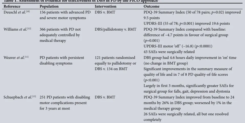
time disability, reduce dyskinesia, improve quality of life and lead to reduction of the levodopa dose[10] (Table 1). However, it was only in
2009 that a randomised controlled trial found DBS to be superior to BMT in improving ‘on’ time, reducing dyskinesias and improving qualityoflife scores.[11] A year later, in a trial of DBS and BMT v.
BMT alone for advanced PD (PD SURG), it was found that DBS and BMT were superior to BMT alone when assessing selfreported qualityoflife scores at 1 year[12] (Table 1). Although DBS is accepted
as an alternative treatment option in patients with advanced PD, the EARLYSTIM study showed a substantial benefit in younger patients (mean age 52 years) with shorterduration PD when comparing DBS with BMT, and found significant improvement in the primary outcome of quality of life[13] (Table 1).
The longterm benefit of DBS has now been shown in 5 and 10year followup studies.[10] DBS is still effective in managing
[image:2.595.50.529.510.751.2]motor symptoms 10 years after implantation. The usefulness of DBS in managing axial features of advanced PD seems to be less
Table 1. Assessment of evidence for effectiveness of DBS in PD by the PICO approach*
Reference Population Intervention Outcome
Deuschl et al.[10] 156 patients with advanced PD
and severe motor symptoms DBS v. BMT PDQ39 Summary Index (50 of 78 pairs; 9.5 points p=0.02) improved UPDRSIII (55 of 78; p<0.001) improved 19.6 points Williams et al.[12] 366 patients with PD not
adequately controlled by medical therapy
DBS/pallidotomy v. BMT PDQ39 Summary Index compared with baseline: difference of –4.7 points in favour of surgical group (p=0.001)
UPDRSIII motor ‘off’ (–16.8) (p<0.0001) 43 SAEs were surgically related
Weaver et al.[11] PD patients with persistent
disabling symptoms
121 patients randomised equally to pallidotomy or DBS v. 134 on BMT
DBS group had 4.6 hours daily improvement in ‘on’ time (no change in BMT group)
Significant improvements in the summary measure of quality of life and in 7 of 8 PD qualityoflife scores (p<0.001)
Largely in first 3 months, significantly greater SAEs for surgical group for falls, gait, depression and dystonia Schuepbach et al.[13] 251 PD patients with disabling
motor complications present for 3 years at most
DBS v. BMT PDQ39 Summary Index improved from baseline to 24 months by 26% in DBS group; worsened by 1% in the medical therapy group
26 SAEs were surgically related, all but one resolved completely
DBS = deep brain stimulation; PD = Parkinson’s disease; PICO = population, intervention, comparison and outcome; BMT = best medical therapy; PDQ39 = 39Item Parkinson's Disease Questionnaire; UPDRS = Unified Parkinson’s Disease Rating Scale; SAEs = serious adverse events.
robust. A neuroprotective effect from DBS could not be demon strated.[14]
DBS surgery is relatively safe, the commonest complications being infection, haemorrhage and transient postsurgical confusion. The reported complication rate varies widely. In a study of 526 consecutive patients with DBS implantation, haemorrhage occurred in 8.4% of all cases.[15] Bleeds occurred at the entry point or
subcortically, but rarely in the target, and more often in hypertensive patients. Asymptomatic haemorrhage occurred in 3.4% of this series of patients, and symptoms were transient in 4.4% of patients and permanent in only 0.6%.[15] Approximately 10% of patients were
reported to have transient postoperative confusion. This has been attributed to intracranial contusion, long surgery duration and dopamine withdrawal.[16]
5. Target site
Three anatomical basal ganglia targets are suitable for stimulation in PD. The target adopted initially was the ventral intermediate (VIM) thalamus, which showed a degree of efficacy that was similar to thalamotomy for PDrelated tremor, but less so for other motor features.[17] Altering the target to the GPi resulted in improvement in
all the major motor features of PD and a reduction in dyskinesia. The GPi was then replaced by the STN based on lesion studies in primates showing possibly greater efficacy.[18]
A debate has persisted for some time about which site is preferable, the STN or the GPi. Randomised controlled trials indicate that compared with bilateral GPi DBS, bilateral STN DBS is equally effective in treating PD motor symptoms.[19,20] The advantages
of targeting the STN include good clinical effect on tremor and bradykinesia, improvement in dyskinesia and motor fluctuations, and the potential to reduce medications.[21] The GPi continues to be
a potential target, since there remain concerns that STN stimulation has a potentially greater effect on cognition and may exacerbate neuropsychiatric conditions, and programming and followup are typically more straightforward for GPi stimulation.[22]
The recommended target in PD is currently the STN. In special circumstances, thalamic targets and the GPi may be considered, e.g. in elderly patients with dominant PD tremor and little progression of akineticrigid symptoms (VIM), or frail patients with prominent dyskinesia, who might not tolerate the complex and lengthy postoperative adjustment of medication and stimulation required in STNDBS.
6. Referral of patients for DBS
The decision when to refer for DBS is not based on level 1 evidence and is currently the subject of ongoing debate and investigation. Most specialists agree that the optimal time to refer is when patients have medicationrefractory motor fluctuations, or dyskinesias or tremor that are resistant to treatment.
It is important to emphasise that delaying referral may cause harm, since it is accepted that the best DBS outcomes are seen in patients who are younger, responsive to dopamine and do not have significant gait problems. PD patients should be considered for DBS if: • PD diagnosis is established
• Age at time of surgery is (ideally) <75 years
• Troublesome motor fluctuations or resistant tremor are present • Dopamine responsiveness is present
• Cognition is normal or minimally impaired
• Gait is impaired, but shows dopamine responsiveness.
Most units will require patients to be known to have PD for >5 years in order to exclude patients with atypical parkinsonism. Patients with
atypical parkinsonism respond poorly to DBS even when there was previously documented response to levodopa.
Although age has not been found to be a specific prognostic indicator in DBS for PD, increasing age is associated with a number of factors that should be considered: medical comorbidity, decreased cognitive ability, levodopa resistance and increased surgical risk. The recommendation is that patients aged >75 years should only be considered in special circumstances.
Accumulating longterm evidence in DBS treatment of PD shows poor response of gait and postural instability to stimulation treatment. Some aspects of gait and posture may improve initially after DBS surgery owing to lengthening of stride and improvement of initiation of gait, but rhythmicity and cadence do not seem to improve. The gait features that do improve are usually responsive to levodopa, and these can be used to predict improvement following surgery. Of note, in some patients, gait may even deteriorate after surgery.[23] In the long term, patients with advanced PD may have
progressive issues with gait, freezing of gait, and balance and posture, and these features are not effectively addressed by STN DBS.[24] DBS should only be considered in patients with gait and
balance impairment where there is proven levodopa response in these domains prior to surgery. Patients should be informed of the possible deleterious effect on gait and balance after surgery, and similarly should be made aware that gait and balance will not respond well to DBS in many cases.
Alternative targets for patients with gaitpredominant PD are being explored, but no alternative target has been shown to be effective.[23]
7. Patient selection/assessment
The patient selection process consists of assessing the patient for suitability for surgery and dopamine responsiveness, and a neurocognitive evaluation (sometimes with psychiatric assessment).
7.1 The DBS team
More than 30% of DBS surgery failures can be attributed to inappro priate patient selection.[25] Each patient needs to be selected according
to their individual riskbenefit profile at an expert centre, by a multidisciplinary team consisting of an experienced neurologist, neurosurgeon and neuropsychologist. This also allows for the patient’s expectations to be managed realistically. An expert centre should be able to produce an audit of results. Other members of the team, but not necessarily seen by every patient, are a neurophysiologist, psychiatrist, anaesthetist and neuroradiologist, and ideally a nurse with experience in movement disorders.[26]
7.2 Dopamine challenge test
The degree of responsiveness to levodopa is considered to be the best predictor for DBS outcomes.[15] The dopamine challenge
test is performed after a period of dopamine withdrawal (at least 12 hours) with the patient in the worst ‘off’ state. Special attention must be paid to dopamine agonist withdrawal, since these drugs have relatively long halflives that may affect the assessment of the response to the dopamine challenge test. A Unified Parkinson’s Disease Rating Scale (UPDRS) III is scored in this state. The patient is then given levodopa orally. The dose can vary from 100 to 250 mg. After at least 1 hour, the UPDRS is scored in the best ‘on’ state. A 30% improvement is generally considered acceptable to proceed to surgery.[27] This has been the operational definition
7.3 Neuropsychological testing
Cognitive dysfunction is common in PD, and ranges from mild cognitive impairment to dementia.[28] As DBS can impact variably
on postsurgical cognitive functioning, and frequently worsens functions that are associated with the frontostriatal areas, cognitive status should be known before surgery.[2932] It is recommended that
all patients should be assessed before deciding about surgery, and that the assessment of cognitive functioning be carried out by a clinical psychologist with specialist knowledge of neuropsychology, or a neuropsychologist. Recommendations for the assessment of cognitive function in PD have been published.[28,33]
In SA, there are few local normal values for standard neuropsychological tests. Education level and not speaking English as a first language can influence the outcome of these tests by as much as two standard deviations. The neuropsychologist should therefore use both quantitative and qualitative approaches where appropriate. The underlying mood of the patient, other medical conditions, and whether the patient is in the treatment ‘on’ or ‘off’ state can all affect the test outcome.[33]
The presence of significant cognitive impairment in a PD patient is an exclusion factor for DBS surgery, particularly if a patient’s test profile on a comprehensive battery of neuropsychological tests indicates impairment of one or more measures of executive function. The presence of mesiotemporal memory impairment (poor encoding rather than impaired recall) should alert the neuropsychologist to cognitive impairment of a different type to that usually observed in executive dysfunction, and suggest further caution regarding a decision to proceed to surgery. Individuals with PD who score in the borderline range are more susceptible to subtle decline in cognitive functioning after surgery,[34] and caution is advised.
8. Surgery
DBS can be done awake with radiological (magnetic resonance imaging (MRI)/computed tomography (CT)), electrophysiological and clinical guidance or asleep with MRIbased guidance and verification of the anatomical target. Both forms of surgery are acceptable and have proven outcomes, and units should use the method with which they are most familiar.
Awake surgery is performed in two phases. The first phase is the stereotactic implantation of the stimulating electrodes in the STN. This is usually performed in the awake state to allow for electrophysiological recording and stimulation to assess for benefits and sideeffects. A stereotactic system that maps image space to physical space is used to deliver the stimulating electrodes to the STN. The trajectory of the electrode is set to avoid critical structures of the brain, including blood vessels, sulci and ventricles. Microelectrode recording (MER) from individual neurons is typically used to identify the neuronal firing patterns during surgery to confirm correct positioning. The second phase is the internalisation of the neurostimulator, performed under general anaesthesia. This involves connecting the stimulating electrodes to the neurostimulator. Both phases can be done in one session.
In MRIguided and verified surgery, the patient receives full anaesthesia throughout the surgery, and planning and verification of lead placement is done by MRI with the stereotactic frame on.[35] In this case, there is no requirement for intraoperative
neurophysiological recordings, resulting in possible benefits in terms of surgical adverse effects and theatre time.
Pre and postsurgical imaging form an essential part of both surgical techniques and are discussed in section 10. Both techniques (awake and asleep) have similar outcomes with regard to motor function and reduction in dyskinesia.[11,35]
9. Neurophysiology (MER and
macroelectrode stimulation (MES))
Despite advances in imaging, most units around the world rely on electrophysiological and clinical localisation to confirm and refine the radiological target.[36] The first phase in intraoperative
electrophysiology is to perform MER. Up to five test electrodes can be advanced simultaneously in micrometric steps to the radiological target and beyond. The different anatomical targets in the basal ganglia each have a unique electrophysiological footprint that defines the boundaries of the target.
The second phase in the determination of the physiological boundaries of the target involves electrical stimulation with a macro test electrode. MES is done to assess clinical effect and stimulation related adverse effects before permanent placement of the DBS lead. Someone trained in movement disorders, such as a neurologist, neurosurgeon or specifically trained PD nurse, should perform the assessments.
Stimulation is performed using a constantcurrent stimulator on the MER system with standard settings. Monopolar stimulation at predefined intervals is done in incrementing current strengths (generally 1 5 mA) to assess the area delineated by MER and imaging.
Clinical benefits of acute intraoperative MES are monitored with contralateral wrist rigidity, tremor and bradykinesia.[37] When there is
little or no clinical effect at amplitude settings of up to 3 mÅ, it should be considered that the position in the STN is not optimal.
Sideeffects secondary to stimulation include contralateral muscle contractions and/or eye deviation. Sideeffects are classified as transient or persistent and mild, moderate or severe. Only stimulationinduced sideeffects after the supramaximal threshold are considered acceptable to choose a trajectory for implanting the permanent DBS lead. A combination of acceptable clinical effects on tremor, rigidity and bradykinesia without the induction of persisting adverse effects is required to assure optimal lead placement.[36]
10. Imaging
10.1 Imaging before surgery
Imaging the brain is essential for the preoperative target planning in DBS. Acceptable imaging modalities for targeting include MRI, CT and ventriculography. T2weighted imaging or susceptibility weighted imaging (SWI) is generally considered useful to delineate the target and to indicate which neighbouring structures to avoid, and at least 1.5 Tesla field strength is recommended. Wholebrain T1weighted imaging with contrast is used for planning and ensuring a safe trajectory to the target and verifying the entire course from cortical entry to target.[26]
10.2 Imaging after surgery
Postsurgical imaging is somewhat controversial. The majority of units agree that some form of imaging is necessary to ensure that the leads are in position. This then acts a reference point in the case of subsequent limited benefit, adverse effects or lead displacement. In a consensus document, a CT scan was judged to be adequate, but MRI was generally seen as superior to CT.[26]
However, there is concern in performing MRI following DBS, as it can result in heating of the leads, magnetic field interactions and movement of the components, induced currents, and functional disruption of the DBS components. [38] Carefully following
manufacturer guidelines permits the use of conditionally safe MRI scans to audit the lead location. Larson et al.[39] demonstrated
the National Parkinson Foundation has scanned >3 304 patients without any complications.[40]
Our recommendation is that postsurgical imaging with at least CT imaging with a presurgical MRI merge is necessary, although post surgical MRI is preferred.
11. Programming
Programming of the device is as important as accurate electrode placement in ensuring successful treatment. To ensure the full benefit of this specialised treatment, programming should be done by a trained specialist. Managing programming cannot be done in isolation from medication adjustments and other treatments, so a neurologist with training or experience in DBS should preferably be in charge of programming.
The programming process consists of the initial programming, followup sessions in the weeks after surgery, and longterm follow up once symptom control and medication adjustment are stable.
The standard DBS electrode consists of four 1.5 mm cylindrical contacts each spaced 0.5 mm from one other. Various programming options can be used to maximise the clinical effect. These include the use of different contacts in mono or bipolar mode and adjusting the voltage/current, pulse width and frequency of stimulation.[41]
The initial programming is patient specific and depends on the microlesion effect (positive surgical effect that can last several weeks), the sideeffects of stimulation, the patient’s stamina and whether bilateral stimulation is required. Initial programming should preferably be done when the patient is in the offmedication phase.[41]
Initial followup programming sessions address habituation of the stimulation effect and the complete resolution of the microlesioning effect after surgery. Increases in stimulation are generally required, although in some patients other parameter changes may prove necessary. Checking of impedance (resistance of the electrode and electrodetissue barrier to electrical stimulation) and battery life is advised. Careful documentation of each programming session is very important for future reference.
Longterm followup includes device and battery checks, and adjustment of stimulation and medication to allow for symptom control with progression of disease. Intervals are patient specific.
12. Managing pharmacological
treatment
The aim of DBS is not to withdraw medication completely, but to improve the patient’s quality of life. Medication reduction and postsurgical management need to be done by an experienced neurologist with an understanding of DBS and medication interactions. Special care in terms of the dopamine agonist withdrawal syndrome is required.[42] When adjusting DBS
programming, medication adjustments must be made in parallel. Stimulation should improve parkinsonian signs; dopaminergic drugs can be slowly reduced if this is seen. Dyskinesias are often increased with STN stimulation for a short time after stimulation is initiated, another reason for medication reduction.[15] Sudden
stopping of dopaminergic therapy can result in withdrawal syndromes.[43] These include motor and delayed nonmotor
symptoms such as apathy and depression. STN stimulation may result in euphoria or even hypomania with a dopamine dysregulation syndrome, which requires dopamine reduction. DBS patients must be assessed regularly to optimise both motor and behavioural states, by titrating stimulation with levodopa to avoid akinesia or dyskinesia, and by careful adjustment of medication to avoid apathy or impulse control disorders.[44]
13. Recommendations
Based on the outcome of the initial workshop, review of the literature, consultation with patient groups and input from an external expert consultant, the following are the key points concerning the management of DBS in PD:
• DBS is a safe and effective treatment for carefully selected patients with PD (section 4).
• Patients should be carefully selected for symptoms that are known to respond to DBS (section 6).
• Patients need to undergo dopamine challenge testing to assess the degree of response to dopamine (section 7.2).
• Patients should undergo neuropsychological testing, and the pres ence of significant cognitive impairment is usually a contraindica tion to DBS (section 7.3).
• Currently, several radiological approaches to selecting an appropriate target may be used, and both intraoperative physio logical recordings and MRIverified approaches are valid methods of performing DBS (sections 8 and 9).
• Careful followup and programming of the DBS system is mandatory, and a major reason for DBS failure is inadequate programming and management of medication (sections 11 and 12).
Acknowledgements. None.
Author contributions. DA, RvC and JC worked jointly on all aspects of the
original and revised submission .
Funding. Medtronic and Amayeza funded the publication of this guideline. Conflicts of interest. DGA has received sponsorship to attend movement
disorder congresses from Boston Scientific and Medtronic as well honorariums from Medtronic for presentations in the movement disorder field. He has attended a neurologists’ training programme for DBS at the National Hospital, London, on two occasions, a collaboration between University College London and Medtronic.
RvC has received payment for talks from Cipla South Africa, and sponsorship for travelling to attend congresses from Medtronic. He is also the recipient of a research grant sponsored by Medtronic.
JC has received payment for talks from Cipla South Africa, and has attended a number of training sessions at Netcare Pretoria East, as well as at ChristianAlbrechtsUniversity, Kiel, Germany (both sponsored by Medtronic). He is a participant in the neurologists’ training programme with attendance at the National Hospital, London, and in Tolochenaz, Switzerland (a Medtronicsponsored programme). He receives an annual stipend from Elsevier for his role as an associate editor.
1. Carr J, Kies B, Fine J. Guideline for the treatment of Parkinson’s disease. S Afr Med J 2009;99(10):755 756, 758.
2. Rascol O, Brooks DJ, Korczyn AD, De Deyn PP, Clarke CE, Lang AE. A fiveyear study of the incidence of dyskinesia in patients with early Parkinson’s disease who were treated with ropinirole or levodopa. 056 Study Group. N Engl J Med 2000;342(20):14841491. https://doi.org/10.1056/ NEJM200005183422004
3. Laitinen LV, Bergenheim AT, Hariz MI. Leksell’s posteroventral pallidotomy in the treatment of Parkinson’s disease. J Neurosurg 1992;76(1):5361. https://doi.org/10.3171/jns.1992.76.1.0053
4. Eggington S, Valldeoriola F, Chaudhuri KR, Ashkan K, Annoni E, Deuschl G. The costeffectiveness of deep brain stimulation in combination with best medical therapy, versus best medical therapy alone, in advanced Parkinson’s disease. J Neurol 2014;261(1):106116. https://doi.org/10.1007/s00415013 7148z
5. Becerra JE, Zorro O, RuizGaviria R, et al. Economic analysis of deep brain stimulation in Parkinson disease: Systematic review of the literature. World Neurosurg 2016;93:4449. https://doi.org/10.1016/j. wneu.2016.05.028
6. Pietzsch JB, Garner AM, Marks WJ. Costeffectiveness of deep brain stimulation for advanced Parkinson’s disease in the United States. Neuromodulation 2016;19(7):689697. https://doi. org/10.1111/ner.12474
7. Dams J, BalzerGeldsetzer M, Siebert U, et al. Costeffectiveness of neurostimulation in Parkinson’s disease with early motor complications. Mov Disord 2016;31(8):11831191. https://doi.org/10.1002/ mds.26740
8. Benabid AL, Benazzous A, Pollak P, Dostrovsky JO, Lozano AM. Mechanisms of deep brain stimulation. Mov Disord 2002;17(Suppl 3):S73S74. https://doi.org/10.1002/mds.10145
9. Limousin P, Pollak P, Benazzouz A, et al. Effect of parkinsonian signs and symptoms of bilateral subthalamic nucleus stimulation. Lancet 1995;345(8942):9195. https://doi.org/10.1016/S0140 6736(95)900624
11. Weaver FM, Follett K, Stern M, et al. Bilateral deep brain stimulation vs best medical therapy for patients with advanced Parkinson disease: A randomized controlled trial. JAMA 2009;301(1):6373.
https://doi.org/10.1001/jama.2008.929
12. Williams A, Gill S, Varma T, et al. Deep brain stimulation plus best medical therapy versus best medical therapy alone for advanced Parkinson’s disease (PD SURG trial): A randomised, openlabel trial. Lancet Neurol 2010;9(6):581591. https://doi.org/10.1016/S14744422(10)700934
13. Schuepbach WMM, Rau J, Knudsen K, et al. Neurostimulation for Parkinson’s disease with early motor complications. N Engl J Med 2013;368(7):610622. https://doi.org/10.1056/NEJMoa1205158
14. RodriguezOroz MC, Moro E, Krack P. Longterm outcomes of surgical therapies for Parkinson’s disease. Mov Disord 2012;27(14):17181728. https://doi.org/10.1002/mds.25214
15. KleinerFisman G, Herzog J, Fisman DN, et al. Subthalamic nucleus deep brain stimulation : Summary and metaanalysis of outcomes. Mov Disord 2006;21(Suppl 14):S290S304. https://doi.org/10.1002/ mds.20962
16. Voon V, Kubu C, Krack P, Houeto JL, Tröster AI, Tro AI. Deep brain stimulation: Neuropsychological and neuropsychiatric issues. Mov Disord 2006;21(Suppl 14):S305S327. https://doi.org/10.1002/ mds.20963
17. Hariz MI, Krack P, Alesch F, et al. Multicentre European study of thalamic stimulation for parkinsonian tremor: A 6 year followup. J Neurol Neurosurg Psychiatry 2008;79(6):694699. https:// doi.org/10.1136/jnnp.2007.118653
18. Benabid AL, Chabardes S, Mitrofanis J, Pollak P. Deep brain stimulation of the subthalamic nucleus for the treatment of Parkinson’s disease. Lancet Neurol 2009;8(1):6781. https://doi.org/10.1016/S1474 4422(08)702916
19. Anderson VC, Burchiel KJ, Hogarth P, Favre J, Hammerstad JP. Pallidal vs subthalamic nucleus deep brain stimulation in Parkinson disease. Arch Neurol 2005;62(4):554560. https://doi.org/10.1001/ archneur.62.4.554
20. Follett KA, Weaver FM, Stern M, et al. Pallidal versus subthalamic deepbrain stimulation for Parkinson’s disease. N Engl J Med 2010;362(22):20772091. https://doi.org/10.1056/NEJMoa0907083.
21. Odekerken VJJ, van Laar T, Staal MJ, et al. Subthalamic nucleus versus globus pallidus bilateral deep brain stimulation for advanced Parkinson’s disease (NSTAPS study): A randomised controlled trial. Lancet Neurol 2013;12(1):3744. https://doi.org/10.1016/S14744422(12)702648
22. Okun MS, Fernandez HH, Wu SS, et al. Cognition and mood in Parkinson’s disease in subthalamic nucleus versus globus pallidus interna deep brain stimulation: The COMPARE trial. Ann Neurol 2009;65(5):586595. https://doi.org/10.1002/ana.21596.
23. PötterNerger M, Volkmann J. Deep brain stimulation for gait and postural symptoms in Parkinson’s disease. Mov Disord 2013;28(11):16091615. https://doi.org/10.1002/mds.25677
24. Maetzler W, Nieuwhof F, Hasmann SE, Bloem BR. Emerging therapies for gait disability and balance impairment: Promises and pitfalls. Mov Disord 2013;28(11):15761586. https://doi.org/10.1002/ mds.25682
25. Okun MS, Tagliati M, Pourfar M, et al. Management of referred deep brain stimulation failures: A retrospective analysis from 2 movement disorders centers. Arch Neurol 2005;62(8):12501255.
https://doi.org/10.1001/archneur.62.8.noc40425
26. Bronstein JM, Tagliati M, Alterman RL, et al. Deep brain stimulation for Parkinson disease. Arch Neurol 2011;68(2):165171. https://doi.org/10.1001/archneurol.2010.260
27. Defer GL, Widner H, Marié RM, Rémy P, Levivier M. Core assessment program for surgical intervention therapies in Parkinson’s disease (CAPSITPD). Mov Disord 1999;14(4):572584. https:// doi.org/10.1002/15318257(199907)14:4<572::AIDMDS1005>3.0.CO;2C
28. Litvan I, Goldman JG, Tröster AI, et al. Diagnostic criteria for mild cognitive impairment in Parkinson’s disease: Movement Disorder Society Task Force guidelines. Mov Disord 2012;27(3):349 356. https://doi.org/10.1002/mds.24893.
29. Mikos A, Zahodne L, Okun MS, Foote K, Bowers D. Cognitive declines after unilateral deep brain stimulation surgery in Parkinson’s disease: A controlled study using reliable change, Part II. Clin Neuropsychol 2010;24(2):235245. https://doi.org/10.1080/13854040903277297
30. RodriguezOroz MC, Obeso JA, Lang AE, et al. Bilateral deep brain stimulation in Parkinson’s disease: A multicentre study with 4 years followup. Brain 2005;128(10):22402249. https://doi. org/10.1093/brain/awh571
31. Temel Y, Blokland A, Steinbusch HWM, VisserVandewalle V. The functional role of the subthalamic nucleus in cognitive and limbic circuits. Prog Neurobiol 2005;76(6):393413. https://doi.org/10.1007/ s0022100501516
32. Witt K, Daniels C, Reiff J, et al. Neuropsychological and psychiatric changes after deep brain stimulation for Parkinson’s disease: A randomised, multicentre study. Lancet Neurol 2008;7(7):605 614. https://doi.org/10.1016/S14744422(08)701145
33. Marras C, Tröster AI, Kulisevsky J, Stebbins GT. The tools of the trade: A state of the art ‘How to Assess Cognition’ in the patient with Parkinson’s disease. Mov Disord 2014;29(5):584596. https:// doi.org/10.1002/mds.25874
34. Smeding HMM, Speelman JD, Huizenga HM, Schuurman PR, Schmand B. Predictors of cognitive and psychosocial outcome after STN DBS in Parkinson’s disease. J Neurol Neurosurg Psychiatry 2011;82(7):754760. https://doi.org/10.1136/jnnp.2007.140012
35. Foltynie T, Zrinzo L, MartinezTorres I, et al. MRIguided STN DBS in Parkinson’s disease without microelectrode recording: Efficacy and safety. J Neurol Neurosurg Psychiatry 2011;82(4):358363.
https://doi.org/10.1136/jnnp.2010.205542
36. Gross RE, Krack P, RodriguezOroz MC, Rezai AR, Benabid A. Electrophysiological mapping for the implantation of deep brain stimulators for Parkinson’s disease and tremor. Mov Disord 2006;21(Suppl S14):S259S283. https://doi.org/10.1002/mds.20960
37. Marks WJ, ed. Deep Brain Stimulation Management. Cambridge: Cambridge University Press, 2010. 38. Bhidayasiri R, Bronstein JM, Sinha S, et al. Bilateral neurostimulation systems used for deep brain
stimulation: In vitro study of MRIrelated heating at 1.5 T and implications for clinical imaging of the brain. Magn Reson Imaging 2005;23(4):549555. https://doi.org/10.1016/j.mri.2005.02.007
39. Larson PS, Richardson RM, Starr PA, Martin AJ. Magnetic resonance imaging of implanted deep brain stimulators: Experience in a large series. Stereotact Funct Neurosurg 2008;86(2):92100. https:// doi.org/10.1159/000112430
40. Tagliati M, Jankovic J, Pagan F, Susatia F, Isaias IU, Okun MS. Safety of MRI in patients with implanted deep brain stimulation devices. Neuroimage 2009;47(Suppl 2):T53T57. https://doi.org/10.1016/j. neuroimage.2009.04.044
41. Volkmann J, Moro E, Pahwa R. Basic algorithms for the programming of deep brain stimulation in Parkinson’s disease. Mov Disord 2006;21(Suppl 1):S284S289. https://doi.org/10.1002/mds.20961
42. Nirenberg MJ. Dopamine agonist withdrawal syndrome and nonmotor symptoms after Parkinson’s disease surgery. Brain 2010;133(11):e155. https://doi.org/10.1093/brain/awq165
43. Thobois S, Ardouin C, Lhommée E, et al. Nonmotor dopamine withdrawal syndrome after surgery for Parkinson’s disease: Predictors and underlying mesolimbic denervation. Brain 2010;133(4):1111 1127. https://doi.org/10.1093/brain/awq032
44. Lhommée E, Klinger H, Thobois S, et al. Subthalamic stimulation in Parkinson’s disease: Restoring the balance of motivated behaviours. Brain 2012;135(5):14631477. https://doi.org/10.1093/brain/ aws078