Review Article
Tissue Engineering: A Biological Solution for Tissue Damage, Loss
or End Stage Organ Failure
Mohammad A. Heidaran
Orquest Inc., 365 Ravendale Drive, Mountain View, CA 94043, USA
ABSTRACT
In recent years the science of tissue engineering has emerged as a powerful tool for the development of a novel set of tissue replacement parts and technologies. Recent advances in the fields of biomaterials, stem cell technologies, growth factor field and biomimetics have created a unique set of opportunities for investigators to fabricate lab-grown tissues from combination of extracellular matrices (scaffolds), cells, and bioactive molecules. Despite these breakthrough advances, the major challenges facing this new emerging field of bioengineering remain unresolved as lab-grown tissues still exhibit a general lack of functional and biomechanical stability needed for transplantation. A successful strategy to develop true human replacement parts requires a multidisciplinary approach that converges recent advances in tissue, matrix, growth factor and developmental biology with recent technological breakthroughs in tissueinformatics, bioinformatics, highthrouput combinatorial chemistry and stem cell technologies. Iran. Biomed. J. 4: 1-5, 2000
Keywords: Extracellular matrices, Stem cells, Biomaterial, Tissue engineering, Growth factors
INTRODUCTION
Medical needs for tissue and organ substitutes result from trauma, age-related diseases, degenerative conditions and end-stage organ failure [1]. Currently, physicians treat organ or tissue loss by transplanting organs from one individual to another. Although these procedures have saved and improved many lives, they remain an imperfect solution. Transplantation is severely limited by critical organ donor shortages, and by difficulties in overcoming immune responses to transplants received from unrelated donors. A true solution to this massive problem can be found through tissue engineering, an interdisciplinary field that applies the principles of life sciences and engineering to the development of biological substitutes that restore, maintain, and improve tissue function.
Broadly defined, tissue engineering is the development of artificial implants, laboratory-grown tissues, cells and/or molecules to replace and support the function of defective or injured parts of the body. Motivated by the potential for curing diseases and
the ability to design custom tissue for implantation, researchers are attempting to engineer virtually every human tissue, for example skin, cartilage, bone, central nervous system tissues, muscles, liver and pancreatic islet cells. Although isolated human cells have been grown (or "cultured") outside of the body for many years, the possibility of growing complex three-dimensional tissues that literally replicate the design and function of actual human tissue has been realized only recently. For example, the first bioengineered internal organ to reach the clinic may be the bladder, which can be made in special bioreactors by co-culturing various cells on a scaffold of synthetic or natural polymers [2, 14].
While several tissue engineering breakthroughs have been made, there remain two important challenges to further progress in generating laboratory-grown tissues and organs: (1) the refinement of polymer scaffolding that mimics the organ architecture, and also supports the growth of appropriate stem cells [3]; and (2) an abundant source of pluripotent stem cells, i.e. those cells having the potential to proliferate and become fully
specialized. Such cells, for example, can form bone, cartilage, muscle or fat, depending on the exact nature of their environment [4, 5]. Currently most, if not all, organs and tissues made in the laboratory are generated using stem cells of animal or in some cases undefined human origin. Unfortunately, tissues made in this manner have very limited clinical use, primarily because they, like donor tissues and organs, are frequently rejected by the recipient’s immune system. A scientifically sound and cost effective strategy to circumvent this problem is to use stem cells isolated from the intended tissue recipient.
Recent advances in cellular and molecular biology have created a window of opportunity for the successful isolation of pluripotent stem cells from embryonic tissue [6, 7], adult bone marrow, [5, 8] pripheral and umbilical cord blood [9]. The advantages of using fetal tissues as a source of stem cells include noninvasive cell collection, low risk of complications, and immunological naiveté. Stem cells can be stored using conventional cryo-preservation techniques, until they are needed by the individual later in life.
The availability of stem cell banks housing samples from each individual will permit the custom fabrication of functional and immunologically compatible tissue grafts for millions of adult patients afflicted by acute or chronic diseases. The availability of stem cells is also expected to facilitate future transplantation of genetically modified cells for the treatment of cancer, or other degenerative conditions. In addition, this approach is also expected to have a wide array of practical applications for the diagnosis and the treatment of genetic abnormalities [10].
The sciences involved: Bioengineering, biomaterials, extracellular matrices and scaffolds, stem cells, and soluble factors that control cell fate.
Tissues and organs consist of specialized living cells arranged within a complex structural and functional framework known generally as extracellular matrix (ECM). The great diversity observed in ECM composition contributes enormously to the properties and function of each organ and tissue: the rigidity and tensile strength of bone, the resilience of cartilage, the flexibility and hydrostatic strength of blood vessels, and the elasticity of skin, are examples of how different ECM compositions contribute to tissue function. Equally important is
role of ECM during growth, development, and wound repair, where it provides a reservoir for soluble signaling molecules, and through its own dynamic composition, a source of additional signals to migrating, proliferating, and differentiating cells. Artificial substitutes for ECM, called scaffolds, can consist of natural or synthetic polymers, or both, and have been used successfully alone and in combination with cells and soluble factors to induce tissue formation or promote tissue repair. Cells are also central to many tissue engineering strategies, and significant efforts have been made to identify and propagate pluripotent stem cells, to identify signaling events important for proper differentiation, and to identify ideal micro-environments for maximum cellular function. These efforts that have led to a convergence of research in bioengineering, biomaterials, ECM, cell growth and differentiation, and soluble factors that control cell fate (Fig. 1).
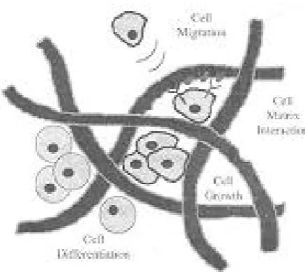
The coordinated function of many cell types is regulated by the integration of extracellular signals derived from soluble factors such as growth factors, and insoluble molecules of the extracellular matrix (ECM) [11]. Indeed, accumulating data suggests that cellular behavior (for example growth, differentiation and cell migration) is regulated by the converging down-stream signaling pathways of receptors for growth factors and ECM molecules [12]. These findings has reinforced the importance of scaffold’s composition and structure in controlling cellular responses in vitro and in vivo and provided a solid scientific foundation for the development of the new generation of biomaterials (Fig. 2).
The highly multidisciplinary nature of tissue engineering. The great diversity in the structure and
function of various tissues and organs is a primary reason for the highly multidisciplinary nature of tissue engineering research. The complexity of biological systems suggests that the manufacture of tissue engineered products will be complicated, and that their operating conditions, design, and specifications are intrinsically multi-factorial. Thus, it is no surprise that professionals trained as material scientists, chemical, mechanical, and electrical engineers, physicians and surgeons, molecular and cellular biologists, immunologists, and biochemists, all contribute meaningfully to research in the field of tissue engineering. The highly multidisciplinary nature of this research creates an unusually great
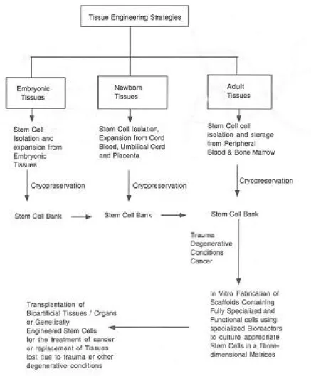
Fig. 1. Schematic representation for ex vivo fabrication of three-dimensional organs or tissues. The approach involves the Following
steps; 1) Isolation of multipotential stem cells from embryonic or adult tissue; 2) Expansion of multipotent stem cells in vitro using defined conditions that maintain their pluripotential phenotype; 3) Fabrication of optimal scaffolds generated from synthetic or natural polymers to promote growth factor assisted cellular recruitment, proliferation and differentiation of stem cells into specialized cell types of interest. 4) Large scale cultivation of stem cells in the optimal scaffolds using special bioreactors. The bioreactors are designed to continously supply the cultured cells with nutrients and to eliminate cellular waste and metabolites. 5) Transplantation of bio-artificial organ into the recipient.
Fig. 2.Schematic representation of molecular mechanism for
extracellular Materix (ECM)/ Growth factor-dependent cellular migration, growth and differentiation. The extracellular matrix is depicted by interlocking black strings. The soluble factors are denoted by green ovals. The ECM components are depicted by red triangles. The cell surface receptors for ECM (integrins) and growth factors (GFR) are shown in red and green respectively (hence the binding of growth factors and ECM molecules to their respective cell surface receptor induces a series of intracellular signaling pathways summarized in references 12, 18). The Cellular migration is defined by directed motion of cells toward the growth factor which is commonly referred to as motogens; Cellular growth is characterized by an increase in cell number stimulated by growth factors or mitogens; and cellular differentiation is denoted by changes in cell shape and morphology.
need for interdisciplinary communication and collaboration among researchers in each of its subdisciplines, a need that has become recognized broadly by government health and technology organizations, academic institutions, and the bio-technological and pharmaceutical companies. These organizations have taken the first steps toward creating an infrastructure that promotes interdisciplinary research, communication, and training of students and young professionals with interests in the future of this science.
The future of tissue engineering research. The
application of information and principles obtained through years of research in several disciplines to the goals of tissue engineering has already resulted in several promising medical advances such as tissue engineered heart valve leaflets, bioartificial
Fig. 3. Schematic representation of different disciplines
involved in the field of tissue engineering. 1) The genomic field focuses on the identification of novel genes encoding for growth factors and extracellular matrix molecules. This is performed by high throughput sequence analysis of the human genome followed by computer aided data analysis using the emerging science of bioinformatics [19]. 2) The combinatorial chemistry and high throughput screening technology involve a series of sophisticated biological and biochemical methods used to measure the cellular response to novel biomaterials. This technology enables scientists to synthesize and rapidly screen for the goodness of large number of candidate biomaterials invitro [20]. 3) The Stem Cell technology is a biotechnology field that focuses on the development of novel methods for isolation, expansion and transplantation of pluripotent cells from embryonic or adult tissues [21]. 4) The biomaterial science focuses on the development of novel scaffolds for the delivery of bioactive factors and cells [1, 2]. The gene therapy is an emerging technology that focuses on the development of appropriate vectors for safe and sustained expression of therapeutic genes that include a) tumor suppressor genes or genes that are mutated or deleted in certain diseases [22, 23].
bladders [14] and bone [15]. There has also
been significant progress in the generation of
artificial liver and pancreas, segments of the
digestive tract, cartilage, small blood vessels
and blood cells, cornea, and in promoting nerve
and muscle regeneration. While the progress in
these and other areas has generated excitement
and public interest, there are several difficult
problems confronting tissue engineering that
require a well-organized, long-term
multi-disciplinary
effort
to
overcome.
The
development of new materials, such as metals,
ceramics, and polymers, are one important
frontier. A better understanding of
immune-logical recognition and rejection,
wound
differentiation, and death, will also contribute
significantly to progress in tissue engineering
research.
In conclusion, the future of tissue engineering
involves the marriage of basic biology with the
development of applicable technologies for 1)
isolation, expansion and storage of embryonic
and adult pluripotent stem cells; 2) development
of bioactive scaffolds for efficient delivery of
cell-based therapeutics; 3) high throughput
biological
and
biochemical
screens
for
evaluation and development of novel materials
and
growth
factor
composites;
and
4)
development of gene-based therapeutics fueled
by recent advances in the field of bioinformatics
and genomics. Ultimately, in the next few
decades,
the
convergence
of
emerging
technologies in Silicon Valley’s computer chip
industry and medical technologies will produce
the first microscopic devices that will remove
the boundaries between the man and machine
[16, 17].
REFERENCES
1. Langer, R. and Vacanti, J.P. (1933) Tissue engineering. Science 260: 920-925.
2. Morgan, J.F. and Yarmush, M.L. (1999) The science
of tissue engineering. Science and
MedicineNovember/DecemberPage 6-7.
3. Nerem, R.M., Sambanis, A. (1995) Tissue
engineering: from biology to biological substitute. Tissue Engineering 1: 3-13.
4. Watt, F.M., Hogan, B.L. (2000) Out of eden: stem cells and their niches. Science 287:1427-1430. 5. Pittenger, M.F., Mackay, A.M., Beck, S.C., Jaiswal,
R.K., Douglas, R., Mosca, J.D., Moorman, M.A., Simonetti, D.W, Craig, S., Marshak, D.R. (1999) Multilineage potential of adult human mesenchymal stem cells. Science284: 143-146.
6. Brustle, O., Jones, K.N., Learish, R.D., Karram, K., Choudhary, K., Wiestler, O.D., Duncan, I.D., McKay, R.G. (1999) Embryonic stem cell-derived glial precursor: a source of myelinating transplants. Science 285: 754-756.
7. Thomason, J.A., Itskovitz-Eldor, J., Shapiro, S.S., Waknitz, M.A., Swiergiel, J.J., Marshall, V.S., Jones, J.M. (1998) Embryonic stem cells lines derived from human blastocysts. Science 282: 1145-1147.
8. Petersen, B.E., Bowen, W.C., Patrene, K.D., Mars, W.M., Sullivan, A.K., Murase, N., Boggs, S.S., Greenberger, J.S., Goff, J.P. (1999) Bone marrow as a potential source of hepatic oval cells. Science 284: 1168-1170.
9. Boyer, M., Townsend, L.E., Vogel, L.M., Falk, J., Reitz-Vick, D., Trevor, K.T., Villalba, M.m.Bendick, P.J., Glover, J.L. (2000) Isolation of endothelial cells and their progenitor cells from human peripheral blood. J. Vasc. Surg. 31: 181-189.
10. Rudolph, K.L., Chang, S., Millard, M., Schreiber-Agus, N., DePinho, Ronals, A. (2000) Inhibition of experimental liver cirrhosis in mice by telomerase gene delivery. Science 287: 1253-1258.
11. Juliano, R.L. and Huskill, S. (1993) Signal transduction from the extracellular matrix. J. Cell Biol. 120: 577-585.
12. Schwartz, M.A. (1997) Integrins, oncogenes and anchorage independence. J. Cell Biol. 139: 575-578. 13. Niklason, L.E., Gao, J., Abbott, W.M., Hirschi, K.K.,
Houser, S., Marini, R., Langer, R. (1999) Functional arteries grown in vitro. Science 284: 489-493. 14. Atala, A. (1999) Creation of bladder tissue in vitro
and in vivo: A system for organ replacement. Adv. Exp. Med. Biol. 462: 31-42.
15. Liu, L.S., Thompson, A.Y., Heidaran, M.A., Poser,
J.W. and Spiro, R.C. (1999) A novel
collagen/hyaluronate bone grafting matrix. Biomaterials 20:1097-1108.
16. Seeman, N.C. (1999) DNA engineering and its application to nanotechnology. Trends Biotechnol. 11: 437-443.
17. Zajichuk, R. (1999) New technologies in medicine: biotechnology and nanotechnology. Dis. Mon. 11: 449-495.
18. Yu, J.C., Li, W., Wang, L.M., Pierce, J.H. and Heidaran, M.A. (1995) Differential requirement of a motif within the carboxy terminal domain of aPDGFR for PDGF-focus forming activity, chemotaxis or growth. J. Biol. Chem. 270: 7033-7036.
19. Zhang, M.Q. (1999) Large scale gene expression data analysis: new challenges to computational biologists. Genome Res. 8: 681-688.
20. Kundu, B., Khare, S.K., Rastogi, S.K. (1999) Combinatorial chemistry: polymer supported synthesis of peptides and non-peptide libraries. Prog. Drug Res. 53: 89-156.
21. Vogel, G. (2000) Can old cells learn new tricks? Science 287: 1419.
22. SoRelle, R. (2000) Gene therapy at a crossroad. Circulation 101:E9001.
23. Sikic, B.I. (1999) New approaches in cancer treatment. Ann. Oncol. 10 Suppl. 6:149-153.