*For correspondence:sdnibley@ gmail.com
Competing interests:The authors declare that no competing interests exist.
Received:25 November 2016 Accepted:08 December 2016 Published:26 July 2017 Author Keywords:ultrasound, general practitioner, GP, pocket, tendon
CopyrightsBJGP Open 2017; DOI:10.3399/
bjgpopen17X100893
GP-confirmed complete Achilles tendon
rupture using pocket-sized ultrasound: a
case report
SJ Davis,
MBChB1*, A Lott,
MBBS2, E Besada,
MD31
GP & University Lecturer, Department of General Practice, Institute of Community
Medicine, University of Tromsø, Tromsø, Norway;
2Junior Radiologist, Department
of Radiology, Institute of Clinical Medicine, University of Tromsø (UiT) The Arctic
University of Norway, Tromsø, Norway;
3Rheumatologist & University Lecturer,
Department of Rheumatology, Institute of Clinical Medicine, University of Tromsø
(UiT) The Arctic University of Norway, Tromsø, Norway
Introduction
The incidence of complete Achilles tendon rupture is 18 per 100 000 patient-years1 and is usually diagnosed clinically by GPs. The extent of clinical misdiagnosis is unknown in Norway, but may be high.2This is important as delayed treatment has unfavourable consequences.1,3We report how a GP, with no clinical ultrasound experience, recorded images with a pocket-sized ultrasound device (PSUD) under supervision to confirm a complete Achilles tendon rupture. This could present a new indication for GP ultrasound.
Case report
A 36-year-old man experienced acute pain above the right heel accompanied by an audible snap while sprinting. He immediately had difficulty walking and 3 hours later consulted an on-call GP. Pos-terior ankle swelling with a tender depression 3 cm proximal to the calcaneum was found. Active plantar flexion against resistance was weak and Simmonds–Thompson test was ‘partially positive’ on applying a strong calf-squeeze. Based on these findings, calf muscle rupture was diagnosed as the Achilles tendon was thought to be intact. The patient was advised to elevate the foot and wait 2 weeks for improvement. Two days later a second GP, who was aware of a history of an audible snap, considered complete tendon rupture and reexamined the patient. Findings included an absent right heel raise due to weakness, minimal active plantar flexion against gravity and lying prone, significant right ankle swelling without bruising, and an altered angle of declination. Palpation elicited no ankle bony tenderness, yet a painful gap was identified 6 cm proximal from the calcaneal attachment, along the line of the Achilles tendon. Simmonds–Thompson’s test was clearly positive. The positive Simmond’s triad indicated a clinical diagnosis of complete rupture of the Achilles tendon.
A 3.4–8 MHz linear array probe PSUD (VScan dual probe, GE Healthcare), set at a depth of 3.5 cm, was used under the supervision of a rheumatologist experienced in ultrasound. The tendon was enlarged from 1 cm to 6 cm above the calcaneal insertion, where a clear gap was seen (Figure 1). Two hours later a radiologist-performed ultrasound (LOGIQ E9, GE Healthcare) and reported an enlarged distal tendon and a complete rupture at 5–6 cm from the calcaneal attach-ment, creating a 2.7 cm blood-filled gap (Figure 2). Surgical exploration 8 days post-injury found a complete Achilles tendon rupture ‘5–10 cm above the ankle joint’.
Discussion
Tromsø Hospital serves a large area with a population of approximately 160 000. Between 2010– 2014 an average of 21 patients per year were referred by their GP for suspected Achilles rupture.
PRACTICE & POLICY
*For correspondence:sdnibley@ gmail.com
Competing interests:The authors declare that no competing interests exist.
Received:25 November 2016 Accepted:08 December 2016 Published:26 July 2017 Author Keywords:ultrasound, general practitioner, GP, pocket, tendon
CopyrightsBJGP Open 2017; DOI:10.3399/
bjgpopen17X100893
GP-confirmed complete Achilles tendon
rupture using pocket-sized ultrasound: a
case report
SJ Davis,
MBChB1*, A Lott,
MBBS2, E Besada,
MD31
GP & University Lecturer, Department of General Practice, Institute of Community
Medicine, University of Tromsø, Tromsø, Norway;
2Junior Radiologist, Department
of Radiology, Institute of Clinical Medicine, University of Tromsø (UiT) The Arctic
University of Norway, Tromsø, Norway;
3Rheumatologist & University Lecturer,
Department of Rheumatology, Institute of Clinical Medicine, University of Tromsø
(UiT) The Arctic University of Norway, Tromsø, Norway
Introduction
The incidence of complete Achilles tendon rupture is 18 per 100 000 patient-years1 and is usually diagnosed clinically by GPs. The extent of clinical misdiagnosis is unknown in Norway, but may be high.2This is important as delayed treatment has unfavourable consequences.1,3We report how a GP, with no clinical ultrasound experience, recorded images with a pocket-sized ultrasound device (PSUD) under supervision to confirm a complete Achilles tendon rupture. This could present a new indication for GP ultrasound.
Case report
A 36-year-old man experienced acute pain above the right heel accompanied by an audible snap while sprinting. He immediately had difficulty walking and 3 hours later consulted an on-call GP. Pos-terior ankle swelling with a tender depression 3 cm proximal to the calcaneum was found. Active plantar flexion against resistance was weak and Simmonds–Thompson test was ‘partially positive’ on applying a strong calf-squeeze. Based on these findings, calf muscle rupture was diagnosed as the Achilles tendon was thought to be intact. The patient was advised to elevate the foot and wait 2 weeks for improvement. Two days later a second GP, who was aware of a history of an audible snap, considered complete tendon rupture and reexamined the patient. Findings included an absent right heel raise due to weakness, minimal active plantar flexion against gravity and lying prone, significant right ankle swelling without bruising, and an altered angle of declination. Palpation elicited no ankle bony tenderness, yet a painful gap was identified 6 cm proximal from the calcaneal attachment, along the line of the Achilles tendon. Simmonds–Thompson’s test was clearly positive. The positive Simmond’s triad indicated a clinical diagnosis of complete rupture of the Achilles tendon.
A 3.4–8 MHz linear array probe PSUD (VScan dual probe, GE Healthcare), set at a depth of 3.5 cm, was used under the supervision of a rheumatologist experienced in ultrasound. The tendon was enlarged from 1 cm to 6 cm above the calcaneal insertion, where a clear gap was seen (Figure 1). Two hours later a radiologist-performed ultrasound (LOGIQ E9, GE Healthcare) and reported an enlarged distal tendon and a complete rupture at 5–6 cm from the calcaneal attach-ment, creating a 2.7 cm blood-filled gap (Figure 2). Surgical exploration 8 days post-injury found a complete Achilles tendon rupture ‘5–10 cm above the ankle joint’.
Discussion
Tromsø Hospital serves a large area with a population of approximately 160 000. Between 2010– 2014 an average of 21 patients per year were referred by their GP for suspected Achilles rupture.
PRACTICE & POLICY
*For correspondence:sdnibley@ gmail.com
Competing interests:The authors declare that no competing interests exist.
Received:25 November 2016 Accepted:08 December 2016 Published:26 July 2017
GP-confirmed complete Achilles tendon
rupture using pocket-sized ultrasound: a
case report
SJ Davis,
MBChB1*, A Lott,
MBBS2, E Besada,
MD31
GP & University Lecturer, Department of General Practice, Institute of Community
Medicine, University of Tromsø, Tromsø, Norway;
2Junior Radiologist, Department
of Radiology, Institute of Clinical Medicine, University of Tromsø (UiT) The Arctic
University of Norway, Tromsø, Norway;
3Rheumatologist & University Lecturer,
Department of Rheumatology, Institute of Clinical Medicine, University of Tromsø
(UiT) The Arctic University of Norway, Tromsø, Norway
Introduction
The incidence of complete Achilles tendon rupture is 18 per 100 000 patient-years1 and is usually diagnosed clinically by GPs. The extent of clinical misdiagnosis is unknown in Norway, but may be high.2This is important as delayed treatment has unfavourable consequences.1,3We report how a GP, with no clinical ultrasound experience, recorded images with a pocket-sized ultrasound device (PSUD) under supervision to confirm a complete Achilles tendon rupture. This could present a new indication for GP ultrasound.
Case report
A 36-year-old man experienced acute pain above the right heel accompanied by an audible snap while sprinting. He immediately had difficulty walking and 3 hours later consulted an on-call GP. Pos-terior ankle swelling with a tender depression 3 cm proximal to the calcaneum was found. Active plantar flexion against resistance was weak and Simmonds–Thompson test was ‘partially positive’ on applying a strong calf-squeeze. Based on these findings, calf muscle rupture was diagnosed as the Achilles tendon was thought to be intact. The patient was advised to elevate the foot and wait 2 weeks for improvement. Two days later a second GP, who was aware of a history of an audible snap, considered complete tendon rupture and reexamined the patient. Findings included an absent right heel raise due to weakness, minimal active plantar flexion against gravity and lying prone, significant right ankle swelling without bruising, and an altered angle of declination. Palpation elicited no ankle bony tenderness, yet a painful gap was identified 6 cm proximal from the calcaneal attachment, along the line of the Achilles tendon. Simmonds–Thompson’s test was clearly positive. The positive Simmond’s triad indicated a clinical diagnosis of complete rupture of the Achilles tendon.
A 3.4–8 MHz linear array probe PSUD (VScan dual probe, GE Healthcare), set at a depth of 3.5 cm, was used under the supervision of a rheumatologist experienced in ultrasound. The tendon was enlarged from 1 cm to 6 cm above the calcaneal insertion, where a clear gap was seen (Figure 1). Two hours later a radiologist-performed ultrasound (LOGIQ E9, GE Healthcare) and reported an enlarged distal tendon and a complete rupture at 5–6 cm from the calcaneal attach-ment, creating a 2.7 cm blood-filled gap (Figure 2). Surgical exploration 8 days post-injury found a complete Achilles tendon rupture ‘5–10 cm above the ankle joint’.
Discussion
Tromsø Hospital serves a large area with a population of approximately 160 000. Between 2010– 2014 an average of 21 patients per year were referred by their GP for suspected Achilles rupture.
PRACTICE & POLICY
Author Keywords:ultrasound, general practitioner, GP, pocket, tendon
CopyrightsBJGP Open 2017; DOI:10.3399/
bjgpopen17X100893
GP-confirmed complete Achilles tendon
rupture using pocket-sized ultrasound: a
case report
SJ Davis,
MBChB1*, A Lott,
MBBS2, E Besada,
MD31
GP & University Lecturer, Department of General Practice, Institute of Community
Medicine, University of Tromsø, Tromsø, Norway;
2Junior Radiologist, Department
of Radiology, Institute of Clinical Medicine, University of Tromsø (UiT) The Arctic
University of Norway, Tromsø, Norway;
3Rheumatologist & University Lecturer,
Department of Rheumatology, Institute of Clinical Medicine, University of Tromsø
(UiT) The Arctic University of Norway, Tromsø, Norway
Introduction
The incidence of complete Achilles tendon rupture is 18 per 100 000 patient-years1 and is usually diagnosed clinically by GPs. The extent of clinical misdiagnosis is unknown in Norway, but may be high.2This is important as delayed treatment has unfavourable consequences.1,3We report how a GP, with no clinical ultrasound experience, recorded images with a pocket-sized ultrasound device (PSUD) under supervision to confirm a complete Achilles tendon rupture. This could present a new indication for GP ultrasound.
Case report
A 36-year-old man experienced acute pain above the right heel accompanied by an audible snap while sprinting. He immediately had difficulty walking and 3 hours later consulted an on-call GP. Pos-terior ankle swelling with a tender depression 3 cm proximal to the calcaneum was found. Active plantar flexion against resistance was weak and Simmonds–Thompson test was ‘partially positive’ on applying a strong calf-squeeze. Based on these findings, calf muscle rupture was diagnosed as the Achilles tendon was thought to be intact. The patient was advised to elevate the foot and wait 2 weeks for improvement. Two days later a second GP, who was aware of a history of an audible snap, considered complete tendon rupture and reexamined the patient. Findings included an absent right heel raise due to weakness, minimal active plantar flexion against gravity and lying prone, significant right ankle swelling without bruising, and an altered angle of declination. Palpation elicited no ankle bony tenderness, yet a painful gap was identified 6 cm proximal from the calcaneal attachment, along the line of the Achilles tendon. Simmonds–Thompson’s test was clearly positive. The positive Simmond’s triad indicated a clinical diagnosis of complete rupture of the Achilles tendon.
A 3.4–8 MHz linear array probe PSUD (VScan dual probe, GE Healthcare), set at a depth of 3.5 cm, was used under the supervision of a rheumatologist experienced in ultrasound. The tendon was enlarged from 1 cm to 6 cm above the calcaneal insertion, where a clear gap was seen (Figure 1). Two hours later a radiologist-performed ultrasound (LOGIQ E9, GE Healthcare) and reported an enlarged distal tendon and a complete rupture at 5–6 cm from the calcaneal attach-ment, creating a 2.7 cm blood-filled gap (Figure 2). Surgical exploration 8 days post-injury found a complete Achilles tendon rupture ‘5–10 cm above the ankle joint’.
Discussion
Tromsø Hospital serves a large area with a population of approximately 160 000. Between 2010– 2014 an average of 21 patients per year were referred by their GP for suspected Achilles rupture.
Davis Set al. BJGP Open 2017;DOI: 10.3399/bjgpopen17X100893 1 of 3
*For correspondence:gc.island@ gmail.com pollybrandon@mac. com
Competing interests:The authors declare that no competing interests exist.
Received:14 August 2016 Accepted:16 August 2016 Published:09 January 2017
This article is Open Access:
https:// creativecommons.org/licenses/ by/4.0/)
sBJGP Open 2017; DOI:10.3399/ bjgpopen17X100557
Primary care in the Calais Jungle
Gerry Clare,
BSc, MSc, PhD, FRCOphth1*, Polly Nyiri,
MBBChir, DTM&H, MA International Health2*
1
Consultant ophthalmologist, Western Eye Hospital, London, UK;
2GP, Health
Inclusion Clinic, Guy’s and St Thomas’ Hospital NHS Foundation Trust, London, UK
Introduction
Last summer our small medical team visited the Calais ’Jungle’. Since that time much has changed and the camp is being demolished and by the time this article is read, it will probably be long gone. Some youngsters are finally being brought to the UK under the ’Dubs’ amendment. However, once this camp is cleared it will not solve the ongoing flight of refugees from war torn areas: other camps are already appearing.
July 2016
A young Afghan man caught his finger on a sharp point while trying to cross a barbed wire fence. The finger was partially degloved. He attended the local hospital, where they placed a few sutures, but now, 2 weeks later, the skin is necrotic and the underlying tissue looks infected. He is in danger of losing his finger.
A middle-aged Sudanese man has been having rigors and is generally unwell. He says it is similar to when he last had malaria.
A young Ukrainian woman complains of lower back pain and urinary frequency.
The paths of these three people may never have crossed; yet here they are, denizens of the Calais Jungle. They turn up to a makeshift primary care ‘clinic’ that we set up in the heart of the unofficial refugee camp one weekend in July 2016.
With only basic medical supplies, we are immediately challenged by what we see. How can we arrange secondary care for the young Afghan in danger of losing his finger? We try to persuade him to return to the original local hospital, but he is reluctant. It was not a good experience for him the first time round.
With the other two patients, it is easier. They can attend the Salam clinic run by a local association during weekdays. Later, we receive word that malaria has been confirmed in our Sudanese patient.
More people arrive, presenting with scabies, rat bites, tinea, chest infections, and wheezing from inhaling smoke from fires lit to cook and keep warm in their tents at night. We examine a severely malnourished 2-year-old boy. We meet several of the camp’s 600 unaccompanied children, at grave risk of sexual exploitation. We learn that there is inadequate safeguarding in place to protect them. A young Eritrean man comes in worried about his eye. He has sustained direct ocular trauma from a rubber bullet, and will never see normally again out of that eye. We see haematomas from police batons, and hear about children being exposed to tear gas again and again (Figure 1).
The reality
These are no ordinary patients. They have travelled far from home to escape war, poverty, and mis-ery. They have endured personal odysseys to get here, experienced untold hardships, and suffered unimaginable privations. Many have survived the loss of their families, torture, and rape. Their jour-neys over, for the moment at least, they must make their homes in the Calais Jungle. Their new shel-ters are in many cases mere tarpaulin covers, and their new beds just rugs on the ground. They own next to nothing. There is little for them to do, besides use their ingenuity to cross the English Chan-nel in search of a better life. They are vulnerable to exploitation, crime, injury, and disease. Poten-tially violent clashes with local police, with other ethnic groups resident in the Jungle, or local far
Clare G and Nyiri P. BJGP Open 2017;DOI: 10.3399/bjgpopen17X100557 1 of 5
PRACTICE & POLICY
CC BY license (
The Authors; *For correspondence:marcela. ewing@rccvast.se
Competing interests:The authors declare that no competing interests exist.
Received:31 August 2017
Accepted:10 November 2017
Published:07 February 2018
Author Keywords:diagnosis, general practice, lung cancer, non-metastatic, primary health care, Sweden
Copyrights2018, DOI:10.3399/ bjgpopen18X101397
Clinical features of patients with
non-metastatic lung cancer in primary care: a
case-control study
Marcela Ewing,
MD1*, Peter Naredi,
PhD, MD2, Chenyang Zhang,
MSc3,
Lars Lindsko¨ld,
PhD4, Jo¨rgen Ma˚nsson,
PhD, MD51
PhD student, Department of Public Health and Community Medicine/Primary
Health Care, Institute of Medicine, Sahlgrenska Academy, University of
Gothenburg, Gothenburg, Sweden;
2Professor, Department of Surgery, Institute of
Clinical Sciences, Sahlgrenska Academy, University of Gothenburg, Sahlgrenska
University Hospital, Gothenburg, Sweden;
3Statistician, Regional Cancer Centre
West, Sahlgrenska University Hospital, Gothenburg, Sweden;
4Senior Lecturer,
Department of Applied Information Technology, University of Gothenburg,
Gothenburg, Sweden;
5Professor, Department of Public Health and Community
Medicine/Primary Health Care, Institute of Medicine, Sahlgrenska Academy,
University of Gothenburg, Gothenburg, Sweden
Abstract
Background:Lung cancer (LC) kills more people than any other cancer globally, mainly due to the late stage of diagnosis.
Aim:To identify and quantify the prediagnostic features of non-metastatic lung cancer (nMLC) and to compare the clinical features in GPs’ chest X-ray referral letters with the clinical features (expressed as diagnostic codes) in medical records.
Design & setting:A population-based case-control study was conducted using diagnostic codes from national and regional healthcare databases in Sweden.
Method: In total, 373 patients diagnosed with LC in 2011 (of which 132 had nMLC) and 1472 controls were selected from the Swedish Cancer Register (SCR) and regional healthcare database, respectively. Diagnostic codes registered in medical records from primary care consultations in the year before LC diagnosis were collected from the regional healthcare database. Odds ratios (OR) were calculated for variables associated with nMLC. The GPs’ referral letters for chest X- ray were retrieved from the regional repository for radiology.
Results: Clinical features with the highest OR were vitamin B12 deficiency anaemia (OR 6.7, 95% confidence interval [CI] = 1.6 to 27.9), dyspnoea (OR 5.0, 95% CI = 2.0 to 12.7), and chronic bronchitis (OR 5.0, 95% CI = 1.3 to 18.6). Clinical features that were GPs’ reasons for requesting chest X-ray were almost three times more frequent in referral letters compared to the corresponding diagnostic codes in the medical records.
Conclusion: Patients with nMLC could not be identified by symptoms. The clinical features in referral letters for X-ray were more frequent than corresponding diagnostic codes from medical records.
How this fits in
Late-stage diagnosis is a main reason for the high mortality of LC. Different risk assessment tools have been developed for GPs in order to detect LC earlier by clinical features. This study shows that
patients with nMLC could not be identified by clinical features. However, despite the lack of this spe-cific knowledge, GPs’ referrals for a chest X-ray resulted in a 40% detection rate of nMLC.
Introduction
LC is one of the deadliest and most common cancers in the world. With an estimated 1.8 million new cases in the world each year, this cancer is responsible for almost one cancer death in five.1LC is the fourth most common cancer in Europe with >410 000 new cases diagnosed in 2012.1The high mortality is due to both late-stage diagnosis and delay in treatment.2–5In the UK, 46 000 new cases of LC were diagnosed in 2014, and half of the patients with known stage were diagnosed at Stage IV (metastatic disease).6In Sweden, 4194 patients were diagnosed with LC in 2015, and 3626 died from it.7,8 Despite having high survival rates for many types of cancer, Sweden has poor survival rates for LC.9The relative 5-year survival rate for LC in Sweden is 18%.10The low survival rate is mainly due to late-stage diagnosis. More than 50% of all Swedish patients with LC are diagnosed at Stage IV, with a relative 5-year survival rate of 2.6%. However, when LC is diagnosed at Stage I, the relative 5-year survival rate is 63.8%.10In order to increase survival rates for patients with LC, the most important factor is being able to identify those with a potentially curable disease. There is value in identifying patients at Stage I–III, whose LC has yet not spread, because they, as a group, have a relative 5-year survival of 36.1% versus 2.6% for Stage IV cancer.10
Screening of target groups has been discussed as a method for early diagnosis of LC. Low-dose computed tomography (LDCT) in a defined population of high-risk persons has shown high sensitiv-ity and acceptable specificsensitiv-ity.11Publications from different LC screening trials show that up to 70% of screen-detected, non-small cell LCs were found in Stage I, compared to around 15% found in rou-tine clinical care.12LDCT is currently being used as screening for LC in the US.12
GPs are important in cancer diagnostics because in countries like Sweden, Norway, Denmark, and France, approximately 70–87% of patients with cancer are diagnosed in a primary care setting.5,13– 15
Because Sweden possesses unique total population-based databases, a case-control study could be conducted using regional databases for healthcare and diagnostic imaging in combination with the national cancer register.
This study aimed to:
. identify the clinical features of nMLC in primary care before the diagnosis is made; and . validate the clinical features from the regional healthcare database with clinical features in
GPs’ referral letters for chest X-rays.
Method
Study design
A total population-based, case-control study was designed, using the SCR and a regional healthcare database in Region Va¨stra Go¨taland (RVG), Sweden. This region, which has 1.6 million inhabitants, is situated in the south-west of the country.
The SCR, which was established in 1958, is one of the oldest disease registers in the world and has high validity.16All physicians, including pathologists, in Sweden are obliged by law to report all incident cases of cancer in both living and deceased patients to the SCR.17Each patient has a unique personal identity number, which all Swedish residents acquire either at birth or when they immigrate to Sweden.
In 2006, the Enterprise Information Archive (EIA), a regional database for radiology information, was established. It allows both textual information and images to be shared (stored and distributed) from every radiology department in the RVG.21Both publicly and privately-financed radiology clinics send information to this database.
Study population
All patients in the RVG with LC diagnosed in 2011 were identified from the SCR. As this study was total population-based, no sample size was calculated.
Patients and matched controls were investigated for primary care diagnostic profiles. The inclu-sion criteria were:
. being diagnosed in RVG with LC;
. being alive at the time of the cancer diagnosis;
. being aged18 years; and
. having visited the GP during the year before cancer diagnosis.
Individuals were excluded from participation if they:
. lacked controls;
. had a previous cancer diagnosis in the SCR (1991–2010); or . had a metastasised, Stage IV LC.
Patients with a previous cancer diagnosis registered in the SCR during the 20-year period before 2011 were deliberately omitted, to avoid consultations in primary care being a control or related to a previous cancer. The controls were selected from the regional healthcare database. They had the same inclusion criteria as the patients with cancer, with the exception of a cancer diagnosis. Only controls from RVG who had visited a GP in primary care between 1 January 2010 and 31 December 2011 were eligible. Four controls were matched to each case for age, sex, and primary care unit.
Data collection and study measurements
The unique personal identity numbers of both cases and controls were linked to the regional health-care database. All the data concerning diagnoses and dates of consultations with a GP between 1 January 2010 and 31 December 2011 were collected. The data extracted included diagnostic codes according to the Swedish version of the ICD-10;22or the Classification of Diseases and Health Prob-lems 1997 Primary Care (KSH97-P). This is an abbreviated version of ICD-10, adapted to Swedish pri-mary care to facilitate diagnostic coding.23,24
The unique personal identity numbers of cases were linked to the EIA database. GPs’ referral let-ters for chest X-ray — containing detailed clinical information with risk factors, symptoms, and signs from physical examinations and pathological laboratory results — were retrieved either from the EIA database or other repositories.
Two medical oncologists and a GP, independently of each other, coded the clinical features in all the referral letters for chest X-ray, using the ICPC-2 codes because these are more symptom-based. Where the codes were not consistent between the three coders, a consensus was reached on the final coding. These codes were then compared with the ICD-10 diagnostic codes from medical records in the healthcare database. As the authors only had access to diagnostic codes, the referral letters provided the reasons for chest X-ray referrals. In addition, because a more symptom-based coding classification was used (ICP-2), a comparison was made between how well the clinical fea-tures in referral letters corresponded to the clinical feafea-tures coded in a less symptom-based classifi-cation (ICD-10) in the regional healthcare database.
Diagnostic codes
Data analyses
The 575 diagnostic codes were used as variables for univariable conditional logistic regression. Those found to be associated with cancer entered multivariable analyses, after which a list of statisti-cally significant variables associated with LC was compiled. All analyses were performed using the statistical software R (version 3.0.1).
Results
Cases and controls
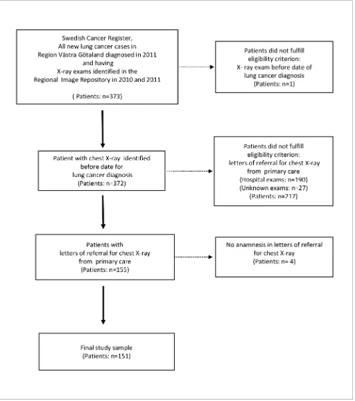
In total, 373 patients with LC were identified in the SCR. Of these, 132 patients had Stage I–III (35%) non-metastatic cancer, and the remaining 241 patients had Stage IV (65%). Although four controls had been matched to each case, 20 had died before their case was assigned a cancer diagnosis, so a total of 1472 controls were generated. The characteristics of the study sample is shown inTable 1. The disease burden for cases and controls was similar regarding the median number of unique diag-nostic codes in the year before cancer diagnosis. Data retrieved from the regional database for radi-ology information (EIA) showed that 151 (40%) out of 373 patients with LC had been referred by a GP for a first chest X-ray in the year prior to cancer diagnosis (Figure 1). Hence, the majority of patients (51%) had been referred for chest X-ray by physicians in secondary care.
Variables
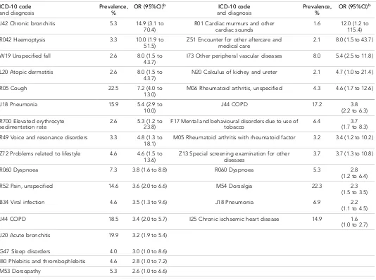
After the univariable conditional regression was done, there were 15 significant variables left (P<0.05) for patients with non-metastatic cancer and 23 for patients with metastatic cancer. The vari-ables with an odds ratio of >1.5 are presented inTable 2. After multivariate conditional regression, several significant variables were found to be independently associated with nMLC, but because there were too few cases for each combination of features, no calculation of positive predictive val-ues could be performed. Even though all the patients included in this study consulted a GP in the year prior to their LC diagnosis, there were differences in their diagnostic profile depending on whether they had been referred for their first chest X-ray by their GP or from secondary care (Table 3). In total, 40% of the patients referred for their first chest X-ray from primary care had nMLC, compared to 30% of those referred from secondary care. The clinical features were 2.7 times more frequent (337 versus 126) in referral letters for chest X-ray than the corresponding features in the healthcare database (Table 4).
Table 1.Sample characteristics of patients with lung cancer and controls
Characteristics
Patients with lung cancer,n= 373
Controls,n= 1472
Median age at diagnosis, years (range) 69 (30–93) 70 (30–93)
Female,n(%) 178 (48) 706 (48)
Male,n(%) 195 (52) 766 (52)
Age <60 years,n(%) 61 (16) 242 (16)
Age 60–80 years,n(%) 264 (71) 1046 (71)
Age >80 years,n(%) 48 (13) 184 (13)
Stage I–III (M0a),
n(%) 132 (35)
Stage IV (M1a),
n(%) 241 (65)
Median number of consultations per patient in year before cancer diagnosis,n(IQR) 5 (3–9) 4 (2–7)
Median number of unique diagnostic codes per patient in year before cancer diagnosis,n (IQR)
6 (4–10) 6 (3–9)
Discussion
Summary
The study identified 12 features that were associated with nMLC, of which eight were also in com-mon with metastatic LC. The features with the highest OR for nMLC were vitamin B12 deficiency anaemia, dyspnoea, and chronic bronchitis. Clinical features that were GPs’ reasons for request for chest X-ray were almost three times more frequent in referral letters compared to the corresponding diagnostic codes in the medical records.
Table 2.Univariable analysis of diagnoses depending on stage with odds ratio >1.5ain patients in primary care during 12 months before lung cancer diagnosis
Stage I–III (M0b) Stage IV (M1b)
ICD-10 code
and diagnosis Prevalence% OR (95%CI)c and diagnosisICD-10 code Prevalence% OR (95% CI) c
D51 Vitamin B12 deficiency anaemia
3.8 6.7 (1.6 to 27.9)
L20 Atopic dermatitis 1.2 12.0 (1.2 to 115.4)
R060 Dyspnoea 8.4 5.0 (2.0 to 12.7)
R042 Haemoptysis 2.1 9.6 (1.9 to 49.7)
J42 Unspecified chronic bronchitis
3.8 5.0 (1.3 to 18.6)
I26 Pulmonary embolism 1.7 8.0 (1.5 to 43.7)
J44 COPD 20.6 4.3
(2.4 to 7.5)
W00 Fall due to ice and snow 1.7 8.0 (1.5 to 43.7)
I73 Other peripheral vascular diseases
4.6 4.2 (1.3 to 13.9)
M05 Rheumatoid arthritis with rheumatoid factor 2.9 4.7 (1.6 to 13.9)
B34 Viral infection of unspecified site
5.4 4.0 (1.4 to 11.4)
N20 Calculus of kidney and ureter 2.1 4.5 (1.2 to 17.1)
R05 Cough 13.8 3.8
(2.0 to 7.5)
W19 Unspecified fall 2.5 4.3 (1.3 to 14.3)
J18 Pneumonia 12.2 3.2 (1.6 to 6.2)
J18 Pneumonia 9.5 3.8 (2.1 to 7.0)
R52 Pain, unspecified 10.7 2.3 (1.1 to 4.7)
G40 Epilepsy and current seizures 2.5 3.7 (1.2 to 11.6)
N30 Cystitis 14.5 2.0 (1.1 to 3.6)
R05 Cough 14.9 3.6
(2.2 to 5.8)
J20 Acute bronchitis 16.0 1.8 (1.1 to to 3.2)
I73 Other peripheral vascular diseases 7.0 3.6 (1.8 to 6.8)
M54 Back pain 18.3 1.8 (1.1 to 3.1)
R22 Localised swelling, mass, and lump of skin and subcutaneous tissue
3.7 3.1 (1.3 to 7.5)
M06 Other rheumatoid arthritis 2.5 3.0 (1.0 to 8.6)
J44 COPD 15.3 3.0
(1.9 to 4.7)
R060 Dyspnoea 5.8 2.5 (1.3 to 4.9)
K51 Diverticular disease of intestine 3.7 2.4 (1.0 to 5.4)
J20 Acute bronchitis 14.9 2.3 (1.5 to 3.5)
R52 Pain, unspecified 11.2 2.0 (1.3 to 3.3)
M54 Back pain 19.8 2.0 (1.3 to 2.8)
aOdds ratio are calculated between cases and controls. Diagnostic codes with OR <1.5 are omitted.bTNM Classification of Malignant Tumours code.cP<0.05. COPD =
Strengths and limitations
The main strength of this study is that it is total population-based. All patients with cancer were identified through the SCR, so there is no selection bias and the completeness of the register is very high.16 The study looked at the clinical features presented during the year before LC diagnosis, because knowing these has major consequences for timelier and earlier LC diagnosis, which in turn affects prognosis. The use of diagnostic codes is another strength of the study. However, this could also be considered a limitation because not all the symptoms for which patients consulted a GP would be recorded as a diagnostic code in their medical record, as other fields of research in pri-mary care databases have shown.25
Most cancer symptoms occur 3–6 months before the cancer diagnosis, but a longer time than the one used in this study may be needed for observation.26The lack of laboratory results to validate the diagnoses of vitamin B12 deficiency anaemia, which had the strongest association with nMLC is another limitation. The absence of smoking status of patients with cancer is a limitation too, as the symptomatology of smokers has more severe implications than that of non-smokers.27
Another limitation is that the authors were unable to design a risk assessment tool for nMLC in primary care. This is due either to the lack of a large enough sample, resulting in the inability to Table 3.Univariate analysis of diagnoses referred from primary or secondary care to the first chest X-ray during 12 months before lung cancer diagnosisa
Primary care chest imaging referral (n= 151) Secondary care chest imaging referral (n= 190)
ICD-10 code
and diagnosis Prevalence,% OR (95%CI)
b ICD-10 code
and diagnosis Prevalence,% OR (95%CI)
b
J42 Chronic bronchitis 5.3 14.9 (3.1 to 70.4)
R01 Cardiac murmurs and other cardiac sounds
1.6 12.0 (1.2 to 115.4)
R042 Haemoptysis 3.3 10.0 (1.9 to 51.5)
Z51 Encounter for other aftercare and medical care
2.1 8.0 (1.5 to 43.7)
W19 Unspecified fall 2.6 8.0 (1.5 to 43.7)
I73 Other peripheral vascular diseases 8.0 5.4 (2.5 to 11.8)
L20 Atopic dermatitis 2.6 8.0 (1.5 to 43.7)
N20 Calculus of kidney and ureter 2.1 4.7 (1.0 to 21.4)
R05 Cough 22.5 7.2 (4.0 to 13.0)
M06 Rheumatoid arthritis, unspecified 4.3 4.6 (1.7 to 12.6)
J18 Pneumonia 15.9 5.4 (2.9 to 10.0)
J44 COPD 17.2 3.8
(2.2 to 6.3)
R700 Elevated erythrocyte sedimentation rate
2.6 5.3 (1.2 to 23.8)
F17 Mental and behavioural disorders due to use of tobacco
6.4 3.7 (1.7 to 8.3)
R49 Voice and resonance disorders 3.3 4.8 (1.3 to 18.1)
M05 Rheumatoid arthritis with rheumatoid factor 3.2 3.4 (1.2 to 10.2)
Z72 Problems related to lifestyle 4.6 4.6 (1.5 to 13.6)
Z13 Special screening examination for other diseases
3.7 3.7 (1.3 to 10.8)
R060 Dyspnoea 7.3 3.8 (1.6 to 8.8) R060 Dyspnoea 5.3 2.8 (1.2 to 6.4)
R52 Pain, unspecified 14.6 3.6 (2.0 to 6.6) M54 Dorsalgia 22.3 2.3 (1.5 to 3.5)
B34 Viral infection 4.6 3.5 (1.3 to 9.6) J18 Pneumonia 6.9 2.2 (1.1 to 4.5)
J44 COPD 18.5 3.4 (2.0 to 5.7) I25 Chronic ischaemic heart disease 14.9 1.6 (1.0 to 2.7)
J20 Acute bronchitis 19.9 3.2 (1.9 to 5.4)
G47 Sleep disorders 4.0 3.0 (1.0 to 8.6)
I80 Phlebitis and thrombophlebitis 4.6 2.8 (1.0 to 7.2)
M53 Dorsopathy 5.3 2.6 (1.0 to 6.6)
capture combinations of features, or to a truly low frequency of combination of features in the non-metastatic population, which may not be detected even with a larger sample size.
The low prevalence of clinical features in the regional healthcare database in comparison to clini-cal features in referral letters for X-ray is probably due to the former consisting mainly of diseases and the latter of symptoms. Another explanation could be that the reimbursement system for pri-mary care providers is partly based on the disease burden of the patients, which favours disease codes over symptom codes.
Comparison with existing literature
To the authors’ knowledge, this is the first study to present the clinical features of LC in patients with a non-metastatic disease. This is also the first study to present vitamin B12 deficiency anaemia as being a risk marker for nMLC. Perhaps this finding is a paraneoplastic phenomenon. However, previously published studies have shown that individuals with vitamin B12 deficiency anaemia are at increased risk for other cancers, such as gastric cancers and blood malignancies.28,29A recent sys-tematic review from the UK has suggested that patients with thrombocytosis in primary care have an increased risk of several cancers, among them LC, which this study was unable to show as it lacked data on blood test results.30Another UK study from primary care has reported association with LC in the first year after presentation with back problems, which is in line with this study’s findings.31In this study, the clinical information in referral letters for chest X-ray was extensive, in contrast to what has been reported in the literature.32
A Danish study showed that patients with LC and chronic obstructive pulmonary disease (COPD) had more contacts in primary care in the 11 months prior to diagnosis than did patients with LC but without COPD. Thus, having COPD can mask symptoms of LC.33This is in line with the findings pre-sented here, that COPD is a risk marker in patients with both non-metastatic and metastatic LC. Table 4.ICPC-2 codesain letters of referral for chest X-ray compared with corresponding ICD-10 codes in the regional healthcare database
ICPC-2 codes in letters of referral for chest X-ray
Codes recorded,
n
Proportion of total ICPC-2 codes, %
ICD-10 codes in the regional healthcare database
Codes recorded,
n
Proportion of total ICD-10 codes, %
A23 Risk factor NOS 70 17.5 F17 Mental and behavioural disorders due to use of tobacco Z72 Problems related to lifestyle
13 1.4
R05 Cough 65 16.3 R05 Cough 34 3.7
A91 Abnormal result investigation NOS
49 12.3 R79 Other abnormal findings of blood chemistryR919 Abnormal findings on
diagnostic imaging of lung
2 0.2
R02 Shortness of breath/ dyspnoea
42 10.5 R060 Dyspnoea 9 1.0
R95 COPD 19 4.8 J44 COPD 28 3.0
A04 Weakness/tiredness 17 4.3 R53 Tiredness 7 0.8
T08 Weight loss 15 3.8 R63 Symptoms and signs concerning food and fluid intake
0 0.0
R01 Pain respiratory system 13 3.3 R07 Pain in throat and chest 6 0.6
R24 Haemoptysis 12 3.0 R042 Haemoptysis 5 0.5
L04 Chest symptom/ complaint
11 2.8 R07 Pain in throat and chest(included in the results of ICPC-2 code R01)
R25 Sputum/phlegm abnormal
9 2.3 R09 Other symptoms and signs involving the circulatory and respiratory system
0 0.0
R03 Wheezing 8 2.0 R060 Dyspnoea(included in the results of ICPC-2 code R01)
R81 Pneumonia 7 1.8 J18 Pneumonia 22 2.4
Total 337 84.7 Total 126 13.6
An LC assessment tool for primary care has been developed and implemented in the UK.27,34As this study did not result in a scoring instrument for LC, the results presented here are not easily com-parable. The UK assessment tool makes no distinction between features depending on tumour stages. Compared to the UK study that found nine clinical features associated with LC, the present authors found only two in common with the nMLC group: dyspnoea and cough. In this study, hae-moptysis was only associated with metastatic LC.
QCancerÒis another risk prediction algorithm.35,36It is designed to estimate the 10-year risk of having 11 common cancers, including LC, and is based on both symptoms and risk factors. The symptoms studied were mostly ’red flag’ symptoms and risk factors associated with LC. The tumour stages at diagnosis were not recorded, and there was no evidence as to whether use of the tool was likely to lead to identification of LC at an earlier stage. The clinical features presented in this study are the result of all symptoms and diseases being registered as diagnostic codes in general practice, and not just features that have been reported to be associated with LC in other studies.
A large UK study has developed and validated a risk prediction model for LC, using a combina-tion of patients’ sociodemographic and early clinical features identified 4–12 months before diagnosis.37 Again, the study was hard to compare with this one, as the clinical features were not associated with different tumour stages. The symptoms cough, dyspnoea, chest infections, and lower respiratory tract infections had similar OR as in this study, while haemoptysis had an OR twice as large compared to that in the present study’s findings.
A recently published systematic review of risk prediction tools for patients with LC based on UK primary care data compared five different tools.38There was not sufficient evidence to recommend any of them because of the lack of external validation, evaluation in clinical practice, and cost impact. Also, none of the tools differentiated between symptoms depending on tumour stage.
Existing risk prediction tools are not designed for identification of early-stage LC. However, LC screening of high-risk target groups with LDCT has shown many promising results in the detection rate of early-stage LC. This screening has been implemented in the US, but the results have been discouraging so far, because <4% of the eligible 6.8 million smokers in the US have received LDCT screening.39
Implications for research
Patients with nMLC cannot be easily identified by symptoms. However, this study showed that refer-rals for chest X-ray from primary care resulted in a detection rate of 40% of patients with nMLC.
Funding
The study was conducted without external funding. The access to the regional healthcare database VEGA was financed by Regional Cancer Centre West, Sahlgrenska University Hospital, Gothenburg, Sweden.
Ethical approval
The Regional Ethical Review Board in Gothenburg has approved the study protocol (252-12).
Provenance
Freely submitted; externally peer reviewed.
Acknowledgements
The authors thank Thomas Bjo¨rk-Eriksson, MD, PhD, Assistant Professor, Department of Oncology, Institute of Clinical Sciences, Sahlgrenska Academy, University of Gothenburg and Regional Cancer Centre West, Gothenburg, Sweden and Andreas Hallqvist MD, PhD, Department of Oncology Sahl-grenska University, Gothenburg, Sweden for their assistance in coding of the letters of referral. The authors also thank Erik Holmberg PhD, statistician at Regional Cancer Centre West, Gothenburg, for the extraction of data from the SCR and Kristina Narbro PhD and Mona–Lis Dalbrekt, Department of Health Care Evaluation, County Council, RVG, for their help in extraction of data from the regional healthcare database.
References
2. Lyratzopoulos G, Wardle J, Rubin G. Rethinking diagnostic delay in cancer: how difficult is the diagnosis?
BMJ2014;349:g7400.doi: 10.1136/bmj.g7400
3. Macleod U, Mitchell ED, Burgess C,et al. Risk factors for delayed presentation and referral of symptomatic cancer: evidence for common cancers.Br J Cancer2009;101(Suppl 2):S92–S101.doi: 10.1038/sj.bjc. 6605398
4. Neal RD, Tharmanathan P, France B,et al. Is increased time to diagnosis and treatment in symptomatic cancer associated with poorer outcomes? Systematic review.Br J Cancer2015;112(s1):S92–S107.doi: 10. 1038/bjc.2015.48
5. Tørring ML, Frydenberg M, Hansen RP, et al. Evidence of increasing mortality with longer diagnostic intervals for five common cancers: a cohort study in primary care.Eur J Cancer2013;49(9):2187–2198.doi: 10.1016/j.ejca.2013.01.025
6. Cancer research UK. Lung cancer incidence statistics.http://www.cancerresearchuk.org/health-professional/ cancer-statistics/statistics-by-cancer-type/lung-cancer/incidence - heading-Three(accessed 23 Jan 2018). 7. Socialstyrelsen [The National Board of Health and Welfare]. Statistics on Cancer Incidence 2015.http://www.
socialstyrelsen.se/publikationer2017/2017-1-20(accessed 23 Jan 2018).
8. Socialstyrelsen [The National Board of Health and Welfare].Statistisk om do¨dsorsaker 2015[Statistics on causes of death 2015].http://www.socialstyrelsen.se/publikationer2016/2016-8-3(accessed 23 Jan 2018). 9. De Angelis R, Sant M, Coleman MP,et al. Cancer survival in Europe 1999-2007 by country and age: results
of EUROCARE-5 — a population-based study.Lancet Oncol2014;15(1):23–34.doi: 10.1016/S1470-2045 (13)70546-1
10. Regionalt Cancercentrum. Lungcancer. A˚rsrapport fra˚n Nationella lungcancerregistret (NLCR) 2015[Lung cancer. Annual report from the National Lung Cancer Register (NLCR) 2015]. 2016.http://cancercentrum.se/ globalassets/cancerdiagnoser/lunga-och-lungsack/kvalitetsregister/rapport/nlcr_rapport_tom2015
_korr161219.pdf(accessed 23 Jan 2018).
11. Moyer VA. US Preventive Services Task Force. Screening for lung cancer: US Preventive Services Task Force recommendation statement.Ann Intern Med2014;160(5):330–338.doi: 10.7326/M13-2771
12. Heuvelmans MA, Groen HJ, Oudkerk M. Early lung cancer detection by low-dose CT screening: therapeutic implications.Expert Rev Respir Med2017;11(2):1–12.doi: 10.1080/17476348.2017.1276445
13. Ma˚nsson J. The diagnostic process of cancer from the general practitioner’s point of view [PhD Thesis].
Go¨teborg: Go¨teborgs Universitet. 1999.
14. Demagny L, Holtedahl K, Bachimont J,et al. General practitioners’ role in cancer care: a French-Norwegian study.BMC Res Notes2009;2(1):200.doi: 10.1186/1756-0500-2-200
15. Ewing M, Naredi P, Nemes S,et al. Increased consultation frequency in primary care, a risk marker for cancer: a case-control study.Scand J Prim Health Care2016;34(2):2015–2212.doi: 10.1080/02813432. 2016.1183692
16. Barlow L, Westergren K, Holmberg L,et al. The completeness of the Swedish Cancer Register: a sample survey for year 1998.Acta Oncol2009;48(1):27–33.doi: 10.1080/02841860802247664
17. Socialstyrelsen [The National Board of Health and Welfare].Cancerincidens i Sverige 2014[Cancer incidence in Sweden 2014].https://www.socialstyrelsen.se/Lists/Artikelkatalog/Attachments/20008/2015-12-26.pdf
(accessed 23 Jan 2018).
18. Bjo¨rck S, Palaszewski B, Friberg L, et al. Atrial fibrillation, stroke risk, and warfarin therapy revisited: a population-based study.Stroke2013;44(11):3103–3108.doi: 10.1161/STROKEAHA.113.002329
19. World Health Organization. International statistical classification of diseases and related health problems. 10th Revision: WHO. 2010.http://apps.who.int/classifications/icd10/browse/2010/en(accessed 26 Jan 2018) 20. Wonca International Classification Committee. International classification of primary care. 2nd edition.
http://www.kith.no/upload/2705/ICPC-2-English.pdf(accessed 23 Jan 2018).
21. Lindsko¨ld L. Designing and using an information infrastructure in radiology [PhD Thesis]. Department of Clinical Science, Intervention and Technology. Karolinska Institutet. 2012.https://openarchive.ki.se/xmlui/ bitstream/handle/10616/41212/Thesis_Lars_Lindskold.pdf?sequence=4(accessed 26 Jan 2018).
22. Socialstyrelsen [The National Board of Health and Welfare). Internationell statistisk klassifikation av sjukdomar och relaterade ha¨lsoproblem-systematisk fo¨rteckning Svensk version (ICD-10-SE)[International statistical classification of diseases and related health problems, 10th revision (ICD-10-SE), Swedish version]. Stockholm: Socialstyrelsen. 2010.
23. Socialstyrelsen [The National Board of Health and Welfare].Klassifikation av sjukdomar och ha€lsoproblem 1997. Prima€rva
˚
rd (KSH97-P)[Classification of Diseases and Health Problems 1997. Primary Care (KSH97-P]. Stockholm: Socialstyrelsen. 1997.24. Nystro¨m M, Vikstro¨m A, Nilsson GH,et al. Enriching a primary health care version of ICD-10 using SNOMED CT mapping.J Biomed Semantics2010;1(1):7.doi: 10.1186/2041-1480-1-7
25. Ford E, Nicholson A, Koeling R, et al. Optimising the use of electronic health records to estimate the incidence of rheumatoid arthritis in primary care: what information is hidden in free text?BMC Med Res Methodol2013;13(1):105.doi: 10.1186/1471-2288-13-105
26. Biswas M, Ades AE, Hamilton W. Symptom lead times in lung and colorectal cancers: what are the benefits of symptom-based approaches to early diagnosis?Br J Cancer2015;112(2):271–277.doi: 10.1038/bjc. 2014.597
28. Murphy G, Dawsey SM, Engels EA,et al. Cancer risk after pernicious anemia in the US elderly population.
Clin Gastroenterol Hepatol2015;13(13):2282–2289.doi: 10.1016/j.cgh.2015.05.040
29. Anderson LA, Gadalla S, Morton LM,et al. Population-based study of autoimmune conditions and the risk of specific lymphoid malignancies.Int J Cancer2009;125(2):398–405.doi: 10.1002/ijc.24287
30. Bailey SE, Ukoumunne OC, Shephard E,et al. How useful is thrombocytosis in predicting an underlying cancer in primary care? A systematic review.Fam Pract2017;34(1):4–10.doi: 10.1093/fampra/cmw100
31. Jordan KP, Hayward RA, Blagojevic-Bucknall M,et al. Incidence of prostate, breast, lung and colorectal cancer following new consultation for musculoskeletal pain: a cohort study among UK primary care patients.
Int J Cancer2013;133(3):713–720.doi: 10.1002/ijc.28055
32. Triantopoulou C, Tsalafoutas I, Maniatis P, et al. Analysis of radiological examination request forms in conjunction with justification of X-ray exposures.Eur J Radiol2005;53(2):306–311.doi: 10.1016/j.ejrad. 2004.02.012
33. Guldbrandt LM, Møller H, Jakobsen E,et al. General practice consultations, diagnostic investigations, and prescriptions in the year preceding a lung cancer diagnosis.Cancer Med2017;6(1):79–88.doi: 10.1002/ cam4.965
34. Hamilton W, Green T, Martins T,et al. Evaluation of risk assessment tools for suspected cancer in general practice: a cohort study.Br J Gen Pract2013;63(606):30–36.doi: 10.3399/bjgp13X660751
35. Hippisley-Cox J, Coupland C. Development and validation of risk prediction algorithms to estimate future risk of common cancers in men and women: prospective cohort study.BMJ Open2015;5(3):e007825.doi: 10.1136/bmjopen-2015-007825
36. Hippisley-Cox J, Coupland C. Identifying patients with suspected lung cancer in primary care: derivation and validation of an algorithm.Br J Gen Pract2011;61(592):e715–e723.doi: 10.3399/bjgp11X606627
37. Iyen-Omofoman B, Tata LJ, Baldwin DR,et al. Using socio-demographic and early clinical features in general practice to identify people with lung cancer earlier.Thorax2013;68(5):451–459.doi: 10.1136/thoraxjnl-2012-202348
38. Schmidt-Hansen M, Berendse S, Hamilton W, et al. Lung cancer in symptomatic patients presenting in primary care: a systematic review of risk prediction tools.Br J Gen Pract2017;67(659):e396–e404.doi: 10. 3399/bjgp17X690917