Harpenden, Herts, AL5 2JQ
Telephone: +44 (0)1582 763133 Web: http://www.rothamsted.ac.uk/
Rothamsted Repository Download
A - Papers appearing in refereed journals
Bawden, F. C. and Pirie, N. W. 1957. Activity of fragmented and
reassembled tobacco mosaic virus. Journal of General Microbiology. 17
(1), pp. 80-95.
The publisher's version can be accessed at:
•
https://dx.doi.org/10.1099/00221287-17-1-80
The output can be accessed at:
https://repository.rothamsted.ac.uk/item/8w4q6
.
© 1 August 1957, Microbiology Society.
80
BAWDEN, F. C. & PIRIE, N. W. (1957). J . gen. Microbiol. 17, 80-95
The Activity of Fragmented and Reassembled Tobacco
Mosaic Virus
BY F. C. BAWDEN AND N. W. PIRIE Rothawsted Experimental Station, Harpenden, Hertfordshire
SUMMARY: Studies of the products obtained when tobacco mosaic virus (TMV) is disrupted with alkali or phenol suggest that immunological specificity is primarily an attribute of the protein and infectivity of the nucleic acid. Although exposing the virus to alkali produces infective fragments, it ‘also causes much inactivation, and much of the nucleoprotein sedimented when preparations are ultracentrifuged at pH 6 is not infective. The unsedimented protein fragments are inhibitors of infection ;
from such unsedimentable material, which at 5 g./l. produced no lesions when in- oculated to Nicotiana glutinosa, some infective nucleoprotein could sometimes be separated by precipitation with ammonium sulphate, followed by ultracentrifuga- tion. The infectivity of fragmented TMV is ephemeral, but it is stabilized when the fragments are reunited. Nucleic acid preparations made by phenol are quickly inactivated by ribonucleases from pancreas or leaves ; pancreatic ribonuclease also inactivates alkali-made fragments, but the infectivity of these is stabilized by leaf ribonuclease. Phenol-made preparations are much less infective per unit of phos- phorus than intact TMV, but measurements of the relative infectivities of the two kinds of inocula are complicated because the two respond differently to dilution and they are not equally able to infect N. glutinosa leaves in different physiological states. Although urea does not inactivate phenol-made preparations, nucleic acid made by exposing TMV to urea has little or no infectivity. The possibility that infective TMV can be reassembled in Witro from previously non-infective components cannot be excluded, but all the results that could be interpreted as suggesting this are also interpretable in other ways, either by the removal of inhibitors of infection or by the stabilization of infective frajpents that otherwise would have become inactive before testing.
The significance of the various types of anomalous particles that occur in extracts from plants infected with tobacco mosaic virus (TMV) is undecided and it has also remained uncertain how the property of infectivity is dis- tributed among them. Preparations that contain the predominant type of particle, a rod 15 mp. wide and about 300 mp. long, are more infective than preparations of the smaller particles, but this does not justify the almost generally accepted conclusion that such rods are the minimum infective units, are uniformly infective and the only biologically important particles. Indeed there has long been some evidence to the contrary. Not only did X-ray analysis show that the rods are made from subunits (Bawden, Pirie, Bernal & Fankuchen, 1936), but rods were produced in vitro by aggregating small particles (Bawden & Pirie, 1945). Also, the fact that filters with pores of
50 mp. diameter gave infective filtrates from sap, whereas membranes with much larger pores failed to do so from purified virus preparations, suggested that sap might contain smaller infective units (Bawden & Pirie, 1937). Further-
Fragmented tobacco mosaic
virus
81 preparations, slowly sedimenting nucleoprotein had some infectivity but no demonstrable rods even approaching 300 mp. in. length (Bawden & Pirie, 1945; Crook & Sheffield, 1946). However, occasional rods are not readily detectable in the presence of many particles of other sizes, especially in unshadowed electron micrographs, and the small infectivity was plausibly explained by postulating contamination with a few 300 mp. rods.Similarly, it was plausible to assume that the infectivity remaining in TMV preparations disrupted by alkali (Best, 1936; Bawden & Pirie, 194Oa; Schramm, 1943) was not a feature of the small particles but of remaining intact rods. Alkali and other disrupting agents, such as sodium dodecyl sulphate (Sreenivasaya & Pirie, 1938) and phenol (Bawden & Pirie, 194Ob), decreased infectivity so rapidly that there was no call to regard them as anything except inactivators. However, recent work with disrupted prepara- tions, stimulated by the current interest in the variety of particles produced when TMV multiplies and in nucleic acids as carriers of specific activities, has established the ability of such agents to produce smaller infective units. Not only can preparations disrupted by sodium dodecyl sulphate (Fraenkel- Conrat, 1956) or phenol (Gierer & Schramm, 1956) be infective when free from intact rods 300 m,u. long, but the infectivity is now clearly associated with less stable particles, for it is lost in a day or so a t 18" and very rapidly in the presence of ribonuclease.
Most treatments that separate TMV into protein and nucleic acid denature the protein, but exposure to glycine buffer at about pH 10.5 decreases in- fectivity while leaving the preparation soluble and still reacting specifically with TMV antiserum (Bawden & Pirie, 1940b). This treatment produces a range of products with different properties (Schramm, Schumacher & Zillig, 1955). Some of the residual infectivity may still be in intact particles and most of it is removed by ultracentrifugation, but a part is associated with smaller particles (Schramm et a,?. 1955; Fraenkel-Conrat & Williams, 1955). The great loss of infectivity, however, shows that much of the easily sedimentable material with the gross structure and composition of TMV particles is no longer infective. The unsedimentable protein is mostly free from nucleic acid and there is no evidence that this is infective.
Various treatments aggregate the protein fragments produced from TMV by alkali (Schramm, 1943), as they also do the small particles present in extracts from infected plants (Bawden & Pirie, 1945, 1 9 5 6 ~ ; Takahashi & Ishii, 1952, 1953; Commoner, Newmark & Rodenberg, 1952; Jeener & Lemoine, 1953), and produce rods resembling usual TMV particles. When the proteins are aggre- gated in solutions containing nucleic acid, whether from TMV (Fraenkel- Conrat, 1956) or other sources (Hart & Smith, 1956), nucleic acid is incorporated in the rods. The 'reconstitution' of particles with the form and gross chemical composition of TMV has been stated to confer infectivity on mixtures of the two components, neither of which previously possessed it (Fraenkel-Conrat &
Williams, 1955; Lippincott & Commoner, 1956; Siegel, Wildman & Ginoza, 1956). The infectivity of the reconstituted preparations is slight, and in none of the quoted papers is there unequivocal evidence that infective particles
82
F.
C .
Bawdenand
N . W. Pirie
were produced from non-infective components. Alternative interpretations have already been advanced by Lippincott & Commoner (1956) and by Bawden & Pirie, (1956b); these include: first, the possibility that the treat- ments which separated aggregated particles from the preparation removed already existing infective material from an environment in which its infectivity was obscured by an inhibitor of infection; secondly, the possibility that aggregation stabilized material which otherwise would have lost its infectivity during the manipulations before and during inoculation.
Commoner, Lippincott, Shearer, Richman & Wu (1956) described conditions that yielded preparations of recombined nucleic acid and protein with much greater infectivities than those previously reported. Some fractions from their preparations had 4 0 % of the infectivity of the starting material, but the
significance of this is difficult to assess, because they also showed that their starting material was heterogeneous with respect to infectivity, as probably are our TMV preparations and most others. Their most infective ‘recon- stituted’ particles, therefore, still carried only a fraction of the maximum possible infectivity.
Inhibitors of infection are usually not difficult to recognize, but it may be less easy to distinguish between stabilization of infectivity and the activation of previously non-infective materials. Thus, if a nucleic acid preparation were so labile that its infectivity was destroyed on the surface of a leaf, or as it entered a cell, and if combination with protein protected it against this inactivation, the result would be operationally indistinguishable from creating infectivity. This illustration could be more than a hypothetical analogy, because leaves contain ribonuclease, which has no permanent effect on intact TMV in vitro,
but which rapidly inactivates nucleic acid preparations made by phenol. The experiments we describe below were made to find how biological activity is distributed among the components when TMV is disrupted in various ways, and to distinguish between alternative explanations for apparent gains in infectivity when the nucleic acid and protein are recombined. The results confirm that fragments of the rods can be infective and they amply demonstrate the difficulties of drawing firm conclusions from infectivity tests when inocula differ in composition or have been exposed to different conditions.
METHODS
Most of our experiments were made with the type strain of TMV propagated in tobacco (Nicotiaaa tabacurn L., var. White Burley), but a strain that infects leguminous plants systemically (Bawden, 1956) was used in some, and this was propagated in French beans (Phaseolus vulgaris, var. Prince). All of the several different preparations of TMV used were made by precipitation with ammonium sulphate and at pH 3.3, followed by incubation with citrate and
sedimentation in the ultracentrifuge. They were in the state defined by Pirie
(1956) as TMV (L), which means they were highly aggregated and free from
Infectivity tests were made by the local- lesion method, using Nicotiana g2utinosa as the host. Usually 8 half-leaves were inoculated with each sample being tested, and the different inocula were distributed over the test plants so that each occurred equally often on any one plant, on left- and right-hand half-leaves, and at each leaf position. Serological tests were made by titrating antigen solutions against TMV antiserum at a constant concentration, using methods already described (Bawden & Pirie, 1956a).
Leaf ribonuclease was made from peas (Pisurn sativurn L.) as described by Holden & Pirie (1955a, b) and its activity is expressed in the unit defined by them. When assayed against yeast nucleic acid, during the first stages of hydrolysis, 1 unit is equivalent to 1 pg. commercial pancreatic ribonuclease (Armour
'
crystalline ').RESULTS Fission by alkali
Between pH 9.5 and 10.5 solutions containing 10 g./l. TMV soon lose their opalescence and anisotropy of flow when kept at 0 to 5". At pH 10 these characters are still perceptible after 10 but not after 80 min. With the change in optical characters of the preparation, there is also a change in the precipita- tion behaviour with antiserum. Flocculation occurs more slowly and, with antiserum diluted l / l O O and antigen solutions decreasing from 1 g./l., there is
now usually a zone of antigen excess in which precipitation is inhibited; the type of floccules also changes from the fluffy open type, typical of flagellar (H-type) antigens, to denser granules that settle to a compact sediment, more resembling precipitates given by somatic (0-type) antigens. The precipitation end-point also diminishes and the whole effect is to change purified TMV preparations so that they behave serologically like the small particles of anomalous protein present in infective sap. The effect is reversible and the serological behaviour reverts to the flagellar type when the alkali-treated preparations are aggregated by exposure to ammonium sulphate or acid.
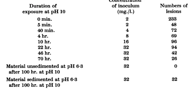
After a few hours at pH 10.5, the serological behaviour changes little, but infectivity continues to decrease during the next few days. Table 1 shows that the decrease is rapid at first and then becomes increasingly slow. After 100 hr. at Oo, the preparation used in the experiment was brought to pH 6.3 and ultracentrifuged for 1 hr. at 70,000 g ; inoculated at 32 mg./l, to Nicotiana
glutinosa the sedimented material produced lesions but the unsedimented material did not. We have similarly centrifuged many TMV preparations after they were exposed to pH 10-10-5 for periods between 1 and 100 hr. and despite large differences in the amount of material that sedimented, it always contained 0.5-0.6
yo
P and had the usual physical properties of TMV. Its infectivity was much decreased, usually to 1-5yo
that of the starting virus preparation, showing that particles with the gross morphology of TMV and similar nucleic acid content can differ greatly in their ability to infect plants.The material remaining in the supernatant fluids after ultracentrifugation at about pH 6 was usually infective when the exposure to alkali was restricted to a few hours but not when it extended to a few days. The infectivity of these
84
F. C . Bawden and
N . W . Pirie
fluids has varied from 0.1-1-0
yo
of that of TMV preparations containing the same amounts of P, and it seems not to be associated with residual TMV rods that failed to sediment, for none could be seen in electron micrographs. Also, when enough control TMV was added to such fluids to double their infectivity, the added virus was readily removed by ultracentrifugation. Further, as described later, these fluids lose their infectivity in conditions where normal TMV does not.Table 1. Effect of alkali on infectivity of T M V
Samples were taken at intervals from a 10 g./l. TMV preparation kept at pH 10 and O",
brought to pH 7 and kept at 0' until the end of the experiment when they were inoculated at the stated concentrations to the same batch of Nicotiana glutinosa. After 100 hr. at pH 10, the preparation was brought to pH 6.3, centrifuged for 1 hr. at 70,000 rev./min., and the material in the supernatant fluid and pellet used as separate inocula. The numbers of lesions are the totals on 6 half-leaves.
Concentration
Duration of of inoculum Numbers of
0 min. 2 233
5 min. 2 48
40 min. 4 72
4 hr. 8 69
10 hr. 16 96
22 hr. 32 94
46 hr. 32 42
70 hr. 32 26
lesions
exposure at pH 10 (mg*/l.)
Material unsedimented at pH 6.3 32 0
after 100 hr. at pH 10
after 100 hr. at pH 10
Material sedimented at pH 6-3 32 32
The infectivity of the supernatant fluids depends not only on the length of exposure to alkali, but also on the pH value at which the fluids are ultra- centrifuged; it is greater at pH values between 7 and 10 than at 6. When supernatant fluids from preparations already ultracentrifuged at pH 9 are brought to pH 6 and ultracentrifuged again, the greater part of the infectivity is now in sedimentable material.
At
different pH values there seems to be a different equilibrium between forces disrupting TMV rods and forces causing the parts to reassociate, and whether the rods reformed when the pH value is brought to 6 are infective or not possibly depends on whether or not a com- ponent fragment was inactivated during the disruption or while free in the alkali.Fragmented
tobacco
mosaicvirus
85this way did not inhibit infection, but the manner of their test would not show the effect. Their proteins and TMV were mixed concentrated and then the solutions were diluted before they were inoculated to test plants, and it is a characteristic of inhibitors that their action is abolished by dilution (Bawden, 1954). Table 2 shows the extent to which two preparations of such material decreased the numbers of lesions produced by purified TMV. When mixed with preparations of infective nucleic acid made by disrupting TMV with phenol, these proteins also decrease the numbers of lesions produced.
Table 2. T M V protein as an inhibitor of infection
Both TMV protein preparations were the material that remained in the supernatant fluids when TMV preparations were ultracentrifuged at pH 6.3 after spending some days at pH 10 and 0". The protein and TMV were mixed immediately before inoculation, and the numbers of lesions are the totals produced on 8 half-leaves.
Numbers of lesions A
I \
Inoculum Protein 1 Protein 2
Protein 5 g./l. 0
Protein 1 g./l., TMV 1 mg./l. Protein 0.2 g./l., TMV 1 mg./l.
Protein 5 g./l., TMV 1 mg./l. 18 79 144)
TMV 1 mg./l. 161
0
46,
117 155 185
The fact that alkali-produced fragments of TMV inhibit infection when present in inocula at concentrations greater than about 0.5 g./l. makes infec- tivity tests with such inocula of doubtful significance. So, too, it greatly com- plicates the interpretation of tests on the
'
reconstitution' of infective virus from apparently non-infective components, for these components might con- tain material with its intrinsic infectivity obscured. From the supernatant fluids of ultracentrifuged, alkali-treated preparations of TMV, which gave no lesions at 5 g./l., we have often separated infective material by again ultra- centrifuging after the preparations were precipitated with ammonium sul- phate and redissolved. The small amount of sedimented material had, weight for weight, about l / l O O the infectivity of control TMV. Our treatment may have produced infective particles by combining previously non-infective components, but the conclusion does not follow of necessity, for the sedimented fraction itself manifested no infectivity when, instead of in water, it was taken up in the non-infective fluid from which it was derived. It is at least as likely that the starting fluid contained infective material and that the treatment merely made possible its removal from the inhibiting action of other materials.Fission by phenol
The solutions produced by disrupting TMV preparations with alkali con- tain both protein and nucleic acid. Exposure to phenol denatures protein, and Bawden & Pirie (1940b) found that 20 hr. in 40 g./l. phenol at 23' de-
86
F .
C.
Bawden
and
N .
W .
Pirie
The concentration of TMV is not critical, but the ratio of water to phenol and the salt concentration both are. Essentially the method involves the partition of denatured protein and nucleic acid between the phenol layer and the water layer; the nucleic acid remains predominantly in the water layer only when the salt concentration is below 0 . 0 4 ~ .
Equal volumes of 900 g./l. aqueous phenol and 5-25 g./l. TMV in 0 . 0 1 7 ~ - sodium citrate were mixed at Oo, shaken briskly by hand for a few seconds and
centrifuged in a tube surrounded by ice for 15 min. at 3000 rev./min. The supernatant fluid was siphoned off, again shaken with a quarter of its volume of the aqueous phenol, and centrifuged in the same way. The supernatant fluid was now shaken twice for a few minutes with 10 vol. of sodium-dried ether to remove most of the phenol and then, still at Oo, the ether was removed
by exhaustion with a filter pump while the fluid was rocked gently. The solution contains about half the nucleic acid of the starting TMV and more can be separated by shaking the combined phenol layers with their own volume of water and repeating the centrifugation and extraction with ether.
The infectivity of freshly made preparations is, as Gierer & Schramm (1956) observed, about that of TMV solutions containing l / l O O O the concentration of P, but relative infectivities of the two types of inocula are difficult to express in such terms for they depend on the plants inoculated and on the concentration of the inocula. No rods can be seen in electron micrographs, and this failure is more significant than with alkali-treated TMV, which contains much con- fusing protein. Rods are readily apparent when TMV is added in amounts needed to give less than the observed infectivity. Also, no pellet forms when the fluids are ultracentrifuged, though one does when TMV is added in amounts to give the observed infectivity. Furthermore, the fluids have con- sistently failed to give a precipitate with TMV antiserum, even when their infectivity was 10 or more times that of TMV preparations diluted to their precipitation end-point. At 80,000 g the infectivity concentrates towards the bottoms of the centrifuge tubes, and electron micrographs show narrow filaments, often intertwined to give a mesh-like appearance, with a size and shape compatible with the slow sedimentation.
After dialysis such preparations consist predominantly of nucleic acid, which is presumably what is resolved by the electron microscope; the pre- parations give the characteristic nucleic acid absorption curve and negative results in colour tests for protein. Electrophoretic examination shows a single small peak, with a mobility in ~ / 1 5 p'H 7 phosphate buffer of 17.5 x cm2
sec-1 v-1, a value consistent with the component being nucleic acid. Gierer &
virus
87Contamination of the nucleic acid preparations by residual TMV is also excluded by the lability of their infectivity; at 18' they often lose their
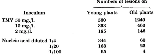
infectivity within a day, whereas TMV is stable indefinitely, The infectivity also differs qualitatively from that of normal TMV both in its behaviour to- wards test plants and in its response to dilution. Table 3 compares the two
types of preparation at three concentrations and on two lots of plants, one young, with the frag.de, light-green leaves typical of
'
shade-grown' plants, and the other older, with tougher and dark-green leaves. The relative infectivity of the two preparations obviously depends on the state of the leaves inoculated and on the concentrations at which they are compared. The decrease in in- fectivity on dilution is greater with the old than with the young plants, but with both lots of plants the decrease is steeper with the nucleic acid prepara- tion. Indeed, the experiment recorded in Table 3 gave a less rapid decreasethan usual. To quote a more typical result with highly susceptible plants, when TMV at 32, 8, 2, 0.5, and 0.12 mg./l. gave 1120, 439, 135, 62 and 25 lesions,
a nucleic acid preparation diluted 1/20, 1/80, 1/320, 1/1280 and 1/5120 gave 469, 100, 18, 4 and 0 respectively. With inocula of the two types that give similar average numbers of lesions per half-leaf when inoculated to plants with 8-10 well-developed leaves, the numbers produced on individual leaves
by the nucleic acid varies much more than with a normal TMV preparation. Also, on such plants TMV usually produces more lesions on leaves on the lower half of the stem, whereas the nucleic acid preparations produce more on the younger leaves, particularly when the lower ones are beginning to
'
harden'.
Table 3. Eflect of age of plant and dilution on relative infectivity of TIM V and nucleic acid preparation
Inoculum TMV 50 mg./l.
10 mg./l. 2 mg./l.
Nucleic acid diluted 1/4
1 /20 1pOo
Numbers of lesions on
-
Young plants Old plants
560 1240
353 460
185 146
3'44 163
65
60 23 4
The local lesions produced in Nicotiana glutinosa by the nucleic acid pre- parations are of the same type as those produced by the virus preparation from which they are derived and they become visible and countable at about the same time. A strain of TMV, which becomes systemic in leguminous plants and when obtained from them produces a characteristic type of lesion in N . glutinosa (Bawden, 1956), was used in a few tests. When a purified
F.
C.
Bawden andN .
W.
Pirie
change in the type of systemic symptoms caused. Some plants inoculated as young seedlings in the winter were much stunted and had their leaves acutely deformed as reported by Commoner et al. (1956) with some of their ‘recon- stituted’ viruses, but so too did comparable plants inoculated at the same time with the TMV from which the nucleic acid was derived.
Fission by urea
The infectivity of the best preparations of the water-soluble products from phenol-treated TMV is much too great to be attributed to contaminations with unaffected virus particles, but it is still too small to be identified certainly with the nucleic acid rather than with some other minor component of the preparations. In an attempt to get more active preparations, which might settle this problem, we tried various other treatments for disrupting the virus. None succeeded in its prime object, but tests with urea demonstrated an interesting fact.
TMV is disrupted and loses infectivity when exposed to neutral or slightly alkaline 5-8M solutions of urea (Bawden & Pirie, 1 9 4 0 ~ ) . The protein, which stays dissolved so long as the urea is concentrated, precipitates on dilution in the presence of some salt, while the nucleic acid remains in solution. When fission is incomplete, either because the exposure is too brief or the TMV too concentrated, some virus remains in solution with the nucleic acid. This can be separated by ultracentrifugation and has the same infectivity, weight for weight, as the starting preparation; hence urea seems not to produce sedi- mentable, non-infective nucleoprotein such as occurs after exposure to alkali. The unsedimented nucleic acid produced very few lesions, and with the idea that infectivity might have been destroyed by oxidation, many variations of the treatment were tried; urea was used in the presence of cysteine, azide, ascorbic acid or thioglycollate, and guanidine or thiourea were used instead of urea, but none gave preparations which, after ultracentrifugation, possessed significant amounts of infectivity.
An obvious explanation for this would be that structures essential for nucleic acid preparations to be infective are destroyed by urea, but this interpretation does not fit well with the fact that preparations made with phenol are not inactivated when later exposed to 8~-urea. Hence it seems that, either the process of separating the nucleic acid by urea damages it in some way avoided by phenol, or infectivity is carried by somethingthat remains in the protein coagulum produced by urea. Disruption of TMV with strontium nitrate (Pirie, 1954) also yielded nucleic acid preparations with too little infectivity to be significant.
EJects of ribonuclease
Fragmented
tobacco mosaic virus
89time of exposure to it. Table 4 shows that 2 hr. at 18" with 0.002 unit enzyme/ ml. removes almost all the infectivity. This exposure does not affect the pre- cipitability of the nucleic acid by acid. After 2 hr. with 0.02 unit enzyme/ml., no soluble P is detectable in preparations treated with uranyl :trichloro- acetic reagent (Holden & Pirie, 1955a) and, by this criterion, with 0.2 unit
enzyme/ml. only 10
yo
is hydrolysed in 2 hr. If, then, as seems likely, ribo-nuclease action is responsible for the loss of infectivity, the loss accompanies the first stages of the action rather than total hydrolysis of the nucleic acid.
Table 4. Inactivation by ribonucleases of nucleic acid prepared by phenol treatment of T M V
The infectivity is expressed as the total numbers of lesions formed on 8 half-leaves. LRase = leaf ribonuclease and PanRase = pancreatic ribonuclease.
Treatment of phenol-made nucleic acid before use as inoculuni Kept at -20"
2 hr. at 18" alone
Mixed with 2 units/l. LRase just before testing 7 min at 18" with 0.2 unit/l. LRase
2 hr. at 18" with 0.2 unit/l. LRase 2 hr. at 18" with 2 units/l. LRase 2 hr. at 18" with 3 mg./l. PanRase
Infectivity 382 310
2440 240 29 7
0
At concentrations above about 5 mg./l. pancreatic ribonuclease strongly inhibits infection of Nicotiana glutinosa by TMV, but exposure to the enzyme has no permanent effect on the virus, which can be recovered unimpaired from non-infective mixtures (Loring, 1942 ; Kleczkowski, 1946). Preparations of the leaf ribonuclease also inhibit infections by TMV, though much less strongly per unit of enzyme activity than the pancreatic enzyme. Inhibition is not apparent until there are about 25 units enzyme/ml., which is 1000 times as
much as was used to destroy the infectivity of the nucleic acid preparation used for the experiment reported in Table 4. When infective nucleic acid prepara-
tions are mixed with leaf enzyme at 5 units/ml., or with pancreatic ribonuclease at 1 mg./l. and immediately inoculated to N . glutinosa, no lesions are produced. With such a susceptible substrate it is operationally impossible to decide whether this is because the enzyme preparations inhibit infection or because they immediately destroy infectivity.
90
F. C .
Bawden
andN . W .
Pirie
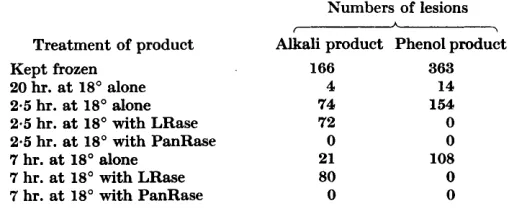
Inactivation that otherwise would have occurred seems to be prevented. It is, though, easy to get the appearance of synthesis unless unchanged starting material is included in tests comparing relative infectivities of fluid at the end of an experiment. We have done this by keeping samples of the starting material frozen at -20°, a condition in which both the alkali- and phenol- made products retain infectivity apparently unimpaired for long periods.
Table 5. The efects of leaf and pancreatic ribonuclease on infective products made from TMV uith alkali or phenol
Aliquots (0.2 ml.) of the two types of infective product were mixed with 0.1 ml. of either
0-02~-sodium citrate (pH 6) or citrate containing either 0.1 units of leaf ribonuclease (LRase) or 0.1 pg. of pancreatic ribonuclease (PanRase), and the mixtures were diluted with 0-7 ml. of water before inoculation to Nicotiana glutinosa.
Treatment of product Kept frozen
20 hr. at 18' alone
2.5 hr. at 18' alone 2.5 hr. at 18' with LRase 2.5 hr. at 18' with PanRase 7 hr. at 18" alone
7 hr. at 18' with LRase 7 hr. at 18" with PanRase
Numbers of lesions A
f -l
Alkali product Phenol product
166 363
4 14
74 154
72 0
0 0
21 108
80 0
0 0
Comparable controls seem not to have been kept in other experiments that have reported the synthesis of infective particles, and some of the reports may therefore refer to stabilization of infectivity rather than to synthesis. The stabilization of infectivity in alkali-made products by preparations of the leaf enzyme may have nothing to do with ribonuclease activity, for these preparations probably contain much more extraneous material than do preparations of the pancreatic enzyme. Experiments with other proteins, and with preparations of leaf ribonuclease after exposure to various types of inactivation, will be needed before any conclusions can be drawn on the part played in the stabilization by the enzyme itself.
Fragmented
tobacco
mosaicvirus
91The ribonuclease activity of our preparations made by disrupting TMV preparations with phenol is so low as to be equivocal when tested for by our methods, and it is uncertain to what extent the ‘depolymerization’ reported by Gierer & Schramm (1956) to accompany the loss of infectivity is a con-
sequence of their products containing some ribonuclease. That such contamina- tions may in part determine the stability of the nucleic acid preparations was suggested by experiments in which we added leaf ribonuclease to one sample of TMV and yeast nucleic acid to another before disrupting with phenol. When the resulting products were exposed to 18”, the one made from TMV
plus ribonuclease lost infectivity more rapidly, and the one from TMV plus yeast nucleic acid, less rapidly than the one from TMV alone.
As TMV is not permanently affected by exposure to ribonuclease in vitro
and the disrupted preparations are inactivated so readily, it is tempting to correlate this difference with the small infectivity of the fragments. All lcaves so far examined contain ribonuclease, and a susceptible substrate might well have difficulty in moving unharmed from where it encounters the leaf to the site where it can become established and effective. We have com- pared the ribonyclease activity of leaves from plants of different ages, and from different positions on the stem, but have not found differences of the same order as those between their relative susceptibility to infection by TMV and phenol-made derivatives. Also, we have added yeast nucleic acid and zinc sulphate to inocula, with the idea that these might protect TMV fragments from attack by ribonuclease, but the additions did not increase the numbers of lesions produced. These negative results are inconclusive, and it is not easy to see how positive evidence can be got for the idea that the poor infectivity of freshly fragmented virus is not intrinsic to it but a consequence of its sensitivity to a host-cell enzyme.
DISCUSSION
Work in several laboratories has shown that fragmented preparations of TMV can be infective, and that their infectivity is too ephemeral to be conferred by contaminations with unchanged virus particles. It is also well established that these fragments can re-associate and produce particles with the form and constitution of the original TMV ; treatments that re-associate fragments stabilize the infectivity and often allow infective material to be separated from preparations which previously seemed to lack this character, These facts raise four obvious questions : have the infective fragments any biological
significance? can the association of non-infective fragments confer infectivity on a reconstituted particle? what is the chemical nature of the infective frag- ments? and what change leads to their loss of infectivity?
The small protein and nucleoprotein fragments produced by disrupting TMV with alkali have obvious similarities with the anomalous particles that fail to sediment or sediment only slowly when infective sap is ultra- centrifuged. Although these anomalous particles are serologically distinguish- able from most of the proteins produced from TMV by alkali (Kleczkowski,
92
F.
C. Bawden
and
N . W. Pirie
and both can be reassembled into the characteristic rods. Alkali, then, might be reversing the process whereby virus particles are synthesized from intermediaries, or duplicating some process in cells whereby particles are naturally disrupted. Evidence from experiments with leaves supplied with
14C02 (van Rysselberge & Jeener, 1955) suggests that the small anomalous particles in sap are more likely to be intermediaries in synthesis than break- down products. The infectivity arid nucleic acid content reported for aggre- gated preparations of these particles has varied (Bawden & Pirie, 1 9 5 6 ~ ) perhaps because they were aggregated by different methods and after the sap had been exposed to different conditions, in which either TMV or other nucleic acid might have been degraded to different extents. The yield of infective, re-aggregated particles from alkali-treated TMV clearly depends on the con- ditions to which the preparations are exposed before they are aggregated.
The effects of alkali may also provide an analogy for what happens when leaves become infected. The amount of virus recoverable from inoculated leaves at first decreases and, as experiments with TMV labelled with 32P show,
P that was in the virus soon appears in other forms. Whether this derives from virus which is causing infections, or from excess inoculum, is impossible to say, but that the virus particles may pass through a temporary phase of suscepti-
bility to pancreatic ribonuclease is suggested by the ability of this enzyme to inhibit infections when leaves are rubbed with solutions of it within 2 hr. of
inoculation with the virus, but not afterwards. Similarly, photoreactivation experiments with potato virus X (Bawden & Kleczkowski, 1955) suggest that these particles undergo changes within 30 min. of being inoculated to leaves and that they are unstable in their changed state. All this combines to suggest that systems in host cells may act rather like alkali and disrupt virus particles to produce an infective component that is more labile than the particles characteristic of in vitro preparations.
The fact that infective material can often be separated from non-infective fragmented preparations of TMV is far from necessarily meaning that infective particles have been created by combining intrinsically non-infective com- ponents. It may mean no more than that an inhibitor of infection has been removed, or that infective fragments, which otherwise would have become non-infective before testing, have been stabilized in vitro. These possibilities are open to experimentation, and all our results that at first sight suggested the synthesis of infective particles became interpretable in other ways.
Fragmented tobacco mosaic virus
stabilized against the hazards encountered during or after inoculation, will still remain in doubt. We got no infections with heated nucleic acid and have failed to increase the infectivity of alkali-treated products of TMV by aggre- gating them with heated nucleic acid, but we do not have the absorbent materials with which Commoner et al. (1956) removed nucleic acid from their alkali-treated preparations and so cannot use their experimental methods.
Study of the fission products of TMV suggests that immunological specificity is primarily an attribute of the protein and infectivity of the nucleic acid. There is no a priori reason why nucleic acid alone should not carry the in- fectivity, and the inactivation by ribonucleases points to it being an essential constituent of the infective unit. Indeed, the simplest interpretation of current results is that nucleic acid is the whole of this unit, and there is no compelling reason to look for any other component. There would be some reason if it should be established unequivocally that combination of non-infective nucleic acid with another substance gave an infective product, but even then it could happen because the product contained the right amount of nucleic acid arranged in the correct way. Before we agree with the conclusion that nucleic acid alone is the vehicle of infectivity, two points need putting. First, the infectivity of nucleic acid preparations is so slight that it could be accounted
for by a 0.1
yo
contamination with some other unit possessing the infectivity of intact TMV, and to establish nucleic acid positively as the bearer would needdemonstrating that the nucleic acid was 9909% pure, which is patently im- possible. Even if the range of possible contaminants is narrowed by postulating a single substance, such as a protein, it would remain difficult to demonstrate convincingly that this was not present to the extent of 0.1
%.
Secondly, our results with urea-treated TMV show that the infectivity of nucleic acid prepara- tions depends on how they are made, and that substances harmless to phenol- made products yield non-infective products, There are many possible explana- tions for this, but one worth some consideration is that phenol-made products contain something other than nucleic acid and that this is lost when the virus is disrupted by urea.We thank Dr A. Kleczkowski for making the electrophoretic measurements and Mr H. L. NIXON for making the electron micrographs.
REFERENCES
BAWDEN, F. C. (1954). Inhibitors and plant viruses. Advanc. Virus Res. 2, 31. BAWDEN, F. C. (1956). Reversible, host-induced changes in a strain of tobacco
mosaic virus. Nature, Lmd. 177, 302.
BAWDEN, F. C. & KLECZKOWSKI, A. (1955). Studies on the ability of light to counter- act the inactivating action of ultraviolet radiation on plant viruses. J . gen.. Microbiol. 13,370.
BAWDEN, F. C. & PIRIE, N. W. (1937). The isolation and some properties of liquid crystalline substances from solanaceous plants infected with three strains of tobacco mosaic virus. Proc. roy. Soc. B, 123, 274.
F .
C .
Bawden
and
N . W .
Pirie
BAWDEN, F. C. & PIRIE, N. W. (19Mb). The inactivation of some plant viruses by urea. Biochem. J. 34, 1258.
BAWDEN, F. C. & PIRIE, N. W. (1945). The separation and properties of tobacco mosaic virus in different states of aggregation. Brit. J . exp. Path. 26, 294. BAWDEN, F. C. & PIRIE, N. W. (1956a). Observations on the anomalous proteins
occurring in extracts from plants infected with strains of tobacco mosaic virus.
J. gen. Microbial. 14, 460.
BAWDEN, F. C. & PIRIE, N. W. (19566). In The Nature of Viruses, p. 37, p. 82 et passim. Ciba Foundation Symp. Ed. G. E. W. Wolstenholme & E. C. P. Millar. London: J, and A. Churchill Ltd.
BAWDEN, F. C., PIRIE, N. W., BERNAL, J. D., & FANKUCHEN, I. (1936). Liquid crystalline substances from virus-infected plants. Nature, Lond. 138, 1051. , BEST, R. J. (1936). The relationship between the activity of tobacco mosaic virus
suspensions and hydrion concentration over the pH range 5 to 10. Austr. J.
exp. Biol. med. Sci. 14, 323.
COMMONER, B., LIPPINCOTT, J. A., SHEARER, G. B., RICHMAN, E. E. & Wu, J. H. ( 1956). Reconstitution of tobacco mosaic virus components. Nature, Lond.
178, 767.
COMMONER, B., NEWMARK, P. & RODENBERG, S. D. (1952). An electrophoretic analysis of tobacco mosaic virus biosynthesis. Arch. Biochem. Biophys.
37, 15.
CROOK, E. M. & SHEFFIELD, F. M. L. (1946). Electron-microscopy of viruses: 1. State of aggregation of tobacco mosaic virus. Brit. J. exp. Path. 27, 328. FRAENKEL-CONRAT, H. (1956). The role of the nucleic acid in the reconstitution of
active tobacco mosaic virus. J. Arner. chem. SOC. 78, 883.
FRAENKEL-CONRAT, H. & Williams, R. C. (1955). Reconstitution of active tobacco mosaic virus from its inactive protein and nucleic acid components. Proc. n.at.
Acad. Sci., Wash. 41, 690.
GIERER, A. & SCHRAMM, G. (1956). Infectivity of ribonucleic acid from tobacco mosaic virus. Nature, Lond. 177, 702.
HART, R. G. & SMITH, J. D. (1956). Interactions of ribonucleotide polymers with tobacco mosaic virus protein to form virus-like particles. Nature, Lond. 178, 739.
HOLDEN, M. & PIRIE, N. W. (1955~). The partial purification of leaf ribonuclease.
Biochem. J. 60, 39.
HOLDEN, M. & PIRIE, N. W. (19553). A comparison of leaf and pancreatic ribo- nuclease. Biochem. J. 60, 53.
JEENER, R. & LEMOINE, P. (1953). Occurrence in plants infected with tobacco mosaic virus of a crystallizable antigen devoid of ribonucleic acid. Nature, London. 171, 935.
KLECZKOWSKI, A. (1946). Combination between different proteins and between proteins and yeast nucleic acid. Biochem. J. 40, 677.
KLECZKOWSKI, A. (1957). A preliminary study of tobacco mosaic virus by the gel diffusion precipitin tests. J . gen. Microbial. 16, 405.
LIPPINCOTT, J. A. & COMMONER, B. (1956). Reactivation of tobacco mosaic virus infectivity in mixtures of virus protein and nucleic acid. Biochim. biophys. Acta,
19, 198.
LORING, H. S. (1942). The reversible inactivation of tobacco mosaic virus by crystal- line ribonuclease. J. gen. PhysioZ. 25, 497.
PIRIE, N. W. (1954). The fission of tobacco mosaic virus and some other nucleo- proteins by strontium nitrate. Biochem. J. 56, 83.
PIRIE, N. W. (1956). Some components of tobacco mosaic virus preparations made in different ways. Biochem. J. 63, 316.
PIRIE, N. W. (1957). Macromolecular nucleoproteins from healthy tobacco leaves.
Fragmented tobacco mosaic virus
RYSSELBERGE, C. VAN & JEENER, R. (19.55). The role of the soluble antigens in the
multiplication of the tobacco mosaic virus. Biochim. biophys. Ada, 17, 158.
SCHRAMM, G. (1943). a e r die Spaltung des Tabakmosaikvirus in niedennolekulare
Proteine und die Ruckbildung hochmolekularen Proteins aus den Spaltstucken. Natumissenschaften, 31, 94.
SCHRAMM, G., SCHUMACHER, G. & ZILLIG, W. (1955). An infectious nucleoprotein
from tobacco mosaic virus. Nature, Lond. 175, 549.
SIEGEL, A., WILDMAN, S. G. & GINOZA, W. (1956). Sensitivity to ultraviolet light of
infectious tobacco mosaic virus nucleic acid. Nature, Lond. 178, 1117.
SREENIVASAYA, M. & PIRIE, N. W. (1938). The disintegration of tobacco mosaic virus
preparations with sodium dodecyl sulphate. Biochem. J. 32, 1707.
TAKAHASHI, W. N. & ISHII, M. (1952). The formation of rod-shaped particles re-
sembling tobacco mosaic virus by polymerization of a protein from mosaic diseased tobacco leaves. Phytopathology, 42, 690.
TAKAHASHI, W. N. & ISHII, M. (1953). A macromolecular protein associated with
tobacco mosaic virus infection : its isolation and properties. A m . J . But. 40, 85.


