M E T H O D O L O G Y A R T I C L E
Open Access
Isolation of primary microglia from the
human post-mortem brain: effects of
ante- and post-mortem variables
Mark R. Mizee
1,2*, Suzanne S. M. Miedema
2†, Marlijn van der Poel
2†, Adelia
1, Karianne G. Schuurman
2,
Miriam E. van Strien
3, Jeroen Melief
2, Joost Smolders
2, Debbie A. Hendrickx
2, Kirstin M. Heutinck
4,
Jörg Hamann
1,4and Inge Huitinga
1,2Abstract
Microglia are key players in the central nervous system in health and disease. Much pioneering research on microglia function has been carried out in vivo with the use of genetic animal models. However, to fully understand the role of microglia in neurological and psychiatric disorders, it is crucial to study primary human microglia from brain donors. We have developed a rapid procedure for the isolation of pure human microglia from autopsy tissue using density gradient centrifugation followed by CD11b-specific cell selection. The protocol can be completed in 4 h, with an average yield of 450,000 and 145,000 viable cells per gram of white and grey matter tissue respectively. This method allows for the immediate phenotyping of microglia in relation to brain donor clinical variables, and shows the microglia population to be distinguishable from autologous choroid plexus macrophages. This protocol has been applied to samples from over 100 brain donors from the Netherlands Brain Bank, providing a robust dataset to analyze the effects of age, post-mortem delay, brain acidity, and neurological diagnosis on microglia yield and phenotype. Our data show that cerebrospinal fluid pH is positively correlated to microglial cell yield, but donor age and post-mortem delay do not negatively affect viable microglia yield. Analysis of CD45 and CD11b expression showed that changes in microglia phenotype can be attributed to a neurological diagnosis, and are not influenced by variation in ante- and post-mortem parameters. Cryogenic storage of primary microglia was shown to be possible, albeit with variable levels of recovery and effects on phenotype and RNA quality. Microglial gene expression substantially changed due to culture, including the loss of the microglia-specific markers, showing the importance of immediate microglia phenotyping. We conclude that primary microglia can be isolated effectively and rapidly from human post-mortem brain tissue, allowing for the study of the microglial population in light of the neuropathological status of the donor.
Keywords:Post-mortem human brain, Primary human microglia, Rapid cell isolation protocol, Primary microglial cell culture, Biobanking
Introduction
Microglia are brain-resident phagocytic cells, which origin-ate from a population of myeloid progenitors from the yolk sac during embryonic development [16, 23, 35] and are maintained through self-renewal without influx of periph-eral cells during adult life [1, 4]. Microglia are key players in
central nervous system (CNS) homeostasis, fulfilling essen-tial roles in neurodevelopment, adult synaptic plasticity, and brain immunity [32, 34]. In the adult brain, microglia act as surveyors of the local environment to sustain homeo-stasis and are therefore highly sensitive to changes associ-ated with damage, inflammation, or infection within and outside the CNS. In order to interact with their environ-ment, microglia exhibit a broad range of sensory mecha-nisms and specific cellular responses, the outcome of which can be both neuroprotective as well as a neurotoxic [22].
During the process of normal aging, the microglial phenotype appears to shift to a primed or more active-* Correspondence:[email protected]
†Equal contributors
1
Netherlands Brain Bank, Netherlands Institute for Neuroscience, Amsterdam, The Netherlands
2Department of Neuroimmunology, Netherlands Institute for Neuroscience,
Amsterdam, The Netherlands
Full list of author information is available at the end of the article
prone state [22, 30], the main reasoning behind micro-glia being linked to pathology in neurodegenerative disorders such as Alzheimer’s disease (AD) [21], Parkin-son’s disease (PD) [33], and multiple sclerosis (MS) [24]. Their role as possible contributors to disease has been complemented by evidence for their involvement in the pathophysiology of developmental and psychiatric disor-ders, such as major depression disorder, bipolar disorder, schizophrenia, and autism [3, 7], either through modula-tion of neuroinflammamodula-tion or neuronal plasticity. How-ever, their role in disease pathology appears ambiguous since microglia also display beneficial and restorative functions [36].
Research on microglia function and their role in health and disease has mostly been carried out ex vivo using im-munohistochemistry and in vivo using murine models. The isolation of microglia from the brains of various genetic mouse models has greatly facilitated our understanding of basic microglia characteristics in health and disease [9]. Nevertheless, these models are of limited value in relation to human CNS disorders. Studies into human microglia function have highlighted similarities but also crucial differ-ences between mice and humans [38]. Added difficulty comes in the form of various CNS disorders for which ani-mal models are not available or fail to reconstitute import-ant human symptoms. Therefore, to investigate the role of microglia in human context it is crucial to study human primary microglia.
In order to specifically study multiple aspects of hu-man microglia, obtaining pure microglia populations from post-mortem human brain samples is essential. To this aim, we have adapted the human microglia isolation method of Dick et al. [12], in turn based on a rat isola-tion protocol [37], for the use of post-mortem human brain tissue. This led to a procedure for the rapid isola-tion of pure human microglia based on cell density sep-aration and capture of CD11b-positive cells using magnetic beads [25]. A major advantage of this isolation procedure in comparison with generally used microglia isolation methods [11] is the omission of effects due to culture and adherence in the procedure, as it allows for direct analysis of isolated microglia. Using this tech-nique, we determined that based on membrane expres-sion of CD45 and CD11b, microglia can be distinguished from autologous peripheral macrophages based on fluor-escence intensity [25]. Furthermore, we demonstrated that microglia show a minimal response to lipopolysac-charide (LPS), indicating a tight regulation of inflamma-tory responses. Finally, we revealed differences in microglial size, granularity, and CD45/CD11b expression in white matter microglia from MS donors, when com-pared to non-MS donors [26], showing that microglial phenotype reflects neuropathological changes. Yet, to ef-fectively study primary human microglia on a larger
scale, there is an urgent need for thorough validation of available protocols and an understanding of the effects of clinical diagnosis and ante- and post-mortem vari-ables on isolated microglia.
Since the development of our procedure for the isola-tion of human microglia in 2012 [25], we performed microglia isolations from over a hundred brain donors from the Netherlands Brain Bank. In addition to our previously published method, we have also developed a faster protocol that reduces the total isolation time, while maintaining similar or higher viable cell yield. Here we set out to validate the practical aspects of hu-man post-mortem microglia isolations and describe the effects of clinical diagnosis and ante- and post-mortem variables on microglial purity and phenotype, such as post-mortem delay (PMD) and cerebrospinal fluid (CSF) pH, and discuss further application possibilities of iso-lated human microglia.
Materials and methods Brain tissue
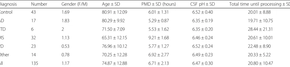
Human brain tissue was obtained through the Netherlands Brain Bank (www.brainbank.nl). The Netherlands Brain Bank received permission to perform autopsies and to use tissue and medical records from the Ethical Committee of the VU University medical center (VUmc, Amsterdam, The Netherlands). On average, the autopsies are performed within 6 h after death. All donors have given informed con-sent for autopsy and use of their brain tissue for research purposes. The pH of the CSF was measured using a fluid-based pH meter (Hanna Instruments, Nieuwegein, The Netherlands), after rapid sampling of the CSF directly from the lateral ventricles at the start of the autopsy. An over-view of the clinical information and post-mortem variables of all brain donors in this study is summarized in Table 1.
Human post-mortem microglia isolation
Lakewood, USA) for 60’(previous method) or with trypsin (Invitrogen) at a final concentration of 0.125% for 45’ (current method) in Hibernate A medium at 37 °C on a shaking platform. Both digestions were incubated in the presence of 33 μg/ml DNAseI (Roche, Basel, Switzerland). The digestion was resuspended 10x with a 10-ml halfway the digestion time. Heat inactivated fetal calf serum (FCS, Invitrogen) was added to quench trypsin activity and the cell suspension was centrifuged for 10 min at 1800 rpm and 4 °C. After discarding the supernatant, the cell pellet was re-suspended in cold DMEM (Invitrogen), supplemented with 10% FCS, 1% Penicillin-Streptomycin (Pen-Strep, Invitro-gen), and 1% gentamycin (InvitroInvitro-gen), and passed through a 100-μm tissue sieve. After the direct addition of 1/3 volume of cold Percoll (GE Healthcare, Little Chalfont, UK) and centrifugation for 30’at 4000 rpm and 4 °C the interphase containing microglia was transferred to a new tube (discard-ing the myelin and erythrocyte layers) and washed two times in DMEM supplemented with 10% FCS, 1% Pen/ Strep, 1% gentamycin, and 25 mM Hepes (Invitrogen). Negative selection of granulocytes (previous method only) and positive selection of microglia with respectively anti-CD15 and anti-CD11b conjugated magnetic microbeads (Miltenyi Biotec, Cologne, Germany) was done by magnetic activated cell sorting (MACS) according to the manufac-turer’s protocol. Briefly, cells were incubated with 10 μl CD15 microbeads for 15 min at 4 °C, washed, resuspended in beads buffer (0.5% BSA, 2 mM EDTA in PBS pH 7.2) and transferred to an MS column placed in a magnetic holder. The flow-through containing unlabeled cells was collected, washed and subsequently incubated with 20 μl CD11b microbeads for 15 min at 4 °C. Cells were then washed and placed on a new MS column in a magnetic holder. The CD11b+cell fraction was eluted from the col-umn by removing the colcol-umn from the magnet, adding beads buffer, and emptying the column with a plunger. Vi-able cells were then counted using a counting chamber and used as described in downstream analyses. The isolation of macrophages was performed using choroid plexus tissue dissected from the lateral ventricle, using the same method as for WM microglia.
Flow-cytometric analysis
The CD11b + cell fraction was evaluated for proper separ-ation of microglia from other cell types by flow cytometry for CD45 (FITC-labeled, Agilent, Santa Clara, USA), CD11b (PE-labeled, eBioscience, San Diego, USA), and CD15 (APC-labeled, Biolegend, San Diego, USA). For CD45 and CD11b, appropriate isotype controls were regu-larly included to assess background levels of fluorescence. Cells were incubated with antibodies in beads buffer, on ice, for 30’. Viability of the cells was analyzed using the fix-able viability dye Efluor 780 or 7-AAD (eBioscience). For spiking the microglia populations, macrophages where la-beled with far red celltracker (Invitrogen) in PBS (1:1000) for 5 min and washed twice with PBS. Fluorescence was measured on either a FACSCalibur or a FACSCanto II machine (both BD biosciences, Franklin Lakes, USA) and analyzed with FlowJo software (Treestar, Ashland, USA). For CD45 and CD11b geometric mean comparisons with post-mortem parameters, only data from the FACS-Calibur was included.
Cell culture
Microglia were cultured in DMEM/F-12 medium (Invitro-gen), supplemented with 10% FCS and 1% Pen-Strep and cultured in plates coated with poly-L-lysine (Invitrogen). Myelin phagocytosis was assessed as described previously [20]. In short, microglia were incubated for 48 h with pHrodo-labeled myelin (10 μg/ml) from a myelin pool containing myelin from 12 donors without neurological abnormalities. All cultures described in the data are de-rived from white matter samples, as cortical microglia did not result in reproducible cultures. To assess the effect of cryogenic storage and subsequent thawing of primary microglia, cells were resuspended in ice-cold mixture of medium and FCS (1:1), containing 10% dimethyl sulfoxide (DMSO, Sigma, St. Louis, USA), placed in a cryogenic container (Nalgene, Thermo Fischer, Waltham, USA) with 2-propanol, and stored overnight in a −80 °C freezer. Cryovials were then transferred to a liquid nitrogen tank. Cells were thawed by slowly adding cold complete RPMI medium (Invitrogen) containing 20% FCS, after 20 min at
Table 1Summary of clinical variables of brain donors used
Diagnosis Number Gender (F/M) Age ± SD PMD ± SD (hours) CSF pH ± SD Total time until processing ± SD
Control 43 1.69 80.91 ± 12.09 6.01 ± 1.31 6.52 ± 0.40 20.01 ± 8.88
AD 17 1.83 80.29 ± 9.92 5.29 ± 0.87 6.35 ± 0.19 19.71 ± 10.75
FTD 6 2 71.50 ± 7.09 5.53 ± 1.62 6.35 ± 0.20 28.44 ± 21.31
MS 32 1.13 65.31 ± 12.15 9.21 ± 1.68 6.46 ± 0.24 20.61 ± 10.01
PD 23 0.53 76.96 ± 10.12 5.77 ± 1.27 6.52 ± 0.24 22.48 ± 8.90
Other 14 0.78 70.25 ± 12.28 6.92 ± 2.77 6.49 ± 0.23 20.33 ± 5.22
All 135 1.17 74.87 ± 12.88 6.71 ± 2.13 6.47 ± 0.30 20.80 ± 10.47
room temperature, cells were washed using warm complete RPMI and either lysed for RNA isolation or ana-lyzed directly using flow cytometry.
RNA isolation and gene expression analysis
Acutely isolated primary microglia were taken up in 1 ml TRIsure (Bioline, London, UK) and stored at −80 °C for further processing. RNA isolation was carried out
according to manufacturer’s protocol using phase separ-ation by addition of chloroform and centrifugsepar-ation, followed by overnight precipitation in isopropanol at−20 ° C. RNA concentration was measured using a Nanodrop (ND −1000; NanoDrop Technologies, Rockland, DE, USA) and RNA integrity was assessed using a Bioanalyzer (2100; Agilent Technologies, Palo Alto, CA, USA). cDNA synthesis was performed using the Quantitect reverse transcription kit (Qiagen, Hilden, Germany) according to manufacturer’s instructions, with a minimal input of 200 ng total RNA. Quantitative PCR (qPCR) was per-formed using the 7300 Real Time PCR system (Applied Biosystems, Foster City, USA) using the equivalent cDNA amount of 1–2 ng total RNA used in cDNA synthesis. SYBRgreen mastermix (Applied Biosystems) and a 2 pmol/ml mix of forward and reverse primer sequences were used for 40 cycles of target gene amplification. An overview of forward and reverse sequences for each gene can be found in Additional file 1: Table S1. Expression of target genes was normalized to the average cycle threshold of GAPDH and EF1a. Cycle threshold values were assessed with SDS software (Applied Biosystems).
Statistical analysis
Data analysis was performed using Graphpad Prism soft-ware (v6 Graphpad Softsoft-ware, La Jolla, CA, USA). Results are shown as mean with standard error of the mean, and statistical analysis was performed using either parametric or non-parametric testing, based on the outcome of the Shapiro-Wilk normality test. The applied test for each calculated value is described in the figure legends.
Results
Isolation and characterization of microglia from post-mortem CNS tissue
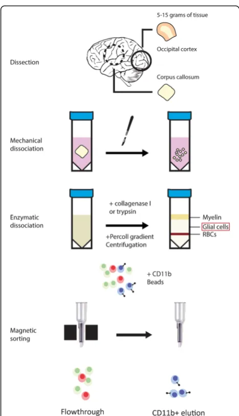
The isolation of viable microglia from post-mortem hu-man CNS tissue has been described by our group previ-ously [25]. For the data used in this study, we have used both the published protocol as well as an adapted ver-sion that is faster (~4 in place of ~5 h) in which collage-nase is replaced by trypsin, and CD15 depletion is omitted. The basic steps of the protocol and the aspects that differ between both protocols are depicted in Fig. 1. The cell capture in both methods relies on the mem-brane expression of CD11b, which is also present on perivascular and infiltrated macrophages in the CNS. To investigate the differences between macrophages and microglia from the same donor, we included choroid plexus (CP) macrophages. To differentiate between the two populations of cells, CP-derived CD11b+ cells were labeled with a fluorescent cell tracker. To ensure that the labeling method did not alter the fluorescence inten-sity of CD45 and CD11b antibodies, unlabeled and la-beled CP macrophages were compared, showing no
Fig. 1Microglia isolation method at a glance Depicted are the two similar methods through which microglia were isolated frompost
-mortembrain tissue. CNS samples were dissected from either occipital
change in CD45 and CD11b fluorescence (Fig. 2a). Fur-thermore, we observed no APC/cell tracker+ cells in the CD11b+ population isolated from WM (Fig. 2b). Repre-sentative FACS plots showing the gating strategy to in-vestigate only viable cells, including assessment of background fluorescence using isotype controls, is shown in Additional file 1: Figure S1. Spiking the WM CD11b+ cells with labeled CP CD11b+ cells enabled us to stain a combined population of WM and CP cells for CD45 and CD11b, while allowing separation of the pop-ulations based on APC+ (Fig. 2c). Comparing the size and granularity of both cell populations in one pool of cells identified CD11b+ cells from WM to have different population characteristics compared to CD11b+ cells from CP, showing the macrophages to be larger and more granular (Fig. 2d). Furthermore, CP-derived mac-rophages clearly showed a higher expression of CD45 and CD11b, when compared to WM-derived cells (Fig. 2e). Quantification of the same analyses from seven different donors with different neurological diagnoses showed that the observations regarding CD45 (avg. 190.8% higher expression levels; Fig. 2f ), and CD11b (avg. 106.4% higher expression levels; Fig. 2g) are con-sistent for all investigated donors. We conclude that microglia can be reliably isolated from post-mortem hu-man CNS tissue, without apparent macrophage contam-ination due to the fact that a large reservoir of macrophages is not present in the CNS parenchyma.
Viable microglia yield from white and grey matter correlates with CSF pH
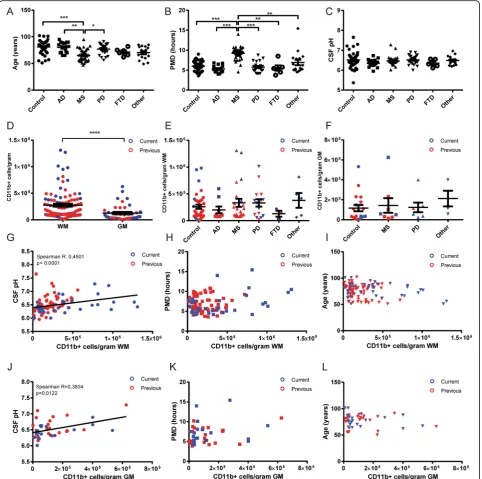
Since post-mortem microglia isolations were performed on brain samples from varying neurological disease and control donors, we first assessed the differences between the various groups of donors with respect to age, PMD, and CSF pH. Only the MS donor group showed a sig-nificant deviation from other groups in age (Fig. 3a) and PMD (Fig. 3b), whereas no significant differences were observed in CSF pH at autopsy between groups. (Fig. 3c). The difference in PMD is explained by the longer aut-opsy protocol for MS donors in which MRI-guided dis-section is needed to separate normal-appearing WM (NAWM) from lesioned areas [10], whereas the differ-ence in age is explained by mortality at a younger age in MS. We then combined data from all isolations, which clearly showed a higher yield of viable microglia per gram WM compared to GM tissue (Fig. 3d). This com-bined graph also shows the high donor-to-donor vari-ability in microglia yield, in both WM and GM isolations. Colors separating the isolations performed using the two described methods showed that the current trypsin method produced the highest yields, al-though the average yield between the two methods is not significantly different (Additional file 1: Figure S2).
Since the region-specific difference in microglia yield could be caused by an inherent difference between WM and GM microglia, we separately analyzed isolations from WM and GM to correlate with donor clinical parameters. We first analyzed the influence of a neurological diagnosis on microglia yield. Although both the AD and FTD groups showed lower WM microglia yield averages compared to the control, MS, and PD groups (Fig. 3e), the average num-ber of microglia isolated from WM and GM (Fig. 3f) was not significantly different between groups. We next ana-lyzed the effect of donor age, PMD, and CSF pH on micro-glia yield. For WM micromicro-glia isolations, we observed a significant correlation of viable microglia yield with CSF pH (Fig. 3g), but no correlation with either PMD (Fig. 3h) or age (Fig. 3i). Although the average yield from GM micro-glia isolations was much lower than those from WM, we observed a similar significant correlation of GM microglia yield with CSF pH (Fig. 3j) and similarly no correlation with either PMD (Fig. 3k) or age (Fig. 3l). Besides investigating PMD, we also included the total time until tissue processing (PMD + time until isolation; averaging 20.8 h over all isola-tions) in our analysis, which did not show any correlation to microglia yield (Additional file 1: Figure S3).
Combined, our data encompassing microglia isolations from over 100 donors clearly shows a robust effect of CSF pH, shown to reflect cortical pH at autopsy [19], on viable microglia yield from post-mortem brain tissue. We have analyzed the clinical information of all donors to determine which variables correlate with CSF pH. In our donor group, the cause of death, often reflecting the agonal state of the donor before passing, is associated with CSF pH (Additional file 1: Figure S4) and shows that the average CSF pH is significantly lower in donors that suffered from cachexia or pneumonia before death, compared to donors that underwent euthanasia.
Changes in microglia expression of CD45 and CD11b are mainly attributable to differences between grey and white matter, and neurological diagnosis
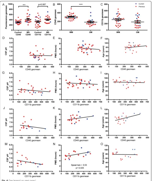
donors showed no difference in mean fluorescence (Additional file 1: Figure S5). Therefore, to exclude any ef-fects of disease-related changes in microglia activation, we
have only included isolations performed on non-demented control donor material in the following analyses. Using CD45 and CD11b immunoreactivity as a readout for
microglial activation state, we analyzed microglia isolated from either WM or GM tissue. Interestingly, we observed a significantly lower membrane expression of CD45 of micro-glia isolated from GM, when compared to WM-derived microglia (Fig. 4b), whereas CD11b expression is not
significantly different (Fig. 4c). Since we also observed a dif-ference in microglia yield from both regions, we separately investigated the effect of clinical and post-mortem parame-ters on microglia from WM and GM isolations. The mem-brane expression of CD45 and CD11b of microglia isolated
from WM tissue did not correlate significantly with either CSF pH, PMD, or age (Fig. 4d-i). The CD45 expression pat-tern for microglia isolated from GM was comparable to that of WM microglia, showing no significant correlation with any of the parameters investigated (Fig. 4j-l). In micro-glia isolated from GM, CD11b expression shows no correl-ation with CSF pH or age (Fig. 4m, o). Differently from WM microglia however, CD11b expression in GM micro-glia significantly correlates with increasing PMD (Fig. 4n). We have also included total time until tissue processing in our analysis, showing no correlation with either CD45 or CD11b expression (Additional file 1: Figure S6).
Taken together, our data show that microglial CD45 expression clearly differs between cells isolated from WM or GM. Average CD45 expression on microglia iso-lated from either WM or GM is unreiso-lated to CSF pH, PMD, age, and population viability. We show a similar absence of correlations for CD11b in both GM and WM microglia, with the only exception being that GM micro-glia showed increasing CD11b expression with increas-ing PMD. By combinincreas-ing data of both microglia isolation methods, we also observed a significant increase in both CD45 and CD11b expression of GM microglia isolated using the current method, compared to the previous protocol (Additional file 1: Figure S7). This difference was not observed for WM microglia.
In vitro applications of primary human microglia and effects of cryogenic storage
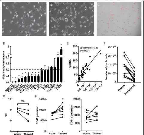
To expand the possible research applications of primary hu-man microglia, we investigated the possibility to cryogeni-cally store microglia for biobanking purposes and their potential for (long-term) in vitro culture. Using poly-L-Lysine as a culture substrate, we found that primary micro-glial cultures show a slightly ramified morphology and can be maintained for 5 days in vitro (DIV) (Fig. 5a) and 10 DIV (Fig. 5b) without apparent signs of proliferation or cell death. Accordingly, immunocytochemistry for proliferation marker Ki-67 only sporadically decorated microglia nuclei (Additional file 1: Figure S8). All microglial cultures were derived from WM samples, as microglia cultures from GM isolations showed no adherence or outgrowth past 2 days in culture. Microglia retain phagocytic function after 5 DIV, as
evidenced by the uptake of pHrodo-labeled myelin (Fig. 5c). How the cultured microglial phenotype compares to the phenotype directly after isolation however, has not been ad-dressed to date. We therefore used microglia isolated from four different WM donors, isolated RNA either directly after isolation or after 4 days of basal culture, and investi-gated the change in gene expression from acute to cultured microglia for each donor (Fig. 5d). Of all investigated genes, only the macrophage marker and lipopolysaccharide co-receptor CD14 was significantly upregulated after 4 days. Interestingly, the microglia/macrophage markers purinergic receptor P2Y12 (P2RY12), fractalkine receptor (CX3CR1), and CD11b were all significantly decreased after 4 days. Moreover, the pro-inflammatory cytokine interleukin 1 beta (IL-1b) showed an increase in expression, but did not reach significance, and immune-activated genes were downregu-lated, including pro-inflammatory tumor necrosis factor (TNF), glutamate aspartate transporter (GLAST), MHC class II subunit HLA-DRA, Fc gamma receptor IIIa (CD16a), and anti-inflammatory interleukin 10 (IL-10) and transforming growth factor beta (TGFβ). Gene expression of interleukin 1 alpha (IL-1α), chemokine C-C motif che-mokine ligand 3 (CCL3), interleukin 6 (IL-6), CD45, and the CD200 receptor (CD200R) was unchanged. Using this selected set of genes, it becomes apparent that microglia undergo phenotypical changes during culture.
Since RNA analysis directly after isolation is important to accurately relate microglial phenotype to the in situ state of the tissue, we analyzed whether RNA yield is constant be-tween donors. We found a significant correlation bebe-tween the number of viable cells used and the RNA yield obtained (Fig. 5e). Finally, we analyzed the potential to cryogenically store acutely isolated microglia, and the effect of a freeze-thaw cycle on RNA integrity and minimal phenotype. The average recovery rate of viable cells from frozen samples was 27%, although highly variable (±22.7%, Fig. 5f). We an-alyzed the RNA integrity (RIN) from RNA extracted from microglia immediately after isolation, and after cryogenic storage, from the same donors. Although RIN values were slightly decreased, we found no significant decrease of RIN values after thawing and RIN values did not drop below 6, reflecting usable mRNA in many applications (Fig. 5g). We furthermore analyzed CD45 and CD11b expression on (See figure on previous page.)
viable microglia before and after thawing. CD11b expres-sion was not significantly affected by cryogenic freezing and thawing (Fig. 5h), but CD45 expression was increased in thawed microglia compared to acutely analyzed cells, pos-sibly reflecting ongoing cell activation or the selective loss of cells with low CD45 expression. Thus, albeit a small sample size, we show that microglia can be cryogenically frozen and stored for biobanking purposes while
maintaining the possibility to phenotype using flow cytome-try or to analyze gene expression. Furthermore, microglia can be cultured for multiple days, but show profound changes in their gene expression profile due to culture.
Discussion
In a time-span of 5 years, over a hundred human primary microglia isolations have been performedon post-mortem
human brain samples. Analyzing the results of these ef-forts, we here confirm that human microglia can be read-ily isolated from post-mortem CNS tissue based on the membrane expression of CD11b, that microglia are distin-guishable from macrophages, and that the yield of viable microglia is linked to the acidification of the CNS at time of autopsy. Strikingly, neither age, PMD, nor neurological diagnosis was correlated with viable microglia yield. The microglia phenotype from control donors, as assessed by CD45 and CD11b expression, was not correlated with brain acidity, donor age, or PMD. We did observe a robust effect of clinical MS diagnosis on CD45 expression, and to a lesser extent on CD11b expression. This finding is of great importance to any study aimed at linking changes in microglial phenotype to a neurological diagnosis. Finally we show that isolated microglia are suitable for culture and cryogenic storage, but provide a cautionary note re-garding the changes in microglia gene expression profile due to culture. In summary, the most important conclu-sion drawn from this study is that after rapid isolation, changes in microglial phenotype can be readily attribut-able to neurological disease parameters, rather than reflecting uncontrollable donor parameters like age, PMD, idle tissue time, or CNS acidity. This finding is of critical importance to published and future studies implementing the characterization of purified microglia.
The use of purified human microglia to study patho-genic mechanisms of various neurological disorders is relatively new. So far, only a small number of publica-tions exist that describe a microglial phenotype, studying acutely isolated cells with flow cytometry or gene ex-pression analysis, in relation to clinical diagnosis. Our group has previously shown that WM microglia isolated from donors with peripheral inflammation [25] and do-nors diagnosed with MS [26] display increased size, granularity, and CD45 expression when compared with microglia derived from control donors. Similar findings exist for glioblastoma-derived microglia [29]. These find-ings clearly demonstrate the potential of purified micro-glia to shed light on neurological disease processes. There is a growing interest in the use of primary glial cells. A protocol was recently described for the acute purification of human astrocytes from human cortex [40], representing the first description of the molecular profiles for human astrocytes from healthy and tumor tissue, as well as showing a clear distinction between cells from human and mouse origin. Although the ad-vent of genetic animal models resulted in valuable tools to study microglia phenotype and function in animal models of neurological disease [39], the use of human primary cells to study human CNS disorders should gain more traction in the near future. Inevitably, studies that make use of purified human microglia will encounter high inter-donor variation in both cellular yield and
experimental read-out. This study, using a relatively large donor sample size is therefore ideally suited to de-scribe donor variables that should be taken into account when analyzing the experimental read-out parameters.
Microglia yield
method should be preferred however, since it is faster and yields similar or higher microglia numbers.
Microglia phenotype
Since CD11b does not discriminate between microglia and macrophages and no specific human extracellular microglia marker has been described to date, we wanted to ensure that the CD11b+populations isolated from both WM and GM samples are indeed microglia and are not reflecting the presence of infiltrated macrophages in the parenchyma. Although we have previously shown that macrophages isolated from CP are distinguishable from microglia by size, granularity, and CD45/CD11b expres-sion [25, 26], these analyses were performed on separately isolated populations of cells. To further strengthen the no-tion that macrophages are not the source of CNS paren-chymal CD11b+ cells, we developed a way to study both macrophages and microglia in one population. By fluores-cently labeling the autologous CD11b+population isolated from CP tissue, and spiking these cells in the parenchymal CD11b+population before minimal phenotyping, we show that microglia and macrophages can be easily distin-guished within the same population of cells, by size, granularity and CD45/CD11b expression, similar to find-ings in murine microglia [22].
The main reason to use an acute and direct purification of microglia from post-mortem CNS samples is to ex-clude phenotypical changes induced in these cells by pro-longed adherence steps used in other isolation protocols [11, 14, 31] as was shown two decades ago by Becher and Antel [2]. Any phenotypical change detected in acutely isolated populations should therefore be relevant to the neuropathological status or CNS location of the samples from which the cells were extracted. We observed a sig-nificant difference in CD45 expression, but not CD11b expression when comparing WM and GM microglia from control donors. This finding is in line with the notion of region-specific microglia phenotypes described recently [13, 18] as well as a recent study showing different ex-pression profiles for human microglia from cortex and WM [27]. We show that microglia isolated from MS WM can be distinguished from microglia from control donors based on CD45 expression, reflecting an alerted state [26], as human microglia are known to increase the ex-pression of specific CD45 isoforms upon immune activa-tion [8]. However, the MS donor group, due to disease characteristics and autopsy protocol respectively, also sig-nificantly deviates from the control group in age and PMD. It is therefore crucial to be aware of any effect of clinical parameters (other than neurological) on microglia phenotype. Our data clearly show that none of the param-eters investigated (PMD, donor age, CSF pH, total time until isolation, and cell viability) had a significant effect on the minimal phenotype. The only exception to these
observations was the CD11b expression of GM microglia, for which a positive correlation with PMD was found. These findings strengthen the notion that microglial changes found in acutely isolated populations can be reli-ably attributed to the neuropathological status of the CNS sample. That being said, clinical parameters in donor groups should be carefully considered, especially for GM microglia comparisons. Furthermore, care should be taken when comparing microglia phenotypes between studies using different isolation methods. We made use of two similar methods where the main difference is the use of either trypsin or collagenase I, both of which are widely used for tissue digestion. Although no differences were apparent in WM microglia phenotype, GM microglia ap-pear to be more sensitive to the choice of method, show-ing increased CD45 and CD11b immunoreactivity with the current method. Although our sample size for this comparison was small, this could reflect a differential sen-sitivity of differentiating markers to enzymatic cleavage in WM and GM microglia.
In vitro culture and cryogenic storage of primary microglia
in the microglial gene expression due to 4 days of basal cul-ture using primary microglia from multiple brain donors. Although no evident pro- or anti-inflammatory profile could be distinguished, it was clear that the widely used microglia markers P2RY12 [5] and CX3CR1 [17] were decreased in expression level. In contrast, the lipopolysac-charide co-receptor CD14 was highly upregulated, as we and others have shown before with increasing culture time [2, 25]. Our data show that cultured human microglia can be readily used in functional experiments, but it should be stressed that cultured microglia can no longer be compared to their acutely analyzed counterparts. Whether specific culture conditions like the addition of TGFβ1 are also able to skew cultured human microglia to a more resting-like phenotype warrants further investigation. An alternative isolation method to obtain pure populations of primary hu-man microglia from autopsy tissue was recently described, relying on the adherent properties of microglia [31]. When purifying microglia for functional experiments, total cell yield becomes especially important, so the choice of isola-tion method must be determined by the downstream appli-cation. Finally, we explored the possibility of cryogenically storing primary microglia for biobanking purposes. This would allow researchers without direct access to unfixed brain autopsy samples to work with primary human micro-glia, enhancing the amount of possible scientific investiga-tion. We here show that although cell recovery after cryogenic storage is variable, high quality RNA can still be extracted after cryogenic storage, and minimal cellular phe-notyping is still possible. Since we do show effects of cryo-genic storage on CD45 expression, acutely analyzed microglia and cryogenically stored microglia should not be compared directly.
Conclusions
In summary, we show that changes in microglial pheno-type, analyzed in an extensive collection of acutely isolated microglia, can be attributed to neurological diagnosis rather than reflect normal variation in ante-and post-mortem parameters. This finding is of critical importance to published and future studies revolving around the characterization of acute cell isolations from human neurological specimens.
Additional file
Additional file 1:Additional file one contains all supplemental information mentioned in this manuscript: supplemental table 1, supplemental figures 1-8, and the detailed microglia isolation protocol description. (PDF 320 kb)
Acknowledgements
We would like to thank the whole team of the Netherlands Brain Bank (www.brainbank.nl) for their work and contributions, and Dr. Corbert van Eden for input during scientific discussions.
Funding
The psychiatry program of the Netherlands Brain Bank (www.nbb-psy.nl) is funded by grant 240-921200 from the Netherlands Organization for Scientific Research (NWO).
Availability of data and materials
The dataset used in the current study containing donor parameters and microglia yield is available from the corresponding author on reasonable request.
Authors’contributions
In order of the author list: MM wrote the manuscript and performed and interpreted all analyses. SM and MP contributed to the writing and proofreading the manuscript. AA performed the culture experiments. KS performed the bulk of the microglia isolations. MS was critical in setting up the new isolation method. JM was critical in setting up the previous isolation method. JS and DH performed myelin and microglia isolations. KH assisted with analysis on the BD FACS-Canto II. JH and IH supervised all of the work and provided feedback on the manuscript. All authors read and approved the final manuscript.
Competing interests
The authors declare that they have no competing interests.
Consent for publication
Not applicable.
Ethics approval and consent to participate
The Netherlands Brain Bank received permission to perform autopsies and to use tissue and medical records from the Ethical Committee of the VU University medical center (VUmc, Amsterdam, The Netherlands). All donors have given informed consent for autopsy and use of their brain tissue for research purposes.
Author details
1Netherlands Brain Bank, Netherlands Institute for Neuroscience, Amsterdam,
The Netherlands.2Department of Neuroimmunology, Netherlands Institute for Neuroscience, Amsterdam, The Netherlands.3Department of Astrocyte Biology and Neurodegeneration, Netherlands Institute for Neuroscience, Amsterdam, The Netherlands.4Department of Experimental Immunology, Academic Medical Center, University of Amsterdam, Amsterdam, The Netherlands.
Received: 5 February 2017 Accepted: 5 February 2017
References
1. Ajami B, Bennett JL, Krieger C, Tetzlaff W, Rossi FMV (2007) Local self-renewal can sustain CNS microglia maintenance and function throughout adult life. Nat Neurosci 10:1538–1543. doi:10.1038/nn2014
2. Becher B, Antel JP (1996) Comparison of phenotypic and functional properties of immediately ex vivo and cultured human adult microglia. Glia 18:1–10. doi:10.1002/(SICI)1098-1136(199609)18:1<1::AID-GLIA1>3.0.CO;2-6 3. Beumer W, Gibney SM, Drexhage RC, Pont-Lezica L, Doorduin J, Klein HC,
Steiner J, Connor TJ, Harkin A, Versnel MA, Drexhage HA (2012) The immune theory of psychiatric diseases: a key role for activated microglia and circulating monocytes. J Leukoc Biol 92:959–975. doi:10.1189/jlb.0212100
4. Bruttger J, Karram K, Wörtge S, Regen T, Marini F, Hoppmann N, Klein M, Blank T, Yona S, Wolf Y, Mack M, Pinteaux E, Müller W, Zipp F, Binder H, Bopp T, Prinz M, Jung S, Waisman A (2015) Genetic cell ablation reveals clusters of local self-renewing microglia in the mammalian central nervous system. Immunity 43:92–106. doi:10.1016/j.immuni.2015.06.012
5. Butovsky O, Jedrychowski MP, Moore CS, Cialic R, Lanser AJ, Gabriely G, Koeglsperger T, Dake B, Wu PM, Doykan CE, Fanek Z, Liu L, Chen Z, Rothstein JD, Ransohoff RM, Gygi SP, Antel JP, Weiner HL (2014) Identification of a unique TGF-β-dependent molecular and functional signature in microglia. Nat Neurosci 17:131–143. doi:10.1038/nn.3599 6. Buttgereit A, Lelios I, Yu X, Vrohlings M, Krakoski NR, Gautier EL, Nishinakamura R,
7. Chung W-S, Welsh CA, Barres BA, Stevens B (2015) Do glia drive synaptic and cognitive impairment in disease? Nat Neurosci 18:1539–1545. doi:10.1038/nn.4142
8. Cosenza-Nashat MA, Kim M-O, Zhao M-L, Suh H-S, Lee SC (2006) CD45 isoform expression in microglia and inflammatory cells in HIV-1 encephalitis. Brain Pathol 16:256–265. doi:10.1111/j.1750-3639.2006.00027.x
9. Crotti A, Ransohoff RM (2016) Microglial physiology and pathophysiology: insights from genome-wide transcriptional profiling. Immunity 44:505–515. doi:10.1016/j.immuni.2016.02.013
10. De Groot CJ, Bergers E, Kamphorst W, Ravid R, Polman CH, Barkhof F, van der Valk P (2001) Post-mortem MRI-guided sampling of multiple sclerosis brain lesions: increased yield of active demyelinating and (p)reactive lesions. Brain 124:1635–1645
11. De Groot CJ, Montagne L, Janssen I, Ravid R, Van Der Valk P, Veerhuis R (2000) Isolation and characterization of adult microglial cells and oligodendrocytes derived from postmortem human brain tissue. Brain Res Brain Res Protoc 5:85–94
12. Dick AD, Pell M, Brew BJ, Foulcher E, Sedgwick JD (1997) Direct ex vivo flow cytometric analysis of human microglial cell CD4 expression: examination of central nervous system biopsy specimens from HIV-seropositive patients and patients with other neurological disease. AIDS 11:1699–1708 13. Doorn KJ, Brevé JJP, Drukarch B, Boddeke HW, Huitinga I, Lucassen PJ,
van Dam A-M (2015) Brain region-specific gene expression profiles in freshly isolated rat microglia. Front Cell Neurosci 9:84. doi:10.3389/fncel.2015.00084 14. Durafourt BA, Moore CS, Blain M, Antel JP (2013) Isolating, culturing, and
polarizing primary human adult and fetal microglia. Methods Mol Biol 1041: 199–211. doi:10.1007/978-1-62703-520-0_19
15. Durrenberger PF, Fernando S, Kashefi SN, Ferrer I, Hauw J-J, Seilhean D, Smith C, Walker R, Al-Sarraj S, Troakes C, Palkovits M, Kasztner M, Huitinga I, Arzberger T, Dexter DT, Kretzschmar H, Reynolds R (2010) Effects of antemortem and postmortem variables on human brain mRNA quality: a BrainNet Europe study. J Neuropathol Exp Neurol 69:70–81. doi:10.1097/ NEN.0b013e3181c7e32f
16. Ginhoux F, Greter M, Leboeuf M, Nandi S, See P, Gokhan S, Mehler MF, Conway SJ, Ng LG, Stanley ER, Samokhvalov IM, Merad M (2010) Fate mapping analysis reveals that adult microglia derive from primitive macrophages. Science 330:841–845. doi:10.1126/science.1194637 17. Goldmann T, Wieghofer P, Müller PF, Wolf Y, Varol D, Yona S, Brendecke SM,
Kierdorf K, Staszewski O, Datta M, Luedde T, Heikenwalder M, Jung S, Prinz M (2013) A new type of microglia gene targeting shows TAK1 to be pivotal in CNS autoimmune inflammation. Nat Neurosci 16:1618–1626. doi:10.1038/nn.3531 18. Grabert K, Michoel T, Karavolos MH, Clohisey S, Baillie JK, Stevens MP,
Freeman TC, Summers KM, McColl BW (2016) Microglial brain region-dependent diversity and selective regional sensitivities to aging. Nat Neurosci 19:504–516. doi:10.1038/nn.4222
19. Hardy JA, Wester P, Winblad B, Gezelius C, Bring G, Eriksson A (1985) The patients dying after long terminal phase have acidotic brains; implications for biochemical measurements on autopsy tissue. J Neural Transm 61:253–264 20. Hendrickx DAE, Schuurman KG, van Draanen M, Hamann J, Huitinga I (2014)
Enhanced uptake of multiple sclerosis-derived myelin by THP-1 macrophages and primary human microglia. J Neuroinflammation 11:64. doi:10.1186/1742-2094-11-64
21. Heneka MT, Carson MJ, El Khoury J, Landreth GE, Brosseron F, Feinstein DL, Jacobs AH, Wyss-Coray T, Vitorica J, Ransohoff RM, Herrup K, Frautschy SA, Finsen B, Brown GC, Verkhratsky A, Yamanaka K, Koistinaho J, Latz E, Halle A, Petzold GC, Town T, Morgan D, Shinohara ML, Perry VH, Holmes C, Bazan NG, Brooks DJ, Hunot S, Joseph B, Deigendesch N, Garaschuk O, Boddeke E, Dinarello CA, Breitner JC, Cole GM, Golenbock DT, Kummer MP (2015) Neuroinflammation in Alzheimer’s disease. Lancet Neurol 14:388–405. doi:10. 1016/S1474-4422(15)70016-5
22. Hickman SE, Kingery ND, Ohsumi TK, Borowsky ML, Wang L, Means TK, El Khoury J (2013) The microglial sensome revealed by direct RNA sequencing. Nat Neurosci 16:1896–1905. doi:10.1038/nn.3554
23. Kierdorf K, Erny D, Goldmann T, Sander V, Schulz C, Perdiguero EG, Wieghofer P, Heinrich A, Riemke P, Hölscher C, Müller DN, Luckow B, Brocker T, Debowski K, Fritz G, Opdenakker G, Diefenbach A, Biber K, Heikenwalder M, Geissmann F, Rosenbauer F, Prinz M (2013) Microglia emerge from erythromyeloid precursors via Pu.1- and Irf8-dependent pathways. Nat Neurosci 16:273–280. doi:10.1038/nn.3318 24. Mahad DH, Trapp BD, Lassmann H (2015) Pathological mechanisms in progressive multiple sclerosis. Lancet Neurol 14:183–193. doi:10.1016/ S1474-4422(14)70256-X
25. Melief J, Koning N, Schuurman KG, Van De Garde MDB, Smolders J, Hoek RM, Van Eijk M, Hamann J, Huitinga I (2012) Phenotyping primary human microglia: tight regulation of LPS responsiveness. Glia 60:1506–1517. doi:10.1002/glia.22370 26. Melief J, Schuurman KG, van de Garde MDB, Smolders J, van Eijk M,
Hamann J, Huitinga I (2013) Microglia in normal appearing white matter of multiple sclerosis are alerted but immunosuppressed. Glia 61:1848–1861. doi:10.1002/glia.22562
27. Melief J, Sneeboer MAM, Litjens M, Ormel PR, Palmen SJMC, Huitinga I, Kahn RS, Hol EM, de Witte LD (2016) Characterizing primary human microglia: a comparative study with myeloid subsets and culture models. Glia. 1–12. doi:10.1002/glia.23023
28. Mittelbronn M, Dietz K, Schluesener HJ, Meyermann R (2001) Local distribution of microglia in the normal adult human central nervous system differs by up to one order of magnitude. Acta Neuropathol 101:249–255
29. Olah M, Raj D, Brouwer N, De Haas AH, Eggen BJL, Den Dunnen WFA, Biber KPH, Boddeke HWGM (2012) An optimized protocol for the acute isolation of human microglia from autopsy brain samples. Glia 60:96–111. doi:10.1002/glia.21251
30. Perry VH, Holmes C (2014) Microglial priming in neurodegenerative disease. Nat Rev Neurol 10:217–224. doi:10.1038/nrneurol.2014.38
31. Rustenhoven J, Park TI-H, Schweder P, Scotter J, Correia J, Smith AM, Gibbons HM, Oldfield RL, Bergin PS, Mee EW, Faull RLM, Curtis MA, Scott Graham E, Dragunow M (2016) Isolation of highly enriched primary human microglia for functional studies. Sci Rep 6:19371. doi:10.1038/srep19371 32. Salter MW, Beggs S (2014) Sublime microglia: expanding roles for the
guardians of the CNS. Cell 158:15–24. doi:10.1016/j.cell.2014.06.008 33. Sanchez-Guajardo V, Tentillier N, Romero-Ramos M (2015) The relation
betweenα-synuclein and microglia in Parkinson’s disease: Recent
developments. Neuroscience 302:47–58. doi:10.1016/j.neuroscience.2015.02.008 34. Schafer DP, Stevens B (2015) Microglia function in central nervous system
development and plasticity. Cold Spring Harb Perspect Biol 7:a020545. doi:10.1101/cshperspect.a020545
35. Schulz C, Perdiguero EG, Chorro L, Szabo-Rogers H, Cagnard N, Kierdorf K, Prinz M, Wu B, Jacobsen SEW, Pollard JW, Frampton J, Liu KJ, Geissmann F (2012) A lineage of myeloid cells independent of Myb and hematopoietic stem cells. Science (80-) 336:86–90. doi:10.1126/science.1219179
36. Schwartz M, Butovsky O, Brück W, Hanisch U-K (2006) Microglial phenotype: is the commitment reversible? Trends Neurosci 29:68–74. doi:10.1016/j.tins.2005.12.005 37. Sedgwick JD, Schwender S, Imrich H, Dörries R, Butcher GW, ter Meulen V (1991) Isolation and direct characterization of resident microglial cells from the normal and inflamed central nervous system. Proc Natl Acad Sci U S A 88:7438–7442
38. Smith AM, Dragunow M (2014) The human side of microglia. Trends Neurosci 37:125–135. doi:10.1016/j.tins.2013.12.001
39. Wieghofer P, Prinz M (2015) Genetic manipulation of microglia during brain development and disease. Biochim Biophys Acta - Mol Basis Dis 1862:299–309. doi:10.1016/j.bbadis.2015.09.019
40. Zhang Y, Sloan SA, Clarke LE, Caneda C, Plaza CA, Blumenthal PD, Vogel H, Steinberg GK, Edwards MSB, Li G, Duncan JA, Cheshier SH, Shuer LM, Chang EF, Grant GA, Gephart MGH, Barres BA (2015) Purification and characterization of progenitor and mature human astrocytes reveals transcriptional and functional differences with mouse. Neuron 89:37–53. doi:10.1016/j.neuron.2015.11.013
• We accept pre-submission inquiries
• Our selector tool helps you to find the most relevant journal
• We provide round the clock customer support
• Convenient online submission
• Thorough peer review
• Inclusion in PubMed and all major indexing services
• Maximum visibility for your research
Submit your manuscript at www.biomedcentral.com/submit