www.ijper.org
Optimizing the Amino Acid Sequences of Peptides
and Improving Their Specificity of Binding to SH3
Domains of Target Proteins
Yanrong Ren
1, Qiang Wang
1, Xiaobo Li
21
School of Biological and Chemical Engineering, Chongqing University of Education, Chongqing 400067, P. R. China
2Human Resources Office, Chongqing University of Education, Chongqing 400067, P. R. China
ABSTRACT
Introduction
: It is always a crucial challenge in biotechnology to avoid promiscuous
binding between an anticancer peptide and multiple SH3 domains, thus reducing potential
toxic effects. In spite of a great deal of experimental efforts, the association between
amino acid sequence and binding specificity of peptide remained largely unknown.
Aim
:
The purpose of this study was to optimize the amino acid sequence of peptide ligands
and render high specificity towards designated therapeutic targets.
Results
: By exploring
peptide ligands in MINT database and utilizing SH3PepInt tool for
in silico
peptide-target
binding, here we investigated how the amino acid sequence of a peptide determined
its specificity of binding to the SH3 domain of c-Src protein. We found that the 5
thand the 6
thresidues of proline-rich motif had large influence on peptide-target binding.
By purposely modifying the amino acid at these two key positons, the overall level
of binding promiscuity was significantly reduced.
Conclusion:
Taken together, these
findings corroborated that the SH3 domain of c-Src protein can discern subtle differences
in the amino acid sequence of ligands, which provided a unique opportunity for rational
design of therapeutic peptides.
Key words
: SH3 domain; c-Src; promiscuity; specificity; peptide rational design
DOI: 10.5530/ijper.50.1.7 Correspondence:
Yanrong Ren,
Address: 9 Xuefu Avenue, Nan'an District, Chongqing University of Education, Chongqing 400067, P. R. China
Ph.no: +86(023)86380026 Fax: +86(023) 62658983 E-mail: [email protected]
INTRODUCTION
As one of the major causes of death
world-wide
1, cancer is characterized by
uncon-trolled division of tumor cells and invasion
into other tissues.
2Chemotherapy is adopted
as one of the major approaches to treat
cancer, by which a cytotoxic agent is
deliv-ered to the cancer cells. However, traditional
chemotherapeutical drugs target tumor cells
by disrupting necessary cellular functions
of normal cells, thus leading to a variety
of adverse effects.
3In addition, multidrug
resistance in patients can also cause failure
of chemotherapy.
4Because of that, cancer
treatment using peptides is emerging as a
more targeted to circumvent the problems
of conventional chemotherapy.
5As molecules
formed by combinations of amino acids
linked by peptide bonds through the
dehy-dration-condensation reaction, peptides are
characterized by many pharmacological
advantages, such as smaller size, ease of
synthesis and modification, high
tumor-penetrating ability, and favorable
biocom-patibility.
6In recent years, a number of
peptide-based therapies have been tested in
both
in vitro
and
in vivo
experimental models
and applied to treat various types of cancer.
7During the process of developing
peptide-based drugs, many proteins have been
selected as potential drug targets due to
their critical roles in the pathogenesis of
cancers. Of the various protein targets
chosen, c-Src is an extensively studied
kinase oncogene
in academia and industry.
8c - Src is a non-receptor
tyro-sine kinase that involved
in intracellular signaling and
Submission Date: 30-09-2016;
Revision Date: 03-11-2016;
regulates
the phosphorylation of
mul-tiple proteins.
9Aberrant activation of
c-Src
is
found to be correlated with transformation,
proliferation, tumor angiogenesis, and malignant
pro-gression of a wide variety of human cancers.
10,11These
properties render c-Src a target for a series of chemical
anticancer drugs. While c-Src inhibitors effectively
arrest the cycle progression of tumor cells
12,13, they also
induce serious adverse reactions as most conventional
chemotherapy agents.
14, 15The c-Src protein is composed of an N-terminal
myris-toylation sequence attached to the SH4 domain, a unique
region followed by SH3 and SH2 domains, a linker region,
a kinase domain SH1 domain, and a C-terminal regulatory
domain.
16, 17While the kinase domain serves as the target
for many chemical anticancer drugs, the SH3 domain
is receiving increasing attention in recent years for its
involvement in multiple important cellular processes,
including signal trans duction, cytoskeleton regulation,
and membrane trafficking.
18In the meantime, SH3
domains generally mediate peptide-protein interactions
through the recognition of proline-rich motifs in the
amino acid sequence of peptide.
19, 20Therefore, the SH3
domain of c-Src protein is considered as a potential
tar-get of therapeutic peptides with antitumor activity.
21However, it must be noticed that the SH3 domains may,
in a sense, be highly versatile in interacting with peptide
ligands. For example, SH3 domains of different proteins
may commonly favor a given consensus motif. As a
result, one peptide ligand may accidentally bind to
multiple protein targets via SH3 domains, thus playing
diverse and unpredictable roles in the cell.
22Therefore, it
is always crucial to avoid promiscuous binding between
a candidate peptide and multiple SH3 domains, in order
to reduce potential toxic effects. Although the consensus
amino acid sequence of proline-rich motif serves as
an anchor for interacting with most SH3 domains, the
specificity may vary between individual peptides, which
is profoundly influenced by residues in the core motif.
20Because of that, the amino acid sequence of a candidate
peptide can be elaborately designed, so as to render high
affinity and specificity against designated therapeutic
targets.
23In spite of a great deal of experimental efforts,
20the
association between amino acid sequence and binding
specificity of peptide remained largely unknown. This
situation motivated us to make efforts on this issue
from a computational perspective. By exploring online
peptide database and utilizing bioinformatics tool, we
investigated how the amino acid sequence of a peptide
may influence its
binding specificity towards
the SH3
domain of c-Src protein. We primarily retrieved a set
of prototype peptides that have already been
experi-mentally validated for binding to the SH3 domain of
c-Src protein, among which we identified several peptides
binding to fewer targets. These peptides with better
binding specificity
exhibited some common features
in amino acid sequence. Based on that, we purposely
optimized the amino acid sequence of all prototype
peptides. The results showed that such optimization
effectively hindered promiscuous peptide-target binding,
which provided a practical way of reducing the safety
risks of therapeutic peptides.
MATERIALS AND METHODS
Preparation of prototype peptides
We primarily searched the Molecular INTeraction
(MINT) database (http://mint.bio.uniroma2.it/mint/
Welcome.do) for experimentally validated molecules
interacting with the SH3 domain of human c-Src
pro-tein, including proteins, drugs and peptides. Among
these molecules, only peptides were retained for further
analysis. The peptides with class I canonical
proline-rich motifs were recognized with ‘stringr’ package
(https://cran.r-project.org/web/packages/stringr/) in
the statistical environment R. The consensus sequence
for class I proline-rich motifs was denoted as +xΦPxΦP,
where x represented any naturally occurring amino acid,
Φ represented a hydrophobic amino acid (i.e., alanine,
isoleucine, leucine, methionine, phenylalanine, proline,
tryptophan, valine or glycine) and + represented a
posi-tively charged amino acid (normally arginine or lysine).
The information of amino acid properties (i.e.,
hydro-phobic and positively charged) was queried in TP53
Database of International Agency for Research on Cancer
(http://p53.iarc.fr/AAProperties.aspx).
Prediction of peptide-target interactions
The prototype peptides were submitted to MoDPepInt
(Modular Domain Peptide Interaction, http://mod
-pepint.informatik.uni-freiburg.de/), an interactive web
server with multiple bioinformatics tools for the prediction
of domain-peptide binding. In this study, we utilized
the SH3PepInt tool of MoDPepInt server, which was
based on efficient and sophisticated graph kernel tech
-nique and did not require pre-alignment of the peptides.
Trained on published peptide-protein interaction data
with support vector machines, SH3PepInt can predict
SH3 domains (including c-Src protein). The specificity/
promiscuity of domain-peptide binding was measured
by the number of SH3 domains predicted to interact
with a certain prototype peptide. The more domains
except for c-Src a prototype peptide was predicted to
bind to, the lower binding specificity (i.e., the higher
binding promiscuity) it indicated.
Domain-peptide docking
The interaction between prototype peptide and target
SH3 domain was modeled using the CABS-dock web
server (http://biocomp.chem.uw.edu.pl/CABSdock),
a highly efficient tool for the flexible docking of peptides
to proteins. Peptide sequence was entered in
single-letter amino acid code. Protein domain structure was
provided as Protein Data Bank (PDB) code along with
the chain identifier. Then, possible structures of the
peptide were generated and randomly placed on the surface
of the target domain. Within the set of resulting docking
models, the top 10 selected models with the highest
accuracy were presented by CABS-dock in detail. The
accuracy of docking models was assessed with the
root-mean-square deviation (RMSD) between predicted and
experimental peptide structures, i.e., lower RMSD value
indicated higher quality of prediction.
RESULT AND DISCUSSION
The association between amino acid sequences
and domain-peptide binding
To investigate the correlation between amino acid
sequence and promiscuous peptide-target binding, we
collected a set of prototype peptides for analysis (see
Materials and Methods). First of all, we searched the
Molecular INTeraction (MINT) database,
24so as to
obtain a list of molecules that have been experimentally
validated to interact with the SH3 domain of human
c-Src protein. These molecules included proteins,
drugs and peptides, among which only 10 peptides
with class I canonical proline-rich motifs were retained
for analysis. Then, the 10 prototype peptides were
uploaded to the MoDPepInt (Modular Domain Peptide
Interaction)
25web server. SH3PepInt was a tool
provided by MoDPepInt,
26which used graph
kernel approach to perform alignment-free prediction
of domain-peptide interaction. Therefore, by querying
the amino acid sequence, we were enabled to identify
SH3 domains potentially interacting with the prototype
peptides (see Materials and Methods).
The output results showed that all prototype peptides
were predicted to bind to the SH3 domain of c-Src
protein, suggesting the
consistency between SH3PepInt
models and experiments. Besides c-Src, other proteins
with SH3 domain were also predicted to interact with
some of the prototype peptides. And the number of
such promiscuous interactions varied greatly between
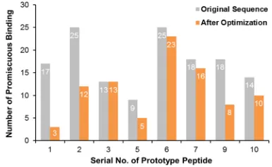
peptides (Table 1). While some peptides only interacted
with a few proteins (e.g., peptide 8 and peptide 5), some
other peptides could bind to up to 25 proteins
(e.g., peptide 6 and peptide 2). Interestingly, we noticed
that those peptides showing relatively higher binding
specificity tended to (1) have leucine (
with symbol L) as
the 5
thresidue of proline-rich motif (e.g., peptides No.
8, No. 5 and No. 4), and (2) have proline (with symbol
P) as the 6
thresidue of proline-rich motif (e.g., peptides
No. 8 and No. 4). The above patterns (Figure 1)
indi-cated that amino acid sequence of proline-rich motif
may be correlated to the degree of promiscuity.
Optimization of amino acid sequences and
improvement of binding specificity
In view of the correlation between the amino acid
sequence
in
proline-rich motif and the promiscuity of
domain-peptide binding, we hypothesized that certain
amino acid residues of the prototype peptides could
be purposely modified to improve
binding specificity
towards
c-Src protein. We tested two parallel schemes
of optimization. First, we substituted leucine for the 5
thresidue of proline-rich motif. Second, we replaced the
6
thresidue of proline-rich motif with proline. Except
for prototype peptides No. 8, No. 5 and No. 4 that
originally had leucine as the 5
thresidue of proline-rich
motif, the first scheme applied to the other 7 prototype
peptides. For 6 out of these 7 peptides, the number of
promiscuous interactions declined after optimization
(Figure 2, Table S1). The second scheme applied to 8
proto type peptides, except for peptides No. 8 and No. 4.
A decline in the number of promiscuous interactions
was observed in 7 out of these 8 peptides (Figure 3, Table
S2).
Such observations demonstrated that the 5
thand the
6
thresidues of proline-rich motif can largely
influence
on peptide-target binding. Therefore, we combined
the above two schemes and modified both the 5
thand
the 6
thresidues. A side-by-side comparison was made
between the original and the modified peptides (Figure 4,
Table S3). One-tailed paired t-test indicated that the
overall level of promiscuity of prototype peptides was
significantly reduced after modification (P-value =
0.0075). These findings came together to suggest that
c-Src binding specificity of peptide ligands can be greatly
oppor-tunity for sequence modification and rational design of
therapeutic peptides.
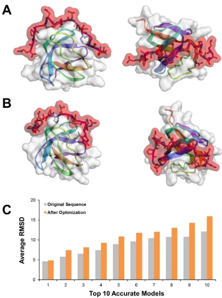
Validating modified peptides with domain-peptide
docking
Molecular docking approach has been widely adopted
to understand ligand-protein interaction.
27So the above
results were validated by reproducing conformation
of docked peptide in crystal structure of target SH3
domain. As a typical example, peptide No. 5 was originally
predicted to bind to the SH3 domain of hematopoietic
cell kinase (HCK). According to the calculation of
MoDPepInt, the probability of such binding significantly
decreased after modifying the 5
thand the 6
thresidues
of proline-rich motif. Therefore, the original and
modi-fied sequences of peptide No. 5, along with the 3D
structure of HCK SH3 domain (PDB ID: 4HCK), were
entered into the CABS-dock web server.
28CABS-dock
performed highly efficient and flexible docking simu
-lation to search for possible binding conformations.
The accuracy of docking models was measured by the
root-mean-square deviation (RMSD). The top 10 models
with the highest accuracy (i.e., the lowest RMSD value)
were selected as final results (see Materials and Methods).
It was shown that the original and modified peptides
had different binding positions and orientations on the
surface of HCK SH3 domain (Figure 5A and 5B). Regarding
the top 10 best models (Figure 5C), the modified peptide
exhibited significantly higher RMSD, namely lower
quality of binding, than the original peptide (P-value =
5.99×10
-5). These results supported the prediction that
modifying both the 5
thand the 6
thresidues of
proline-rich motif may prevent peptide No. 5 from binding to
HCK SH3 domain, thus lowering the promiscuity of
peptide No. 5.
Implication of the current results
Peptides are ideal molecules for drug development
because of low molecular weight and good cellular
uptake. Over these years, the application of peptides is
rapidly growing in a variety of therapeutic areas, with the
number of peptide drugs under clinical trials increasing
steadily. The number has climbed from 1.2 per year in
the 1970s to 16.8 per year in the 2000s.
6Currently, more
than 60 peptide drugs have been approved for marketing
and several hundreds of novel therapeutic peptides are
under preclinical or clinical development.
5The key
contributor to the success of these peptides is their
potent and specific, yet safe, modes of action.
As a class of promising anticancer agents, peptides bind
to key proteins in tumor cells with low toxicity to normal
tissues.
29This tumor-targeting ability of peptides is
Figure 1: The association between the 5th and the 6th
residues of proline-rich motif and binding specificity of
prototype peptides.
Figure 2: Most peptides, except for peptide No. 9, exhibited
better binding specificity after optimizing the 5th residue of
proline-rich motif.
Figure 3: Most peptides, except for peptide No. 6, exhibited
better binding specificity after optimizing the 6th residue of
proline-rich motif.
Figure 4: Most peptides, except for peptide No. 3, exhibited
better binding specificity after optimizing the 5th and the 6th
determined by their complementary binding to a variety
of key proteins in tumor cells. For instance, the positively
charged arginine and lysine in peptide can selectively
form hydrogen bonds with the negatively charged
components of target protein domains.
30Nonetheless,
these complementary properties may not always
guar-antee perfect specificity of peptide-target binding. For
example, SH3 domains have become a well-known and
promising anti-cancer target of peptide ligands with
proline-rich motifs. But SH3 domains are one of the
most abundant domain families encoded in eukaryotic
genomes. So far, at least 300 SH3 domains have been
identified in the human proteome.
31As a result of
highly conserved amino acid sequence and structure
of different SH3 domains, one proline-rich peptide can
be recognized by multiple proteins with SH3 domain.
The promiscuous nature of SH3 domains in binding to
proline-rich peptides may lead to unexpected adverse
reactions due to impact on various cell-signaling pathways
and biological functions.
32So it is an open challenge
in biotechnology to design peptide ligands with a high
specificity of binding to the SH3 domain of designated
target (e.g., c-Src) and without interacting with other
proteins.
In the present study on
c-Src protein, we demonstrated
that subtle alterations in the amino acid sequence could
significantly change the specificity of binding to the
target SH3 domain. We primarily searched MINT
data-base for a set of prototype peptides, which have been
experimentally validated for binding to the SH3 domain
of c-Src protein. Relying on the SH3PepInt tool, we
predicted the interaction between the peptides and various
SH3 domains. Then, by comparing the amino acid
sequence of prototype peptides with relatively high and
low specificity of binding to c-Src protein, we found
that leucine as the 5
thresidue and proline as the 6
thresidue of proline-rich motif could render prototype
peptide a reduced promiscuity. In the last step, we
purposely modified the 5
thand the 6
thresidues of relevant
peptides, which led to significantly better specificity of
peptide-target binding. The above results showed that
promiscuous binding of peptide molecule can be
effec-tively ameliorated by rational design.
Based on various public crystallographic data,
33-38the SH3 domain has been thoroughly researched and
broadly recognized as one of the best available systems
for the examination of ligand-protein interactions. For
example, Larson
et al
. constructed a diverse alignment
of SH3 domain sequences.
39By analyzing conservative
structural features within this alignment, several
posi-tions in the domain were identified for mediating
the peptide-binding function. The existence of such
key positions implied that the recognition of peptide
ligands might be systematically explained,
40,41which
effectively facilitated experiments of rational ligand
designing. Alexandropoulos
et al
. found specific
proline-rich sequences prone to bind to Fyn, Lyn,
and Hck SH3 domains, respectively.
20Pisabarro
et al
.
designed mutations in peptide, so that the affinity for
Abl SH3 domain was selectively increased by 20-fold.
42And Ferguson
et al
. used phage display for ligand
optimi-zation and obtained a peptide 1000-fold increased affinity
for the SEM-5 SH3 domain.
43Here we provided a novel
clue as to increasing the specificity of binding to SH3
domain. The current results will inspire more
subse-quent work on optimization of amino acid sequence, so
as to improve the safety of therapeutic peptides.
Despite of useful information provided by the present
study, more efforts are required to address some
limita-tions of the current results. First, the basis of this study
was the prototype peptides retrieved from MINT
data-base, with which we found the important role played
by the 5
thand the 6
thresidues of proline-rich motif.
However, the number of available prototype remained
Figure 5: Domain-peptide binding validated by molecular
docking. (A) The best fitted binding model of original peptide
No. 5 and HCK SH3 domain. (B) The best fitted binding model
of modified peptide No. 5 and HCK SH3 domain. (C) The
docking RMSD values of original and modified sequences of
relatively low for higher power of statistical tests. More
work, such as literature searching and text mining, will
be required to collect more information about
proto-type peptides proved to bind to the SH3 domain of
human c-Src protein. Second, with a larger set of
proto-type peptides, more patterns of amino acid sequences
related to peptide-target binding specificity can be
explored. Apart from the 5
thand the 6
thresidue, other
residues in or flanking the proline-rich motif should
also be investigated using our method. Third, the current
results of peptide-target binding were mainly based on
in silico prediction, which often led to alternate results in
reality. An inconsistent track record of docking-based
ligand discovery raised the concern about the reliability.
For instance, it was reported that fewer than 20% of
docking screens were eventually supported by crystal
structure identified with experiments.
44In particular,
those small molecules, such as peptides composed of a
few amino acids, are found to be docked less reliably.
45Therefore, besides calculating the binding orientations
and free energies. the optimized peptides need to be
artificially synthesized and experimentally assayed for
binding affinity against targets of interest.
46CONCLUSION
Taken together, by analyzing the promiscuity of
peptide-target binding, we corroborated the ability of the SH3
domain of c-Src protein to discern subtle differences in
the amino acid sequence of peptide ligands. Based on
that, we virtually optimized the proline-rich motifs of
relevant peptides and improved the binding specificity.
Further computational and experimental efforts will be
required to validate and expand current results, which
can be applied to the rational design of peptide-based
anticancer drugs.
ACKNOWLEDGEMENT
Front and Applied Fundamental Research Plan of
Chongqing Science & Technology Commission (No.
cstc2014jcyjA10104); the Program for Innovative
Research Team in Chongqing University of Education
(No. KYC-cxtd03-20141002) and the Project Foun
-dation of Chongqing University of Education (No
16kjpt08) supported this work.
REFERENCES
1. Jemal A, Bray F, Center MM, Ferlay J, Ward E, Forman D. Global cancer statistics. CA: a cancer journal for clinicians. 2011;61:69-90. https://doi. org/10.3322/caac.20107.
2. Vogelstein B, Kinzler KW. Cancer genes and the pathways they control. Nature medicine. 2004;10:789-99.https://doi.org/10.1038/nm1087 PMid:15286780.
3. Narayan V, Vaughn D. Pharmacokinetic and toxicity considerations in the use of neoadjuvant chemotherapy for bladder cancer. Expert opinion on drug metabolism & toxicology. 2015;11:731-42.https://doi.org/10.1517/17425255. 2015.1005600 PMid:25604887.
4. Huang W, Seo J, Willingham SB, Czyzewski AM, Gonzalgo ML, Weissman IL, et al. Learning from host-defense peptides: cationic, amphipathic peptoids with potent anticancer activity. PloS one. 2014;9:e90397. https://doi. org/10.1371/journal.pone.0090397 PMid:24587350 PMCid:PMC3938723. 5. Fosgerau K, Hoffmann T. Peptide therapeutics: current status and future
directions. Drug discovery today. 2015;20:122-8. https://doi.org/10.1016/j. drudis.2014.10.003 PMid:25450771.
6. Borghouts C, Kunz C, Groner B. Current strategies for the development of peptide-based anti-cancer therapeutics. Journal of peptide science : an
official publication of the European Peptide Society. 2005;11:713-26. https://
doi.org/10.1002/psc.717 PMid:16138387.
7. Boohaker RJ, Lee MW, Vishnubhotla P, Perez JM, Khaled AR. The use of therapeutic peptides to target and to kill cancer cells. Current medicinal chemistry. 2012;19:3794-804.https://doi.org/10.2174/092986712801661004 PMid:22725698 PMCid:PMC4537071.
8. Okamoto W, Okamoto I, Yoshida T, Okamoto K, Takezawa K, Hatashita E, et al. Identification of c-Src as a potential therapeutic target for gastric cancer
and of MET activation as a cause of resistance to c-Src inhibition. Molecular cancer therapeutics. 2010;9:1188-97. https://doi.org/10.1158/1535-7163. MCT-10-0002 PMid:20406949.
9. Kim LC, Song L, Haura EB. Src kinases as therapeutic targets for cancer. Nature reviews Clinical oncology. 2009;6:587-95. https://doi.org/10.1038/ nrclinonc.2009.129 PMid:19787002.
10. Yeatman TJ. A renaissance for SRC. Nature reviews Cancer. 2004;4:470-80. https://doi.org/10.1038/nrc1366 PMid:15170449.
11. Irby RB, Yeatman TJ. Role of Src expression and activation in human cancer. Oncogene. 2000;19:5636-42.https://doi.org/10.1038/sj.onc.1203912 PMid:11114744.
12. Johnson FM, Saigal B, Talpaz M, Donato NJ. Dasatinib (BMS-354825) tyrosine kinase inhibitor suppresses invasion and induces cell cycle arrest and apoptosis of head and neck squamous cell carcinoma and non-small
cell lung cancer cells. Clinical cancer research : an official journal of the
American Association for Cancer Research. 2005;11:6924-32. https://doi. org/10.1158/1078-0432.CCR-05-0757 PMid:16203784.
13. Nautiyal J, Majumder P, Patel BB, Lee FY, Majumdar AP. Src inhibitor dasatinib inhibits growth of breast cancer cells by modulating EGFR signaling. Cancer letters. 2009;283:143-51.https://doi.org/10.1016/j. canlet.2009.03.035 PMid:19398150.
14. Conchon M, Freitas CM, Rego MA, Braga Junior JW. Dasatinib - clinical trials and management of adverse events in imatinib resistant/intolerant chronic myeloid leukemia. Revista brasileira de hematologia e hemoterapia. 2011;33:131-9. https://doi.org/10.5581/1516-8484.20110034 PMid:23284261 PMCid:PMC3520638.
15. Brunner AM, Costa DB, Heist RS, Garcia E, Lindeman NI, Sholl LM, et al. Treatment-related toxicities in a phase II trial of dasatinib in patients with squamous cell carcinoma of the lung. Journal of thoracic oncology:
official publication of the International Association for the Study of Lung
Cancer. 2013;8:1434-7.https://doi.org/10.1097/JTO.0b013e3182a47162 PMid:24128713 PMCid:PMC3801424.
16. Sen B, Johnson FM. Regulation of SRC family kinases in human cancers. Journal of signal transduction. 2011;2011:865819. https://doi. org/10.1155/2011/865819PMid:21776389 PMCid:PMC3135246.
17. Kaneko T, Li L, Li SS. The SH3 domain--a family of versatile peptide- and protein-recognition module. Frontiers in bioscience : a journal and virtual library. 2008;13:4938-52. https://doi.org/10.2741/3053 PMid:18508559. 18. Cesareni G, Panni S, Nardelli G, Castagnoli L. Can we infer peptide recognition
specificity mediated by SH3 domains? FEBS letters. 2002;513:38-44. https://
doi.org/10.1016/S0014-5793(01)03307-5.
19. Feng S, Chen JK, Yu H, Simon JA, Schreiber SL. Two binding orientations for peptides to the Src SH3 domain: development of a general model for SH3-ligand interactions. Science. 1994;266:1241-7.https://doi.org/10.1126/ science.7526465 PMid:7526465.
20. Alexandropoulos K, Cheng G, Baltimore D. Proline-rich sequences that bind
National Academy of Sciences of the United States of America. 1995;92:3110-4. https://doi.org/10.1073/pnas.92.8.3110 PMid:7536925 PMCid:PMC42114. 21. Vidal M, Gigoux V, Garbay C. SH2 and SH3 domains as targets for
anti-proliferative agents. Critical reviews in oncology/hematology. 2001;40:175-86. https://doi.org/10.1016/S1040-8428(01)00142-1.
22. Li SS. Specificity and versatility of SH3 and other proline-recognition
domains: structural basis and implications for cellular signal transduction. The Biochemical journal. 2005;390:641-53.https://doi.org/10.1042/BJ20050411 PMid:16134966 PMCid:PMC1199657.
23. Geng L, Wang Z, Yang X, Li D, Lian W, Xiang Z, et al. Structure-based
Design of Peptides with High Affinity and Specificity to HER2 Positive
Tumors. Theranostics. 2015;5:1154-65.https://doi.org/10.7150/thno.12398 PMid:26284145 PMCid:PMC4533098.
24. Licata L, Briganti L, Peluso D, Perfetto L, Iannuccelli M, Galeota E, et al. MINT, the molecular interaction database: 2012 update. Nucleic acids research. 2012;40:D857-61. https://doi.org/10.1093/nar/gkr930 PMid:22096227 PMCid:PMC3244991.
25. Kundu K, Mann M, Costa F, Backofen R. MoDPepInt: an interactive web server for prediction of modular domain-peptide interactions. Bioinformatics. 2014;30:2668-9. https://doi.org/10.1093/bioinformatics/btu350 PMid:24872426 PMCid:PMC4155253.
26. Kundu K, Costa F, Backofen R. A graph kernel approach for alignment-free domain-peptide interaction prediction with an application to human SH3 domains. Bioinformatics. 2013;29:i335-43.https://doi.org/10.1093/ bioinformatics/btt220 PMid:23813002 PMCid:PMC3694653.
27. Rohit H M, Ashok D T, Vijaykumar R, Kashniyal K. Molecular Docking Study of Cassia tora, Brassica campestris and Calotropis procera as Acetylcholinesterase Inhibitor. Indian Journal of Pharmaceutical Education and Research. 2016;50:116-22. https://doi.org/10.5530/ijper.50.1.15. 28. Kurcinski M, Jamroz M, Blaszczyk M, Kolinski A, Kmiecik S. CABS-dock web
server for the flexible docking of peptides to proteins without prior knowledge
of the binding site. Nucleic acids research. 2015;43:W419-24. https://doi. org/10.1093/nar/gkv456 PMid:25943545 PMCid:PMC4489223.
29. Wu D, Gao Y, Qi Y, Chen L, Ma Y, Li Y. Peptide-based cancer therapy: opportunity and challenge. Cancer letters. 2014;351:13-22. https://doi. org/10.1016/j.canlet.2014.05.002 PMid:24836189.
30. Farkhani SM, Valizadeh A, Karami H, Mohammadi S, Sohrabi N, Badrzadeh F.
Cell penetrating peptides: efficient vectors for delivery of nanoparticles,
nanocarriers, therapeutic and diagnostic molecules. Peptides. 2014;57:78-94. https://doi.org/10.1016/j.peptides.2014.04.015 PMid:24795041.
31. Karkkainen S, Hiipakka M, Wang JH, Kleino I, Vaha-Jaakkola M, Renkema GH, et al. Identification of preferred protein interactions by phage-display of
the human Src homology-3 proteome. EMBO reports. 2006;7:186-91. https:// doi.org/10.1038/sj.embor.7400596 PMid:16374509 PMCid:PMC1369250. 32. Agrawal V, Kishan KV. Promiscuous binding nature of SH3 domains to
their target proteins. Protein and peptide letters. 2002;9:185-93. https://doi. org/10.2174/0929866023408760 PMid:12144515.
33. Camara-Artigas A, Ortiz-Salmeron E, Andujar-Sanchez M, Bacarizo J, Martin-Garcia JM. The role of water molecules in the binding of class I and II peptides to the SH3 domain of the Fyn tyrosine kinase. Acta crystallographica Section F, Structural biology communications. 2016;72:707-12.https://doi. org/10.1107/S2053230X16012310. PMid:27599862.
34. Bhatt VS, Zeng D, Krieger I, Sacchettini JC, Cho JH. Binding Mechanism of the N-Terminal SH3 Domain of CrkII and Proline-Rich Motifs in cAbl. Biophysical journal. 2016;110:2630-41.https://doi.org/10.1016/j. bpj.2016.05.008 PMid:27332121.
35. Bacarizo J, Martinez-Rodriguez S, Camara-Artigas A. Structure of the c-Src-SH3 domain in complex with a proline-rich motif of NS5A protein from the hepatitis C virus. Journal of structural biology. 2015;189:67-72.https://doi. org/10.1016/j.jsb.2014.11.004 PMid:25447263.
36. Bacarizo J, Martinez-Rodriguez S, Martin-Garcia JM, Andujar-Sanchez M, Ortiz-Salmeron E, Neira JL, et al. Electrostatic effects in the folding of the SH3 domain of the c-Src tyrosine kinase: pH-dependence in 3D-domain swapping and amyloid formation. PloS one. 2014;9:e113224.https://doi. org/10.1371/journal.pone.0113224 PMid:25490095 PMCid:PMC4260792. 37. Tzeng SR, Lou YC, Pai MT, Jain ML, Cheng JW. Solution structure of
the human BTK SH3 domain complexed with a proline-rich peptide from p120cbl. Journal of biomolecular NMR. 2000;16:303-12.https://doi. org/10.1023/A:1008376624863 PMid:10826882.
38. Horita DA, Baldisseri DM, Zhang W, Altieri AS, Smithgall TE, Gmeiner WH,
et al. Solution structure of the human Hck SH3 domain and identification of
its ligand binding site. Journal of molecular biology. 1998;278:253-65. https:// doi.org/10.1006/jmbi.1998.1690 PMid:9571048.
39. Larson SM, Davidson AR. The identification of conserved interactions within
the SH3 domain by alignment of sequences and structures. Protein science: a publication of the Protein Society. 2000;9:2170-80.https://doi.org/10.1110/ ps.9.11.2170 PMid:11152127 PMCid:PMC2144485.
40. Rickles RJ, Botfield MC, Zhou XM, Henry PA, Brugge JS, Zoller MJ. Phage
display selection of ligand residues important for Src homology 3 domain
binding specificity. Proceedings of the National Academy of Sciences of
the United States of America. 1995;92:10909-13. https://doi.org/10.1073/ pnas.92.24.10909 PMid:7479908 PMCid:PMC40540.
41. Lee CH, Leung B, Lemmon MA, Zheng J, Cowburn D, Kuriyan J, et al. A single
amino acid in the SH3 domain of Hck determines its high affinity and specificity
in binding to HIV-1 Nef protein. The EMBO journal. 1995;14:5006-15. PMid:7588629 PMCid:PMC394604.
42. Pisabarro MT, Serrano L. Rational design of specific high-affinity peptide
ligands for the Abl-SH3 domain. Biochemistry. 1996;35:10634-40. https://doi. org/10.1021/bi960203t PMid:8718852.
43. Ferguson MR, Fan X, Mukherjee M, Luo J, Khan R, Ferreon JC, et al. Directed discovery of bivalent peptide ligands to an SH3 domain. Protein science : a publication of the Protein Society. 2004;13:626-32.https://doi. org/10.1110/ps.03470504 PMid:14978303 PMCid:PMC2286729.
44. Kolb P, Irwin JJ. Docking screens: right for the right reasons?
Current topics in medicinal chemistry. 2009;9:755-70. https://doi. org/10.2174/156802609789207091 PMid:19754393 PMCid:PMC3383315. 45. Tovchigrechko A, Wells CA, Vakser IA. Docking of protein models. Protein
science : a publication of the Protein Society. 2002;11:1888-96. https://doi. org/10.1110/ps.4730102 PMid:12142443 PMCid:PMC2373684.